the endocrine system
advertisement
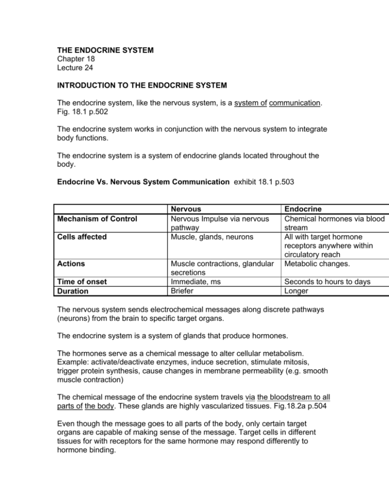
THE ENDOCRINE SYSTEM Chapter 18 Lecture 24 INTRODUCTION TO THE ENDOCRINE SYSTEM The endocrine system, like the nervous system, is a system of communication. Fig. 18.1 p.502 The endocrine system works in conjunction with the nervous system to integrate body functions. The endocrine system is a system of endocrine glands located throughout the body. Endocrine Vs. Nervous System Communication exhibit 18.1 p.503 Mechanism of Control Cells affected Actions Time of onset Duration Nervous Nervous Impulse via nervous pathway Muscle, glands, neurons Muscle contractions, glandular secretions Immediate, ms Briefer Endocrine Chemical hormones via blood stream All with target hormone receptors anywhere within circulatory reach Metabolic changes. Seconds to hours to days Longer The nervous system sends electrochemical messages along discrete pathways (neurons) from the brain to specific target organs. The endocrine system is a system of glands that produce hormones. The hormones serve as a chemical message to alter cellular metabolism. Example: activate/deactivate enzymes, induce secretion, stimulate mitosis, trigger protein synthesis, cause changes in membrane permeability (e.g. smooth muscle contraction) The chemical message of the endocrine system travels via the bloodstream to all parts of the body. These glands are highly vascularized tissues. Fig.18.2a p.504 Even though the message goes to all parts of the body, only certain target organs are capable of making sense of the message. Target cells in different tissues for with receptors for the same hormone may respond differently to hormone binding. We will not discuss local hormones- autocrines, paracrines. Actions of Hormones Specificity Hormones only act on specific target cells that have receptors on their membrane or inside the cell. Generally a target cell will have 2000-100,000 receptors/cell. Target tissues can regulate their responsiveness to their specific hormone by increasing the number of receptors (up-regulation) or decreasing the number of receptors (down-regulation). Blocking of these receptors will prevent the action of the hormone. Example: box p.504 RU486 (mifepristone) binds to progesterone receptors and prevents the effects of the hormone. If given to a pregnant women, the uterus cannot maintain the proper environment for embryonic development. Actions There are 2 main chemical classes of hormones: Exhibit 18.2 p.505 1. Amino acid based (water soluble) . Simple amino acids to peptides to proteins. Most hormones are this type. 2. Steroid based (lipid soluble). Synthesized from cholesterol. Gonadal and adrenocortical hormones. Each type has its own mechanism of triggering cell metabolism changes. 1. Water soluble hormones cannot penetrate the lipid membrane, so it must bind to a receptor in the membrane (first message). This triggers a secondary messenger in the cell to deliver the message and change cellular activities. Fig.18.4 p.507. Example: FSH, LH 2. Lipid soluble hormones can and do diffuse through the lipid membrane and enter the nucleus of the target cell where it binds to its receptor, forming a hormone-receptor complex. This complex then turns on or off a gene(s) to produce or inhibit protein synthesis (usually an enzyme). Fig. 18.3 p.506 Example: Testosterone In either case, the hormone message is greatly amplified by the target cell, where 1 hormone binding to its receptor can produce changes in 100’s or 1000’s of substrate molecules. Enzyme-Substrate Model The endocrine system message can be explained by something called the enzyme-substrate model. A B C D HORMONES (ENZYMES) MANY PRODUCED BY THE BODY B B TARGET ORGAN (SUBSTRATE) CAN ONLY UNDERSTAND THE MESSAGE OF HORMONE A In the enzyme-substrate model many hormones are secreted by the body and sent throughout the body via the blood. The target organ or target tissue can only understand the message of one of the hormones, in this case, hormone A. Endocrine Vs. Exocrine Glands There are actually 2 kinds of glands in the body. 1. Endocrine glands- are ductless and release hormones directly into the blood. Examples: pituitary, thyroid, parathyroid, adrenal, pineal, and thymus glands. 2. Exocrine glands- are ducted glands that secrete their products into body cavities or onto its surface. Examples: sweat glands, mammary glands, and pancreas (in part) to name a few. The ovaries, testes, hypothalamus and pancreas also have endocrine function and produce important hormones. The placenta serves as a temporary endocrine gland. Pituitary Gland Fig. 18.5 p. 510 Pituitary gland- located at the base of the brain is also known as the master gland as it sends out 8 different hormones that affect many other endocrine glands and basic body functions. The pituitary gland is also known as the hypophysis. Of the 9 hormones secreted by the pituitary gland: 7 are secreted by the anterior pituitary (adenohypophysis) 2 hormones are secreted by the posterior pituitary (neurohypophysis) through its connection with the hypothalamus. The posterior pituitary hormones are actually produced in the hypothalamus, stored in and secreted by the posterior pituitary. The hypothalamus is considered part of both the nervous system and the endocrine system. Anterior Pituitary (adenohypophysis) Hormones Fig. 18.6 p511 1. Growth Hormone (GH) - stimulates growth in bone and muscle by: a. b. c. increasing protein synthesis in cells increasing blood sugar levels increasing availability of fat as an energy source for growth Hyposecretion (undersecretion) : causes pituitary dwarfism. Slow bone growth. Hypersecretion (oversecretion): causes pituitary giantism, acromegaly in adulthood Exhibit 18.3 p.516 Fig. 18.9 p514 2. Thyroid-Stimulating Hormone (TSH) - acts upon the thyroid gland stimulating it to produce thyroxine. 3. Adrenocorticotrophic Hormone (ACTH) - stimulates the adrenal cortex to release various hormones (glucocorticoids) 4. Follicle-Stimulating Hormone (FSH) - acts upon the ovaries of the female to stimulate development of the ova and to cause to ovaries to produce estrogen. In males FSH serves to stimulate the testes to produce spermatozoa. FSH is classified as a gonadotrophic hormone as it acts upon the gonads (ovaries and testes). 5. Leutinizing Hormone (LH) in females and Interstitial Cell Stimulating Hormone (ICSH) in males are the same hormone. In females it promotes ovulation and stimulates the ovaries to produce progesterone. In males it stimulates the interstitial cells of the testes to produce testosterone. LH, like FSH, is a gonadotrophic hormone. 6. Prolactin (Lactogenic Hormone) - initiates and maintains milk secretion by the mammary glands. 7. Meloncyte-stimulating hormone- increases skin pigmentation. Posterior Pituitary (neurohypophysis) Hormones Fig. 18.10 p.517 exhibit 18.4 p.520 1. Antidiuretic Hormone (ADH) or Vasopressin - acts on the kidneys to cause increased water reabsorption. Hyposecretion: causes diabetes insipidus which causes individual to secrete large amounts of urine. Hypersecretion: causes excessive water reabsorption and retention. 2. Oxytocin - stimulates uterine contractions during labor and intercourse. The contractions aid sperm travel through the reproductive tract to improve chances for fertilization. Oxytocin also serves to initiate milk ejection by contracting smooth muscle in mammary gland. The pituitary gland is totally regulated by the hypothalamus which is part of the central nervous system. The hypothalamus is able to monitor levels of hormones and substances in the blood to control the pituitary by a mechanism called negative feedback. Through negative feedback the hypothalamus will alter activity in the different regions of the pituitary gland responsible for each of the hormones. Example: fig. 18.12 p.519 ADH secretion and control by negative feedback. Thyroid Gland Exhibit 18.5 p.525 Thyroid gland - located in the neck and is controlled by the pituitary gland. The thyroid gland secretes 2 hormones: 1. Thyroxine (T3 and T4)- a mixture of tetraiodo- and triiodo-thyronine, together called thyroid hormones. 2. Calcitonin Thyroxine - serves to regulate basal metabolic rate through increased cellular respiration rate by: 1. increasing amount of glucose absorbed by digestive tract 2. increasing fat mobilization 3. increasing cellular protein synthesis thereby increasing heat production fig. 18.16 p.524 Hypersecretion (hyperthyroid or Graves’ disease (most common form)) leads to increased BMR, heart action, and blood pressure. Often recognized by a bulging of the eyes due to swelling (edema) behind them and weight loss. Hyposecretion (hypothyroid) during the growing years leads to cretinism (dwarfism and retardation). Adults = myxedema- edema, slow heart, low body temp., sensitivity to cold. Goiter is an enlarged thyroid gland and is a symptom of a disease where the thyroid is over stimulated. It can be caused by a lack of iodine salts in the diet. Iodine is added to table salt to prevent this condition. Iodized salts are important in the synthesis of thyroxine. Goiter can be recognized by the considerable swelling that occurs in the thyroid gland and neck. Calcitonin (CT)- serves to lower blood calcium and phosphate levels by moving the calcium into bone tissue. Parathyroid Glands Exhibit 18.6 p.528 The 4 small parathyroid glands are located on the posterior side of the thyroid gland. Parathyroid Hormone (PTH) - serves with calcitonin in controlling blood calcium levels. Parathyroid hormone serves to increase blood calcium levels by removing calcium from bone and is very important in the breakdown and restructuring of bone. Adrenal Glands Exhibit 18.7 p.534 Each adrenal gland is found on top of each kidney and consists of 2 major regions: Fig. 18.19a,b p.528-9 1. Adrenal cortex 2. Adrenal medulla Cortex – Aldosterone, cortisol, androgens Medullaepinephrine/norepinephrine (Increased Sympathetic Effects ) The adrenal medulla is controlled by the sympathetic division of the autonomic nervous system and secretes epinephrine and norepinephrine. The adrenal cortex secretes aldosterone (a mineralcorticoid), cortisol (cortisone)(a glucocorticoid), and small amounts of sex hormones (androgens). Medulla hormones Epinephrine /Norepinephrine- increases blood pressure(increase heart rate and constrict blood vessels) and respiratory rate, dilates bronchioles, increase blood glucose, and stimulates metabolic activity. Interestingly, this hormone is inactivated within three minutes of release and represents a short term stress response. Cortex hormones Aldosterone (a mineralcorticoid)- serves to increase reabsorption of Na+ and water, decrease K+. Cortisol (a glucocorticoid)- helps regulate metabolism and aids the body in dealing with stress. Stimulates gluconeogenesis, lipolysis, and protein catabolism. Decreases vasodilatation and edema associated with inflammation (anti-inflammatory), depresses immune response. Hyposecretion of cortical hormones leads to Addison’s disease which is characterized by hypoglycemia, dehydration, weakness, weight loss, and anemia. Hypersecretion results in Cushing’s disease which includes hyperglycemia with a redistribution of fat. Fig. 18.22 p.532 Androgens- Inconsequential to males as their testes produce great amounts. In females to sex drive and libido. Pancreas Fig. 18.23 p.535 exhibit18.8 p.538 Pancreas- Located adjacent to the duodenum, the first portion of the small intestine. The pancreas is both an endocrine and exocrine organ. The hormones secreted by the pancreas are: Insulin and glucagon which are important in controlling blood sugar levels. Glucagon - increases blood sugar levels by stimulating the breakdown of glycogen in hepatocytes (storage form in liver) to glucose in the blood. Insulin - serves to decrease blood sugar levels by moving sugar from the blood to the cells throughout the body that need it as an energy source (especially skeletal muscle). Hyposecretion causes sugar diabetes (Diabetes mellitus). Hallmarks are the 3 P’s: polyuria (excessive urine production), polydipsia (excessive thirst), polyphagia (excessive eating). Acidosis occurs as ketones (organic acids produced as byproduct of fatty acid catabolism) accumulate due to use of lipids for ATP production which can result in death. Ovary Exhibit 18.9 p.539 Ovaries- located within the pelvic cavity and produce 2 gonadal hormones (hormones produced by the gonads): 1. Estrogen, and 2. Progesterone. Estrogen and Progesterone serve to promote the development and maintenance of female sex reproductive structures, changes in the breasts and size of the pelvis to prepare the female for child birth, and other secondary characteristics (adipose distribution, hair distribution pattern). The gonadal hormones work in conjunction with the gonadotrophic hormones of the pituitary to regulate the female menstrual cycle, maintain pregnancy, and ready the mammary glands for lactation. Testes Exhibit 18.9 p.539 Testes- Located outside the body cavity within the scrotum. They produce the gonadal hormone testosterone. Testosterone - promotes the development and maintenance of male sexual characteristics. These include increased facial and body hair, muscular build, size of the larynx that lowers the voice, and sex drive (libido). Pineal Gland The pineal gland also known as the pineal body or epithalamus is a portion of the thalamus of the brain. The pineal gland secretes a hormone called melatonin. Melatonin - is important in the timing of the body’s biological rhythms . It is governed by a diurnal (daily) dark-light cycle. It is involved in the timing of sexual maturation, the menstrual cycle, and daily activity patterns. Secretion stimulates sleepiness. Seasonal Affective Disorder (SAD)- a type of depression arises during the winter months when day length is short and an overproduction of melatonin. Bright light therapy may be useful. Small oral doses 2 hours before bed, may be useful for shift workers to synchronize their ‘sleep’ clock. Thymus Gland Thymus gland- Located in the neck, inferior to the thyroid gland. Interestingly, the thymus reaches maximum size at puberty, then it begins to atrophy. The thymus produces hormones that promotes proliferation and maturation of T cells (a type of white blood cell) against invaders (foreign antigens) of the body. We will discuss immunity when we discuss the lymphatic system. Other Hormones Human Chorionic Gonadotropin (hCG)- Stimulates the corpus luteum in the ovary to continue production of estrogens and progesterone to maintain pregnancy. Can be detected about a week following conception. Produced in the placenta. Erythropoietin (EPO)- Increases rate of red cell production. Produced in the kidney. How is the Secretion of Hormones Controlled? 1. Hormone Feedback Mechanisms - that serve to monitor hormone levels in the blood. Gonadal and gonadotrophic hormones. Usually a negative feedback. 2. Other Humoral Feedback Mechanisms – chemicals other than hormones are monitored in the blood, glucose or calcium for example. 3. Direct Nervous Innervation - through nervous system control. Example: adrenal medulla and epinephrine secretion. 4. Indirect Nervous Control - the nervous system controls activity of the gland through the production of neurohumors (neurotransmitters) that act on the gland to cause it to produce hormones. Example: the influence of the hypothalamus by hypothalamic neurosecretory cells secreting releasing and inhibitory hormones in the hypophyseal portal veins to the anterior pituitary. The hypothalamus is of extreme importance in controlling the activity of glands and their production of hormones. This is accomplished via mechanisms 1, 2, and 4 above. Remember that the hypothalamus is an autonomic center concerned with homeostatic control many body function and well as an integrating center for biological rhythms and emotions. So, you can now see why a single external stimulus (e.g. acute blood loss) can be followed by body wide neuroendocrine adjustments. See fig. 18.26 p.544 for an example of the General Adaptation Syndrome (GAS), a wide ranging set of body responses orchestrated by the hypothalamus.
