ORDERED MECHANISM OF BINDING OF PHOSPHORYLASE
advertisement
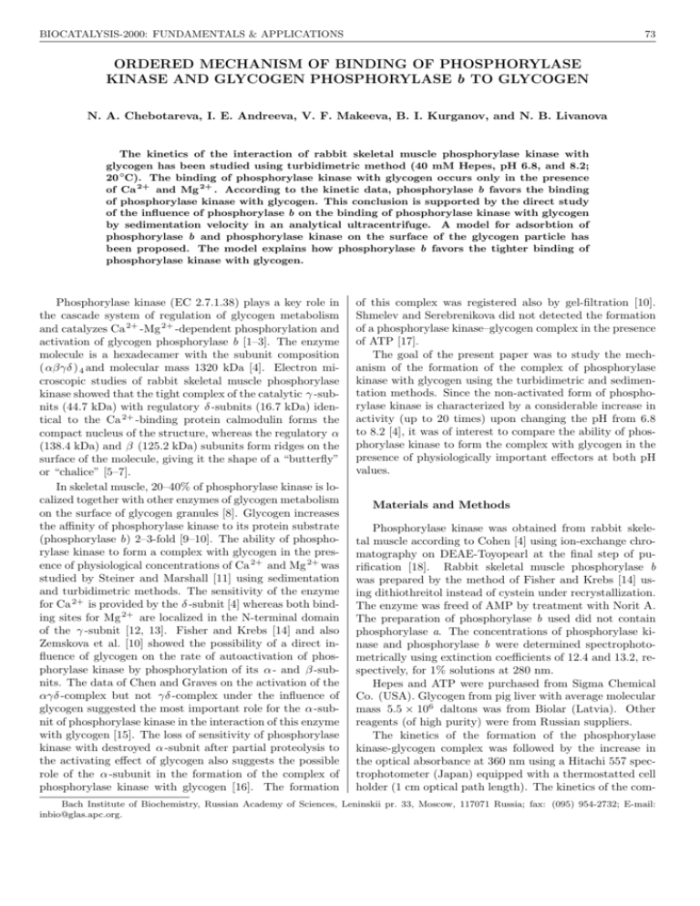
BIOCATALYSIS-2000: FUNDAMENTALS & APPLICATIONS 73 ORDERED MECHANISM OF BINDING OF PHOSPHORYLASE KINASE AND GLYCOGEN PHOSPHORYLASE b TO GLYCOGEN N. A. Chebotareva, I. E. Andreeva, V. F. Makeeva, B. I. Kurganov, and N. B. Livanova The kinetics of the interaction of rabbit skeletal muscle phosphorylase kinase with glycogen has been studied using turbidimetric method (40 mM Hepes, pH 6.8, and 8.2; 20 ◦C). The binding of phosphorylase kinase with glycogen occurs only in the presence of Ca 2+ and Mg 2+ . According to the kinetic data, phosphorylase b favors the binding of phosphorylase kinase with glycogen. This conclusion is supported by the direct study of the influence of phosphorylase b on the binding of phosphorylase kinase with glycogen by sedimentation velocity in an analytical ultracentrifuge. A model for adsorbtion of phosphorylase b and phosphorylase kinase on the surface of the glycogen particle has been proposed. The model explains how phosphorylase b favors the tighter binding of phosphorylase kinase with glycogen. Phosphorylase kinase (EC 2.7.1.38) plays a key role in the cascade system of regulation of glycogen metabolism and catalyzes Ca 2+ -Mg 2+ -dependent phosphorylation and activation of glycogen phosphorylase b [1–3]. The enzyme molecule is a hexadecamer with the subunit composition (αβγδ ) 4 and molecular mass 1320 kDa [4]. Electron microscopic studies of rabbit skeletal muscle phosphorylase kinase showed that the tight complex of the catalytic γ -subnits (44.7 kDa) with regulatory δ -subnits (16.7 kDa) identical to the Ca 2+ -binding protein calmodulin forms the compact nucleus of the structure, whereas the regulatory α (138.4 kDa) and β (125.2 kDa) subunits form ridges on the surface of the molecule, giving it the shape of a “butterfly” or “chalice” [5–7]. In skeletal muscle, 20–40% of phosphorylase kinase is localized together with other enzymes of glycogen metabolism on the surface of glycogen granules [8]. Glycogen increases the affinity of phosphorylase kinase to its protein substrate (phosphorylase b) 2–3-fold [9–10]. The ability of phosphorylase kinase to form a complex with glycogen in the presence of physiological concentrations of Ca 2+ and Mg 2+ was studied by Steiner and Marshall [11] using sedimentation and turbidimetric methods. The sensitivity of the enzyme for Ca 2+ is provided by the δ -subnit [4] whereas both binding sites for Mg 2+ are localized in the N-terminal domain of the γ -subnit [12, 13]. Fisher and Krebs [14] and also Zemskova et al. [10] showed the possibility of a direct influence of glycogen on the rate of autoactivation of phosphorylase kinase by phosphorylation of its α - and β -subnits. The data of Chen and Graves on the activation of the αγδ -complex but not γδ -complex under the influence of glycogen suggested the most important role for the α -subnit of phosphorylase kinase in the interaction of this enzyme with glycogen [15]. The loss of sensitivity of phosphorylase kinase with destroyed α -subnit after partial proteolysis to the activating effect of glycogen also suggests the possible role of the α -subunit in the formation of the complex of phosphorylase kinase with glycogen [16]. The formation of this complex was registered also by gel-filtration [10]. Shmelev and Serebrenikova did not detected the formation of a phosphorylase kinase–glycogen complex in the presence of ATP [17]. The goal of the present paper was to study the mechanism of the formation of the complex of phosphorylase kinase with glycogen using the turbidimetric and sedimentation methods. Since the non-activated form of phosphorylase kinase is characterized by a considerable increase in activity (up to 20 times) upon changing the pH from 6.8 to 8.2 [4], it was of interest to compare the ability of phosphorylase kinase to form the complex with glycogen in the presence of physiologically important effectors at both pH values. Materials and Methods Phosphorylase kinase was obtained from rabbit skeletal muscle according to Cohen [4] using ion-exchange chromatography on DEAE-Toyopearl at the final step of purification [18]. Rabbit skeletal muscle phosphorylase b was prepared by the method of Fisher and Krebs [14] using dithiothreitol instead of cystein under recrystallization. The enzyme was freed of AMP by treatment with Norit A. The preparation of phosphorylase b used did not contain phosphorylase a. The concentrations of phosphorylase kinase and phosphorylase b were determined spectrophotometrically using extinction coefficients of 12.4 and 13.2, respectively, for 1% solutions at 280 nm. Hepes and ATP were purchased from Sigma Chemical Co. (USA). Glycogen from pig liver with average molecular mass 5.5 × 10⁶ daltons was from Biolar (Latvia). Other reagents (of high purity) were from Russian suppliers. The kinetics of the formation of the phosphorylase kinase-glycogen complex was followed by the increase in the optical absorbance at 360 nm using a Hitachi 557 spectrophotometer (Japan) equipped with a thermostatted cell holder (1 cm optical path length). The kinetics of the com- Bach Institute of Biochemistry, Russian Academy of Sciences, Leninskii pr. 33, Moscow, 117071 Russia; fax: (095) 954-2732; E-mail: inbio@glas.apc.org. 74 VESTNIK MOSKOVSKOGO UNIVERSITETA. KHIMIYA. 2000. Vol. 41, No. 6. Supplement plex formation was studied at 20 ◦C in 40 mM Hepes containing 1 mM β -mercaptoethanol at pH 6.8 and 8.2. Autophosphorylation of phosphorylase kinase was registered by measuring on the incorporation of 32 P into the protein as described in Ref. [19]. Sedimentation was studied in a Spinco model E analytical ultracentrifuge (Beckman) equipped with a photoelectric scanner and a monochromator. Sedimentation was monitored by measuring the absorbance of the enzyme at 280 nm. Results Kinetics of the binding of phosphorylase kinase with glycogen at pH 6.8. Since each glycogen particle of the preparation used (with molecular mass 5.5 × 10⁶ daltons) is rather large, it could probably bind several molecules of phosphorylase kinase. Also, since phosphorylase kinase contains specific sites for glycogen binding on its α -subunits [15], it may be able to interact with several glycogen particles. To determine the stoichiometry of the phosphorylase kinase–glycogen complex at the initial stage of its formation, we studied the initial rates of the interaction of the enzyme with polysaccharide at different concentrations of the two components. The typical kinetic curve characterizing the interaction of phosphorylase kinase with glycogen (pH 6.8) is presented in Fig. 1 (curve 1). It was shown that the initial rate of the interaction of phosphorylase kinase with glycogen is directly proportional to the concentration of each component. Thus, we suppose Fig. 1. The kinetics of the interaction of phosphorylase kinase with glycogen (pH 6.8). The kinetic curves of the change in the absorbance at 360 nm ( ∆A360 ) obtained at various concentrations of ATP (mM): 0 (1 ); 0.015 (2 ); 0.06 (3 ); 0.15 (4 ), and 1.5 (5 ). Phosphorylase kinase preincubated with 0.1 mM CaCl 2 , and 10 mM MgCl 2 was added to the solution containing glycogen (0.7 mg/ml), 0.1 mM CaCl 2 , 10 mM MgCl 2 , and various concentrations of ATP. The final concentration of phosphorylase kinase was 80 µ g/ml. that the stoichiometry of the phosphorylase kinase-glycogen complex at the initial stage of their interaction is 1:1. Influence of phosphorylase b on complex formation. A large increase in the initial rate of phosphorylase kinase binding with glycogen (v0 ) is observed in the presence of the substrate of kinase reaction—phosphorylase b. The dependence of v 0 on phosphorylase b concentration remains linear up to phosphorylase b concentration 400 µg/ml at pH 6.8. These data indicate that phosphorylase b favors the binding of phosphorylase kinase with glycogen. Direct support of an increase in the strength of phosphorylase kinase binding with glycogen in the presence of phosphorylase b was obtained by sedimentation analysis. In sedimentation experiments we determined the part of free phosphorylase kinase after sedimentation of the complex of enzyme with glycogen. In the presence of phosphorylase b, 0.1 mM Ca 2+ , and 10 mM Mg 2+ the fraction of non-bound phosphorylase kinase was decreased (Table 1). It should be noted that at lower concentrations of Mg 2+ (0.5 mM and 2 mM) in the presence of 0.1 mM Ca 2+ phosphorylase kinase was capable of binding with glycogen, but phosphorylase b had no influence on this binding (20 ◦C, pH 6.8). Similar sedimentation analysis has been carried out at pH 8.2 in the presence of 0.1 mM Ca 2+ . Under such conditions phosphorylase b causes tighter binding of phosphorylase kinase with glycogen at lower concentration of Mg 2+ than at pH 6.8, in particular, at 2 mM Mg 2+ , but has no influence at 0.5 mM Mg 2+ . The kinetic investigations at pH 8.2 showed that the rate of complex formation of phosphorylase kinase with glycogen is substantially higher than that at pH 6.8 under the same conditions. When Mg 2+ was absent or ATP was added, we observed some decrease in the rate of complex formation at pH 8.2. The significant difference in the process of complex formation at pH 8.2 versus pH 6.8 is the possibility for the enzyme to bind with glycogen in the absence of Mg 2+ . Like the experiments at pH 6.8, at pH 8.2 the rate of phosphorylase kinase binding with glycogen is increased in the presence of phosphorylase b. Thus, the presence of phosphorylase b in the system containing calcium and magnesium ions causes the tighter binding of phosphorylase kinase with glycogen both at pH 6.8 and 8.2. Influence of ATP on complex formation. Shmelev and Serebrenikova [17] showed that 3 mM ATP suppressed the binding of phosphorylase kinase with glycogen. We studied the influence of ATP on complex formation in detail. We found that in the presence of ATP a lag period appears on the curves of the time dependence of the in- Table 1 Influence of phosphorylase b on binding of phosphorylase kinase (PhK) with glycogen according to sedimentation velocity analysis (40 mM Hepes, 0.1 mM Ca 2+ , 10 mM Mg 2+ , pH 6.8, 20 ◦C) Sample composition PhK (0.6 mg/ml) PhK (0.6 mg/ml) + glycogen (0.6 mg/ml) PhK (0.6 mg/ml) + glycogen (0.6 mg/ml) + Phb (0.2 mg/ml) Fraction of non-bound PhK 1.0 0.5 0.25 BIOCATALYSIS-2000: FUNDAMENTALS & APPLICATIONS 75 crease in the optical absorbance (Fig. 1, curves 2–5 ). At the rather long times the kinetic curvers reach the linear part, whose slope decreases as ATP concentration increases. In these experiments phosphorylase kinase was added to the solution containing glycogen and ATP. It is known that incubation of phosphorylase kinase with ATP, Ca 2+ , and Mg 2+ causes the autophosphorylation of the enzyme at the α - and β -subnits [19, 20]. Usually, in experiments on autophosphorylation of phosphorylase kinase, 3 mM ATP, 0.1 mM CaCl 2 , and 10 mM MgCl 2 are used [19]. A study of the kinetics of 32 P incorporation into the phosphorylase kinase molecule in the system containing phosphorylase kinase (60 µg/ml), 0.1 mM CaCl 2 , 10 mM MgCl 2 , and 0.15 mM ATP showed that time–course of autophosphorylation correlated with reaching the stationary part of the kinetic curve of the formation of the glycogen–phosphorylase kinase complex in the presence of ATP. The binding of ATP with phosphorylase kinase, probably, induces certain changes in the conformation of the enzyme molecule which caused the loss of the ability to bind with glycogen. Our results indicate that, the inhibitory action of ATP gradually disappears in the course of autophosphorylation. Discussion As was shown by us previously [21], the time of half-transformation for the process of binding of phosphorylase b with glycogen comprises tens of milliseconds. Thus, phosphorylase b binds with glycogen much more quickly than phosphorylase kinase. Therefore, one might expect that phosphorylase b would prevent the adsorption of phosphorylase kinase on the glycogen particle. However, our data suggest that phosphorylase b favors the formation of the complex of phosphorylase kinase with glycogen. A model for adsorption of phosphorylase b and phosphorylase kinase on the surface of the glycogen particle proposed by us (Fig. 2) explains how phosphorylase b favors the tighter binding of phosphorylase kinase with glycogen. The molecular dimensions of the dimer of phosphorylase b (5.5 × 6.5 × 11 nm) are taken from [22]. According to the electron microscopic data [7], the phosphorylase kinase molecule more often has a “butterfly” form with dimensions 22.2 × 29 nm. The “butterfly” wings are formed by the α and β -subnits and are connected with bridges consisting of the γ - and δ -subnits. In the enzyme–substrate complex formed by phosphorylase kinase and phosphorylase b, the phosphorylase molecule is situated between the “butterfly” wings in such a way that the phosphorylating residues of Ser-14 are turned to the catalytic γ -subnits of phosphorylase kinase. According to X-ray data [23], glycogen-binding sites in dimeric phosphorylase b (glycogen storage sites) are situated on the opposite side relative to Ser-14 residues undergoing phosphorylation. Keeping in mind that the glycogen-binding sites of phosphorylase kinase are situated on its α -subnits [15], it is completely reasonable to propose a structure for the ternary glycogen-phosphorylase b-phosphorylase kinase complex where phosphorylase b is enclosed in a cavity formed by phosphorylase kinase and the sur- Fig. 2. The model of the binding of phosphorylase kinase and phosphorylase b on the glycogen particle. Gs are the glycogen storage sites in the dimeric molecule of phosphorylase b. The locations of the residues Ser-14 which could be phosphorylated are indicated by asterisks. face of the glycogen particle. It is important to note that the formation of such a complex is strongly ordered: first, on the surface of the glycogen particle, phosphorylase b dimer is bound; then the adsorbed phosphorylase molecule is “covered” by the phosphorylase kinase molecule. In such a complex the phosphoryase kinase is held on the surface of the glycogen particle not only by its direct contacts with glycogen, but also by contacts with the adsorbed phosphorylase b molecule. This pattern could explain the tighter binding of phosphorylase kinase with glycogen in the presence of phosphorylase b and sensitivity of this process to the presence of Ca 2+ and Mg 2+ . Mg 2+ has an influence on the conformation of the γ -subnit and via this subunit on the conformation of the whole molecule of phosphorylase kinase [24]. The conformation of the phosphorylase kinase molecule influences its interaction with phosphorylase b and glycogen. In the case when the system contains ATP, the ordered binding of phosphorylase kinase and phosphorylase b on the surface of the glycogen particle is possible, we suppose, only after autophosphorylation of phosphorylase kinase. Because in vivo autophosphorylation is unlikely [25], we suppose that in the cell the formation of such an ordered ternary complex proceeds after phosphorylation of phosphorylase kinase by a cAMP-dependent protein kinase in response to a hormonal signal. The formation of the complex of phosphorylase and phosphorylase kinase on the surface of the glycogen particle proceeds only in the presence of calcium ions which, as known, are functioning as second messenger in the cell, 76 VESTNIK MOSKOVSKOGO UNIVERSITETA. KHIMIYA. 2000. Vol. 41, No. 6. Supplement and simultaneously as activator of phosphorylase kinase. Thus, Ca 2+ , besides its regulatory function, probably, can act as a structure-forming agent providing the assembly of the polyenzyme complexes. This work was financially supported by the Russian Foundation for Basic Research (grants 99-04-48536 and 00-15-97787). References 1. Krebs, E.G., Graves, D.J., and Fisher, E.H. (1959) J. Biol. Chem., 234, 2867–2873. 2. Hayakawa, T., Perkins, J.P., Walsh, D.A., and Krebs E.G. (1973) Biochemistry, 12, 567–573. 3. Livanova, N.B. (1993) Biochemistry (Moscow), 58, 1677– 1684. 4. Cohen, P. (1973) Eur. J. Biochem., 34, 1–14. 5. Schramm, H.J. and Jennissen, H.P. (1985) J. Mol. Biol., 181, 503–516. 6. Wilkinson, D.A., Marion, T.N., Tillman, D.M., Norcum, M.T., Hainfeld, J.F., Seyer, J.M., and Carlson, G.M. (1994) J. Mol. Biol., 235, 974–982. 7. Norcum, M.T., Wilkinson, D.A., Carlson, M.Ch., Hainfeld, J.F., and Carlson, G.M. (1994) J. Mol. Biol., 241, 94–102. 8. Meyer, E., Heilmeyer, L.M.G., Hashke, R.H., and Fisher, E.H. (1970) J. Biol. Chem., 245, 6642–6648. 9. Morange, M. and Buc, H. (1979) Biochimie, 61, 633–643. 10. Zemskova, M.A., Shur, S.A., Skolysheva, L.K., and Vulfson, P.L. (1989) Biochemistry (Moscow), 54, 662–668. 11. Steiner, R.F. and Marshall, L. (1982) Biochim. Biophys. Acta, 707, 38–45. 12. Huang, C.-Y.F., Yuan, C.-J., Luo, S., and Graves, D.J. (1994) Biochemistry, 33, 5877–5883. 13. Huang, C.-Y.F., Yuan, C.-J., Livanova, N.B., and Graves, D.J. (1993) Mol. Cell. Biochem. 127/128, 7–18. 14. Fisher, E.H. and Krebs, E.G., (1962) Methods in Enzymology. Academic Press, N.Y., 5, 369–374. 15. Chan, K.F. and Graves, D.J. (1982) J. Mol. Biol., 257, 5939–5947. 16. Shur, S.A., Pegova, A.N., Skolysheva, L.K., and Vulfson, P.L. (1982) Biochemistry (Moscow), 47, 266–271. 17. Shmelev, V.K. and Serebrenikova, T.P. (1997) Biochem. Mol. Biol. Int., 43, 867–872. 18. Morozov, V.E., Eronina, T.B., Andreeva, I.E., Silonova, G.V., Solovjeva, N.V., Schors, E.I., Livanova, N.B., and Poglazov, B.F. (1989) Biochemistry (Moscow), 54, 448–455. 19. Wang, J.H., Stull, J.T., Huang, T.-S., and Krebs, E.G. (1976) J. Biol. Chem., 251, 4521–4527. 20. Carlson, G.M. and Graves, D.J. (1976) J. Biol. Chem., 251, 7480–7486. 21. Klinov, S. V., Davydov, D.R., Lisovskaya, N.P., and Kurganov, B.I. (1980) Mol. Biol. (Moscow), 14, 348–356. 22. Valentine, R.C. and Chignell, D.A. (1968) Nature, 218, 950–953. 23. Barford, D. and Johnson, L.N. (1989) Nature, 340, 609– 616. 24. Wilkinson, D.A., Fitzgerald, T.J., Marion, T.N., and Carlson, G.M. (1999) J. Protein Chem., 18, 157–164. 25. Cohen, P. (1974) Biochem. Soc. Symp., 39, 51–73.
