Zoology 409, Histology
advertisement
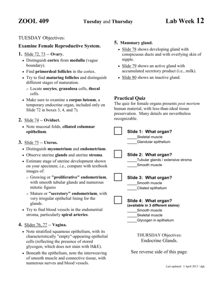
ZOOL 409 Lab Week 12 Tuesday and Thursday TUESDAY Objectives: Examine Female Reproductive System. 1. Slide 72, 73 -- Ovary. Distinguish cortex from medulla (vague boundary). Find primordeal follicles in the cortex. Try to find maturing follicles and distinguish different stages of maturation. o Locate oocytes, granulosa cells, thecal cells. Make sure to examine a corpus luteum, a temporary endocrine organ, included only on Slide 72 in boxes 3, 4, and 7). 2. Slide 74 -- Oviduct. Note mucosal folds, ciliated columnar epithelium. 5. Mammary gland. Slide 78 shows developing gland with conspicuous ducts and with overlying skin of nipple. Slide 79 shows an active gland with accumulated secretory product (i.e., milk). Slide 80 shows an inactive gland. Practical Quiz The quiz for female organs presents post mortem human material, with less-than-ideal tissue preservation. Many details are nevertheless recognizable. □ 3. Slide 75 -- Uterus. Distinguish myometrium and endometrium. Observe uterine glands and uterine stroma. Estimate stage of uterine development shown on your specimen; i.e., compare with textbook images of: o Growing or "proliferative" endometrium, with smooth tubular glands and numerous mitotic figures o Mature or "secretory" endometrium, with very irregular epithelial lining for the glands. Try to find blood vessels in the endometrial stroma, particularly spiral arteries. □ □ □ Slide 1: What organ? ____Skeletal muscle ____Glandular epithelium Slide 2: What organ? ____Tubular glands / extensive stroma ____Smooth muscle Slide 3: What organ? ____Smooth muscle ____Ciliated epithelium Slide 4: What organ? (available in 3 different stains) ____Smooth muscle ____Skeletal muscle ____Glycogen in epithelium 4. Slides 76, 77 -- Vagina. Note stratified squamous epithelium, with its characteristically "empty"-appearing epithelial cells (reflecting the presence of stored glycogen, which does not stain with H&E). Beneath the epithelium, note the interweaving of smooth muscle and connective tissue, with numerous nerves and blood vessels. THURSDAY Objectives: Endocrine Glands. See reverse side of this page. Last updated: 3 April 2013 / dgk ZOOL 409 Tuesday and Thursday TUESDAY Objectives: See reverse side. THURSDAY Objectives: Examine Endocrine Glands. 1. Slides 52, 53 -- Pancreatic islets. Pancreatic islets of Langerhans are distinct patches of endocrine cells, usually less intensely stained and with a cell arrangement (irregular, curving cords) that is distinctly different from the exocrine acini. 2. Slides 84, 85 -- Thyroid and Parathyroid. (Slide 36 in Boxes 1, 2, 4, 9 also also includes thyroid gland.) Thyroid gland is characterized by distinct follicles, lined by simple cuboidal epithelium and filled with colloid (containing stored thyroglobulin). The parathyroid consists of small masses of cells, arranged in cords rather like the cells which comprise pancreatic islets. Lab Week 12 5. Slide 81 -- Pituitary gland. Cells of the adenohypophysis (anterior pituitary, pars distalis) are epithelial in origin, form irregular clumps, and include several different cell types (each associated with a specific pituitary hormone. We can't identify specific hormonal types with routine light microscopy, but we can notice cells with diverse staining properties (acidophils, basophils, chromophobes). The neurohypophysis (posterior pituitary, pars nervosa) is nervous tissue with a fibrous appearance, consisting of axons from the hypothalamus. 6. Slides 72, 73 -- Ovary. Follicular cells of the ovary (granulosa cells and thecal cells) comprise endocrine tissue, with the characteristic appearance of steroidsecreting cells. After an ovum is released, follicular cells grow and differentiate into the corpus luteum. 3. Slides 82, 83-- Adrenal gland. Note distinct difference between cortex (epithelial cells arranged in cords separated by sinusoids) and medulla (very different cell arrangement, derived from neural tissue). In the cortex, you may be able to see three zones (zona glomerulosa nearest the capsule, zona fasciculata, and zona reticularis nearest the medulla), vaguely distinguished by subtle variation in staining and in cell arrangement. Cells of the zona fasciculata should have the distinctive spongy appearance characteristic of steroid manufacture and secretion, resulting from presence of numerous lipid droplets. 4. Slides 67, 68 -- Testis. The endocrine cells are the Leydig cells in testicular stroma, with round euchromatic nuclei and with the numerous lipid droplets (giving a foamy appearance) that characterize steroid-secreting cells. Practical Quiz The quiz again presents post mortem human material, with less-than-ideal tissue preservation. □ □ □ Slide 1: What organ? ____Cuboidal epithelium ____Colloid in follicle Slide 2: What organ? ____Cortex ____Medulla Slide 3: What organ? ____Endocrine tissue ____Exocrine tissue ____Duct Last updated: 3 April 2013 / dgk
