WG-09_2007-11-09_Min_NewOrleans
advertisement
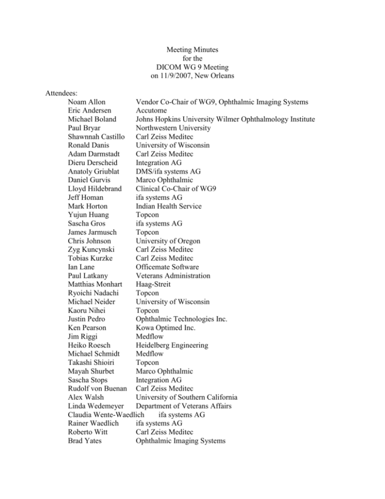
Meeting Minutes for the DICOM WG 9 Meeting on 11/9/2007, New Orleans Attendees: Noam Allon Vendor Co-Chair of WG9, Ophthalmic Imaging Systems Eric Andersen Accutome Michael Boland Johns Hopkins University Wilmer Ophthalmology Institute Paul Bryar Northwestern University Shawnnah Castillo Carl Zeiss Meditec Ronald Danis University of Wisconsin Adam Darmstadt Carl Zeiss Meditec Dieru Derscheid Integration AG Anatoly Griublat DMS/ifa systems AG Daniel Gurvis Marco Ophthalmic Lloyd Hildebrand Clinical Co-Chair of WG9 Jeff Homan ifa systems AG Mark Horton Indian Health Service Yujun Huang Topcon Sascha Gros ifa systems AG James Jarmusch Topcon Chris Johnson University of Oregon Zyg Kuncynski Carl Zeiss Meditec Tobias Kurzke Carl Zeiss Meditec Ian Lane Officemate Software Paul Latkany Veterans Administration Matthias Monhart Haag-Streit Ryoichi Nadachi Topcon Michael Neider University of Wisconsin Kaoru Nihei Topcon Justin Pedro Ophthalmic Technologies Inc. Ken Pearson Kowa Optimed Inc. Jim Riggi Medflow Heiko Roesch Heidelberg Engineering Michael Schmidt Medflow Takashi Shioiri Topcon Mayah Shurbet Marco Ophthalmic Sascha Stops Integration AG Rudolf von Buenan Carl Zeiss Meditec Alex Walsh University of Southern California Linda Wedemeyer Department of Veterans Affairs Claudia Wente-Waedlich ifa systems AG Rainer Waedlich ifa systems AG Roberto Witt Carl Zeiss Meditec Brad Yates Ophthalmic Imaging Systems Staff: Flora Lum American Academy of Ophthalmology I. Welcome and Introductory Remarks; Introduction of Participants Dr Lloyd Hildebrand and Noam Allon welcomed the participants. Everyone introduced themselves and described their interest in a special interest workgroup. II. Approval of Previous Meeting Minutes The minutes of the November 2006 WG9 meeting in Las Vegas were approved. III. Summary of Activities of DICOM and SNOMED Dr. Hildebrand summarized some of the important progress in DICOM and IHE Eye Care. About twelve years ago, the Academy began its standard setting activities. This was a chicken-and-egg situation because vendors did not perceive a market demand, and customers (ophthalmologists) could not imagine the benefits of standards. In 1996, digital imaging standards setting opened up to other specialties in medicine. The Academy realized that digital imaging standards could not be done in isolation, but instead be accompanied by standards for terminology. The Academy employed ophthalmologists to model and add new concepts to SNOMED. The governance of SNOMED has changed over the years from the College of American Pathologists to an International Health Standards Development Organization. This organization is governed by national governments which license SNOMED on a national basis, such as the United States and the United Kingdom. In September, the Academy’s Board of Trustees passed a resolution affirming that SNOMED has been adopted as its official clinical terminology. There also have been significant steps made in incorporating SNOMED into the infrastructure of the Academy’s educational website, known as Ophthalmic News and Education In DICOM, there has been significant progress made on two supplements. Supplement 110, Ophthalmic Tomography, was approved in September 2007 and is posted on the DICOM website. This is due to the hard work and persistent efforts of Justin Pedro, Adam Darmstadt and others. Supplement 130, Refractive Data, was sent out for a second letter ballot and should hopefully be approved by November 13th. This supplement covers the following devices: lensometers, keratometers, autorefractors, autoprojectors and a structured report for prescription data. Linda Wedemeyer, M.D. did a yeoman’s job in championing this supplement and attending meetings to support this supplement. Existing DICOM supplementst that are applicable to eye care include: ultrasound, CT, MRI, visible light such as for surgical microscope and ophthalmic photography with stero representation. IHE has provided a real-life demonstration of standards and brought the standards to life. We have come a long way in these several years. Now, DICOM, SNOMED and IHE are all in the daily vocabulary. IV. Summary of Activities in IHE Eye Care The previous year was a successful Year 1 of IHE Eye Care Connectathon and Showcase, with 10 vendors. This year, there were 12 vendors who participated in the October Connectathon and Showcase, including 3 new vendors. There are educational materials on the Academy’s website to provide guidance to ophthalmologists on IHE compliance and DICOM conformance statements. For Year 3, there will be an emphasis on implementing new standards (refractive data and ophthalmic tomography). V. Visual Field Subgroup The visual field subgroup has already accomplished a lot during previous teleconferences prior to the Annual Meeting. At the Meeting, they discussed the list of data elements that has been designated by clinicians as relevant and which appear to be commonly reported by the major visual field device vendors. Dr. Paul Bryar will create a spreadsheet which summarizes the discussion. In the spreadsheet, each data element, its definition, its data type (1--mandatory; 2--encouraged but optional; 3--optional), and units are listed. Next to each item is a column labeled "comments." Dr. Bryar will circulate this after the Meeting and highlight issues where we need vendor input specifically related to their particular devices. Dr. Paul Bryar will be responsible for the future steps. VI. Macular Thickness Report Subgroup The group discussed at length the various data elements that needed to be included in the proposed supplement. They will model their work after the recently approved Supplement 130 for Refractive Data. The group thought that they could write up case studies that would help explain the clinical circumstances for when macular thickness reports were needed. Mr. Michael Neider will be responsible for the future steps and Dr. Ronald Danis will work with other clinicians to develop the case studies which will be circulated to the larger group. VII. Ophthalmic Mapping Subgroup The group discussed at length the advantages and disadvantages of combining different modalities, corneal topography, wavefront technology, retinal nerve fiber layer thickness analyzer and optic nerve head analyzer. After much deliberation, the group decided that the advantages outweighed the disadvantages and it would be advantageous to provide a generic module to cover the function of mapping: a set of measures that are distributed topographically and are typically presented in pseudo-color representation. The group developed a listing of vendors and clinicians that are involved with these four technologies. The group also discussed different data elements to be included or deleted in the ophthalmic mapping image module and the ophthalmic mapping acquisition parameters module. The next steps are for Flora to edit the draft supplement based on the group’s discussion and to send to the group for additional discussion. The group plans to meet by conference call in January 2008. VIII. Biometry Subgroup The group discussed at length the various data elements that needed to be included in the proposed supplement. They will model their work after the recently approved Supplement 130 for Refractive Data. The group thought that they could develop a draft version soon to be able to be circulated to a larger group of vendors. Dr. Linda Wedemeyer will be responsible for the future steps. IX. Next Meetings of the Working Group 9 A. Possible venues o ASCRS o ARVO (April 28 – May 1) B. Friday, November 8, Atlanta, Academy Annual Meeting Reviewed by legal counsel December 21, 2007
