Final from 2013 - Molecular and Cell Biology
advertisement
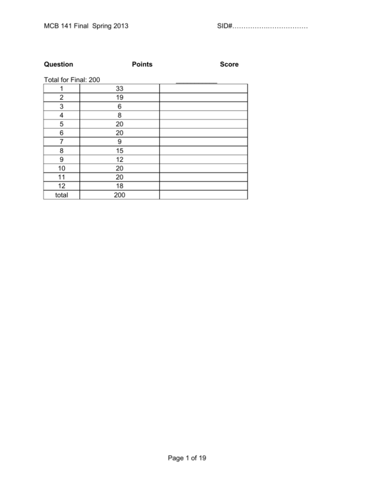
SID#…………….……………… MCB 141 Final Spring 2013 Question Total for Final: 200 1 2 3 4 5 6 7 8 9 10 11 12 total Points Score ___________ 33 19 6 8 20 20 9 15 12 20 20 18 200 Page 1 of 19 SID#…………….……………… MCB 141 Final Spring 2013 Question 1 (33 points) 1a) (10 points) Name that phenotype! Assuming tissue-specific knockouts are possible when needed, what is the consequence of knocking out the following genes in Drosophila? BAM, in the GSCs bicoid, in the nurse cells Antp Scr and abd-A PolyComb repressor binding sites that regulate Labial 1b) (8 points) You suspect Dpp, the Drosophila homolog of BMP, is acting as a morphogen in the wing imaginal disc, turning on spalt at high concentrations, fictional (fic) at intermediate concentrations, and optomotor-blind (omb) at intermediate and lower concentrations. i) Propose a complete model for how different concentrations of Dpp can induce different expression patterns of these target genes. Careful! Page 2 of 19 MCB 141 Final Spring 2013 SID#…………….……………… ii) Design one key experiment (you don’t need to explain technical details, since we didn’t talk much about fly genetic tools) to test whether Dpp is acting as a morphogen or through a relay mechanism, and predict the results for each case. 1c) (12 points) You swap the Bicoid binding sites that regulate transcription of giant and hunchback (i.e., you’ve modified the endogenous genomic loci), without changing the coding sequences of these genes. Answer the following questions about the consequence for this mutant Drosophila embryo. Use the axes here as the questions below indicate. i) What will the expression patterns of Giant and Hunchback be? Draw and label curves representing protein concentration from Anterior to Posterior on the axes above. Explain why you drew the curves where you did here. Page 3 of 19 MCB 141 Final Spring 2013 SID#…………….……………… ii) What will the expression patterns of Kruppel and Knirps be? Draw and label curves representing protein concentration from Anterior to Posterior on the axes above. Explain why you drew the curves where you did here. iii) What will the expression pattern be of a reporter gene (lacZ, in this case) driven by the even skipped stripe 2 enhancer (eve 2) be? As usual, this enhancer is activated by Bicoid and Hunchback, and repressed by Giant and Kruppel. Shade in the bar above the axes labeled “eve2::lacZ” wherever lacZ will be expressed. If lacZ will not be expressed, write “not expressed” below. Either way, explain your answer here. iv) What will the expression pattern of a reporter gene (GFP, in this case) driven by the eve 3 enhancer be? As usual, this enhancer is activated by DSTAT and repressed by Hunchback and Knirps. Shade in the bar above the graph labeled “eve3::GFP” wherever GFP will be expressed. If GFP will not be expressed, write “not expressed” below. Either way, explain your answer here. 1d) (3 points) Name three key transcription factors required for the induction of pluripotent stem cells (iPS cells). Propose a mechanism for how they might function synergistically to activate gene expression. Page 4 of 19 MCB 141 Final Spring 2013 SID#…………….……………… Question 2 (19 points total) 2a. (6 points) Below is shown a schematic drawing of a 128-cell mouse blastocyst shortly before it hatches and implants in the wall of the uterus. Into each box, put the appropriate letter or letters from the list to best identify each region. A letter may be used in one box, more than one box, or not used. A. blastocyst cavity (also called “blastocoel”) B. cells that will later develop into extraembryonic endoderm C. cells of the mural trophoblast D. cells of the polar trophoblast E. cells of the epiblast F. zona pellucida (must be destroyed before implantation occurs) G. cells that later develop into the mouse H. cells that first invade the uterine wall I. cells of the hypoblast (or visceral endoderm) J. cells that proliferate as embryonic stem cells when cultured in a Petri dish K. cells derived from the outermost cells of the 32-64 cell stage embryo L. cells derived from inner cells of the 3264 cell stage embryo Page 5 of 19 MCB 141 Final Spring 2013 2b. (5 points) By the 32-64 cell stages, the mouse embryo contains irreversibly different populations of trophoblast and inner cell mass cells expressing different genes, such as cdx2 only in the trophoblast cells. Briefly describe how compaction (which begins at the late 8-cell stage, see figure) and ongoing cleavage contribute to the formation of these two populations. SID#…………….……………… 2c. (2 points) It has been recently found that cdx2 gene expression is activated in trophoblast cells by the Hippo signaling pathway, which is itself activated by tight junction proteins and adhesion junction proteins. Is this consistent with your explanation above? Indicate the consistency or inconsistency. Page 6 of 19 MCB 141 Final Spring 2013 SID#…………….……………… 2d. (6 points) By the 128 cell stage, the mouse blastocyst contains irreversibly different epiblast cells and hypoblast cells (“visceral endoderm”) expressing different genes, such as gata6 in the hypoblast cells. Briefly describe the cell activities leading to blastocyst cavity formation, and tell how it and cell sorting contribute to the formation of the epiblast and hypoblast cell populations: Question 3: (6 points) As you know, Chordin is a neural inducer released by the organizer of Xenopus, and other chordates, at gastrula and neurula stages. Predict outcomes for the following experimental interventions: 3a) An animal cap is cut from an early gastrula stage embryo developing from an egg injected in the animal hemisphere with chordin mRNA, and this cap is allowed to develop in isolation. It will form (circle one letter): a) epidermis b) posterior enural tissue c) anterior neural tissue 3b) Chordin mRNA is injected in the ventral equatorial region of a 4-cell Xenopus embryo, which is then allowed to develop. The ventral ectoderm of this embryo develops as (circle one letter): a) epidermis b) posterior neural tissue c) anterior neural tissue Explain your answer briefly: Page 7 of 19 SID#…………….……………… MCB 141 Final Spring 2013 3c) An anti-sense morpholino to chordin mRNA is injected in the dorsal equatorial region of a 4cell Xenopus embryo, which is then allowed to develop. Even though the production of Chordin protein is effectively knocked down by the morpholino, the embryo develops a nearly normal nervous system. Suggest why, and suggest what you might do to interfere more completely with neural induction. Question 4. (8 points) The notochord is a distinguishing trait of members of the chordate phylum. Summarize your knowledge of notochord development by writing T (true) or F (false) next to each of the following statements: The notochord will develop in a Xenopus embryo depleted for maternal beta-catenin mRNA. The notochord will develop in a Xenopus embryo depleted for maternal VegT mRNA. The notochord will not develop in a Xenopus embryo injected with anti-sense morpholinos to Nodals (anti-xnr1,2,4,5,6, and derriere). A second notochord will develop in a Xenopus embryo injected on the ventral side with Wnt mRNA at the 4 cell stage. The trunk-tail organizer of the amphibian embryo is composed of notochord precursor cells. The head organizer of the amphibian embryo is composed of notochord precursor cells. Hensen’s node of the chick embryo, and of the mouse embryo, contains notochord precursor cells. Notochord precursor cells release Bmp antagonists such as Chordin, Noggin, and Follistatin in the Xenopus embryo. Notochord precursor cells release Wnt antagonists such as Frzb, Dkk, and Crescent in the Xenopus embryo. Notochord precursor cells engage in convergent extension morphogenesis. Notochord precursor cells engage in spreading migration morphogenesis. The differentiated notochord is located beneath the neural tube and between the left and right rows of somites. The notochord of the chick embryo is laid down behind the regressing Hensen’s node. Roofplate formation in the Xenopus neural tube depends on signals released by the notochord. Floorplate formation in the Xenopus neural tube depends on signals released by the notochord. Sclerotome formation from the somite depends on signals released by the notochord. Page 8 of 19 SID#…………….……………… MCB 141 Final Spring 2013 Question 5 (20 points) Below is a diagram of the somites in three vertebrates, along with the position of the large spinal nerves that innervate the forelimb (black bars - the brachial plexus) and the expression of hoxc6 (shading). 5a) (2 Points)In the age of dinosaurs, some plesiosaurs had 76 cervical (neck) vertebrae, compared the standard mammalian number of 7. At what somite would you expect hoxc6 expression to begin in the plesiosaur? 5b) (4 points)The forelimb (brachial) plexus contains far more cell bodies than the spinal nerves and ganglia in the neighboring somites. However, at the early stage of development these structures contain similar numbers of cell bodies. What mechanisms might account for the difference in cell number between the brachial plexus and the neighboring nerves? What might be the principal regulator? Page 9 of 19 MCB 141 Final Spring 2013 SID#…………….……………… 5c) (6 points) The brachial nerves emerge periodically from the spinal cord. What determines the periodicity? Name two ligand-receptor pairs that are involved in this process, and where they may be expressed. 5d) (8 points) Compared to a chicken, ducks have an A/P axis of similar length, but have more somites, and each of these is smaller than chicken somites. What single change to the duck somite segmentation “clock and wavefront” might have caused the changes in somite number and size? Describe this change conceptually, and propose a potential underlying molecular mechanism. Page 10 of 19 SID#…………….……………… MCB 141 Final Spring 2013 Question 6 (20 points) You are interested in determining what signals induce placode formation in the epidermis during lens development. You have sequenced the transcriptomes of the retina and lens, and notice several interesting features: 1) Wnt3, BMP, and Shh are expressed in the retina, though they are also expressed in other regions of the embryo. 2) Tbx7 and FoxH1 are expressed exclusively in the retina at this stage, and nowhere else. 3) Nodal receptor type II subunit, Frizzled8, Cdx9, and Ret are expressed exclusively in the region of the epidermis that is competent to form the lens placode at this stage. 6a) 4 points Which of these genes might you expect to be a signal that induces lens placode formation, and why? 6b) (8 points) How would you test this gene’s activity? Address the following points: Expression of this gene, by the retina, is required for lens placode induction. This gene acts directly on the presumptive lens placode. (You don’t have to show it’s a morphogen). Page 11 of 19 MCB 141 Final Spring 2013 SID#…………….……………… 6c) (4 points) What two tools or assays could you use to determine whether lens placode formation occurs in the experiment you proposed in part B? You only have to explain what tools you will use (you don’t have to describe how they work, or mention particular gene names, but use whole sentences!). 6d) (4 points) As part of your experiments, you determine that an early step in lens placode induction is activation of the protein Shroom3. What morphogenetic activity would you expect to see in the lens placode cells, and why would you expect this? Page 12 of 19 MCB 141 Final Spring 2013 SID#…………….……………… Question 7 (9 points) Describe 3 instances of Delta/Notch signaling we encountered this semester. What happens in each system if Notch is knocked out or knocked down in that tissue? Page 13 of 19 MCB 141 Final Spring 2013 SID#…………….……………… Question 8 (15 points) Describe the steps you would take to reprogram cells isolated from a muscular dystrophy patient to generate a pool of muscle cells. Include gene names. Page 14 of 19 MCB 141 Final Spring 2013 SID#…………….……………… Question 9 (12 points) You are working in Professor Levine’s lab, and think you may have found a neural crest-like population of cells in a non-vertebrate chordate, the sea squirt Ciona intestinalis. Based on what you know about NC cells in vertebrates, describe 2 experiments you might do to determine exactly how similar these are to authentic NC cells. Page 15 of 19 SID#…………….……………… MCB 141 Final Spring 2013 Question 10 (20 points) Provide ten examples where BMP signaling is used in development. What is the target tissue and what is the result? Page 16 of 19 MCB 141 Final Spring 2013 SID#…………….……………… Question 11 (20 points) The enhancer of the Nodal gene in mouse contains several enhancers. One of them is upstream of the gene, and is called the proximal epiblast enhancer. Another, containing FoxH1 binding sites, is within the first intron of the gene. The proximal epiblast enhancer is initially active radially around the proximal epiblast, but its activity becomes amplified and restricted to the prospective primitive streak, on one side of the embryo. a) (4 points) At the time of primitive streak formation, what region/tissue prevents amplification in the prospective head region, and how? b) (4 points) What two positive feedback events are important for amplifying the expression of Nodal in the primitive streak region? c) (3 points) what transgene would you construct to monitor the activity of the FoxH1 enhancer from the Nodal gene d) (2 points) How would you expect the activity of this enhancer to compare to the FoxH1 enhancer from lefty2? Page 17 of 19 SID#…………….……………… MCB 141 Final Spring 2013 e) (4 points) If you used the lefty2 FoxH1 enhancer to drive the expression of Nodal, what phenotype would you expect? f) (3 points) How would you test if the lefty2 gene is required cell-autonomously? Page 18 of 19 SID#…………….……………… MCB 141 Final Spring 2013 Question 12 (18 points) Shown below is a classic drawing of the skeleton of the Coelacanth, a lobe-finned fish, that is thought to be similar to the fish ancestors of the tetrapods. The fish has some paired lobe fins, and some in the midline (indicated by the asterisks). Assuming that you are easily able to obtain embryos at any stage for these fish, and can isolate any gene sequence that you want, what experiments (at least three) would you do to test whether the outgrowth of these midline fins is similar in developmental mechanism to the tetrapod limb? Restrict your answer to a discussion of FGF signaling. Page 19 of 19
