Four Microscope Mini Labs
advertisement
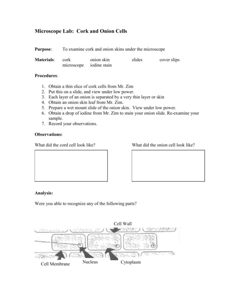
Microscope Lab: Cork and Onion Cells Purpose: To examine cork and onion skins under the microscope Materials: cork microscope onion skin iodine stain slides cover slips Procedures: 1. 2. 3. 4. 5. 6. Obtain a thin slice of cork cells from Mr. Zim Put this on a slide, and view under low power. Each layer of an onion is separated by a very thin layer or skin Obtain an onion skin leaf from Mr. Zim. Prepare a wet mount slide of the onion skin. View under low power. Obtain a drop of iodine from Mr. Zim to stain your onion slide. Re-examine your sample. 7. Record your observations. Observations: What did the cord cell look like? What did the onion cell look like? Analysis: Were you able to recognize any of the following parts? Cell Wall Cell Membrane Nucleus Cytoplasm Microscope Lab: Live Elodea Plant Purpose: To examine live cells of plants. Plants contain chlorophyll. The chlorophyll is located in structures inside the cell called chloroplasts. Question: 1. What is the name of the process that allows plants to make their own food? 2. What does the term transparent mean? 3. What does the term translucent mean? Materials: Slides, cover slip, dropper, tweezers, Elodea plants (aka Anacharis) Procedures: 1. 2. 3. 4. 5. The tip of the elodea plant is the most transparent. Select a suitable leaf from the elodea plant and place it on the slide. Using the proper technique create a wet mount slide. Examine your slide under both high and lower power. Eventually you may see the chloroplasts moving inside the cytoplasm of the cell. Observations: What color do the chloroplasts appear to be? membrane cytoplasm vacuole chloroplast Cell Wall Nucleus (if visible) Analysis: 1. What do the vacuoles appear to be? 2. Why do you think is significant about the color of chloroplasts when compared with the color of the elodea plant. Is there any correlation (connection) between the two? SALT, SUGAR, SAND and the MICROSCOPE Purpose: To compare the crystalline structures of salt, sugar, and sand using a microsope. Question: Materials: What does dissolving mean? What does cohesion mean? sand, salt, sugar, slides, cover slip, dropper, water Procedures: 1. Place a few crystals of salt on a slide and examine it under low power. 2. Try moving the diaphragm of microscope to vary the light intensity. 3. Record a drawing of your observation for the dry salt in your data section 4. THIS IS DIFFICULT: We want to try to observe salt dissolving in water. Place a cover slip over a few salt crystals. Drop a few drops of water on the side of the cover slip Because of cohesion the water will gradually slide under the cover slip. Observe what happens as the water and salt mix. Record your observations. 5. Repeat with the sugar and the salt. Data: Salt (dry) Salt (wet) Sugar (dry) Sugar (wet) Sand (dry) Sand (wet) Analysis: 1. Explain the differences or similarities in the shapes of the salt, sugar, and sand when they are dry. Your paragraph should be at least 5 sentences. 2. Explain the difference or similarities of what happens to the salt, sugar and sand when they are mixed with water. This paragraph should be at least 5 sentences. Microscope Lab: Working with Textiles Purpose: To compare a variety of textile fibers using a microscope Question: 1. What does textile mean? Materials: A variety of textile fibers: cotton, wool, silk, polyester, rayon, linen, etc. Microscope, tweezers, slides, cover clips, water, straight pins, dropper Procedures: 1. Obtain the various textile samples. 2. Each sample will need its own slide. You may share slides with other groups. 3. To prepare the slide: Use the tweezers if necessary to hold the textile fiber on the slide. 4. Using a tweezers, pin, or similar objects: try to separate the fibers of your sample. 5. Using the dropped add a drop of water to your sample and cover with the slip. 6. Observe each material under low and high power; and draw what you observe. Data: Low Power High Power Sample 1 Sample 2 Sample 2 _______________ ________________ ________________
