II. An Overview of Cell Signaling
advertisement
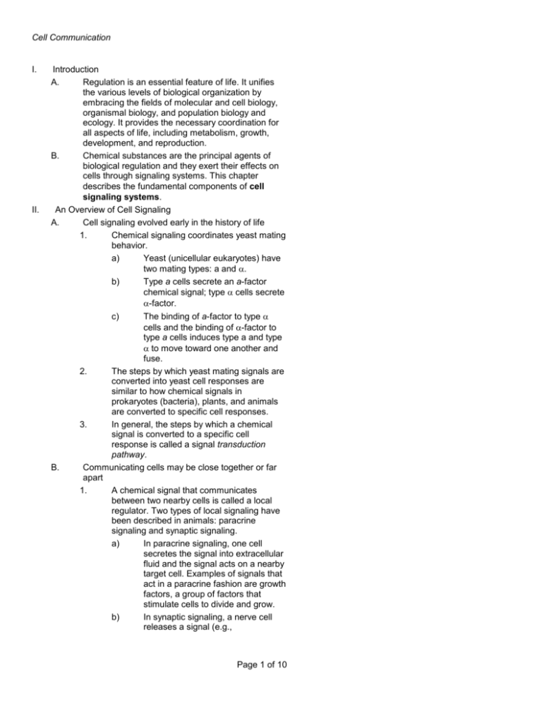
Cell Communication I. II. Introduction A. Regulation is an essential feature of life. It unifies the various levels of biological organization by embracing the fields of molecular and cell biology, organismal biology, and population biology and ecology. It provides the necessary coordination for all aspects of life, including metabolism, growth, development, and reproduction. B. Chemical substances are the principal agents of biological regulation and they exert their effects on cells through signaling systems. This chapter describes the fundamental components of cell signaling systems. An Overview of Cell Signaling A. Cell signaling evolved early in the history of life 1. Chemical signaling coordinates yeast mating behavior. a) Yeast (unicellular eukaryotes) have two mating types: a and . b) Type a cells secrete an a-factor chemical signal; type cells secrete -factor. The binding of a-factor to type cells and the binding of -factor to type a cells induces type a and type to move toward one another and fuse. 2. The steps by which yeast mating signals are converted into yeast cell responses are similar to how chemical signals in prokaryotes (bacteria), plants, and animals are converted to specific cell responses. 3. In general, the steps by which a chemical signal is converted to a specific cell response is called a signal transduction pathway. Communicating cells may be close together or far apart 1. A chemical signal that communicates between two nearby cells is called a local regulator. Two types of local signaling have been described in animals: paracrine signaling and synaptic signaling. a) In paracrine signaling, one cell secretes the signal into extracellular fluid and the signal acts on a nearby target cell. Examples of signals that act in a paracrine fashion are growth factors, a group of factors that stimulate cells to divide and grow. b) In synaptic signaling, a nerve cell releases a signal (e.g., c) B. Page 1 of 10 Cell Communication C. neurotransmitter) into a synapse, the narrow space between the transmitting cell and a target cell, such as another nerve cell or muscle cell. 2. A chemical signal that communicates between cells some distance apart is called a hormone. 3. Hormones have been described in both plants (e.g., ethylene, a gas that promotes growth and fruit ripening) and animals (e.g., insulin, a protein that controls various aspects of metabolism, including the regulation of blood glucose levels). 4. The distinction between local regulators and hormones is for convenience. A particular chemical signal may act both as a local regulator and as a hormone. 5. Insulin, for example, may act in a paracrine fashion on adjacent cells (e.g., other insulin cells in the pancreas, acting to inhibit the further release of insulin in a negative feedback manner) and in a hormonal fashion on distant cells (e.g., liver cells, which store carbohydrate as glycogen). 6. Cells also may communicate by direct contact. Some plant and animal cell possess junctions though which signals can travel between adjacent cells. The three stages of cell signaling are reception, transduction, and response 1. In order for a chemical signal to elicit a specific response, the target cell must possess a signaling system for the signal. Cells which do not possess the appropriate signaling system do not respond to the signal. 2. The signaling system of a target cell consists of the following elements: a) Signal reception. The signal binds to a specific cellular protein called a receptor, which is often located on the surface of the cell. b) Signal transduction. The binding of the signal changes the receptor in some way, usually a change in conformation or shape. The change in receptor initiates a process of converting the signal into a specific cellular response; this process is called signal transduction. The transduction system may have one or many steps. Page 2 of 10 Cell Communication c) III. Cellular response. The transduction system triggers a specific cellular response. The response can be almost any cellular activity, such as activation of an enzyme or altered gene expression. D. The critical features of the target cell signaling system were elucidated by Earl Sutherland (awarded the Nobel Prize in 1971 for his contributions to the understanding of signal transduction) and colleagues who were working on how the hormone, epinephrine, affects carbohydrate metabolism (e.g., glycogen breakdown to glucose1-phosphate) in liver cells. 1. Epinephrine stimulates glycogen breakdown by stimulating the cytosolic enzyme, glycogen phosphorylase (cellular response). 2. Epinephrine could only stimulate glycogen phosphorylase activity when presented to intact cells, suggesting that: a) The plasma membrane is critical for transmitting the signal (reception) b) Activation of glycogen phosphorylase required the presence of an intermediate step or steps in side the cell (signal transduction) E. The mechanisms of the cell signaling process help ensure that important processes occur in the right cells, at the right time, and in proper coordination with other cells of the organism. Signal Reception and the Initiation of Transduction A. A chemical signal binds to a receptor protein, causing the protein to change shape 1. Chemical signals bind to specific receptors. a) The signal molecule is complementary to a specific region of the receptor protein; this interaction is similar to that between a substrate and an enzyme. b) The signal behaves as a ligand, a term for a small molecule that binds to another, and larger molecule. 2. Binding of the ligand to the receptor can lead to the following events: a) Alteration in receptor conformation or shape; such alterations may lead to the activation of the receptor which enables it to interact with other cellular molecules b) Aggregation of receptor complexes Page 3 of 10 Cell Communication B. C. D. Most signal receptors are plasma-membrane proteins 1. Many signal molecules cannot pass freely through the plasma membrane. The receptors for such signal molecules are located on the plasma membrane. There are three families of plasma-membrane receptors - G-protein-linked receptors, tyrosine kinase receptors, and ion channel receptors. G-protein-linked receptors 1. A single polypeptide chain that is threaded back and forth through the plasma membrane in such a way as to possess seven transmembrane domains characterizes the structure of a G-proteinlinked receptor. An example of a G-proteinlinked receptor is the epinephrine receptor. 2. The receptor propagates the signal by interacting with a variety of proteins on the cytoplasmic side of the membrane called Gproteins, so named because they bind guanine nucleotides, GTP and GDP. a) The function of the G-protein is influenced by the nucleotide to which it is bound: (1) G-proteins bound to GDP are inactive. (2) G-proteins bound to GTP are active. b) When a ligand binds to a G-proteinlinked receptor, the receptor changes its conformation and interacts with a G-protein. This interaction causes the GDP bound to the inactive G-protein to be displaced by GTP, thereby activating the G-protein. c) The activated G-protein binds to another protein, usually an enzyme, resulting in the activation of a subsequent target protein. d) The activation state of the G-protein is only temporary, because the active G-protein possesses endogenous GTPase activity, which hydrolyzes the bound GTP to GDP. G-protein-Iinked receptors and G-proteins mediate a host of critical metabolic and developmental processes (e.g., blood vessel growth and development). Defects in the G-protein signaling system form the bases of many human disease states (e.g., cholera). Page 4 of 10 Cell Communication E. F. Tyrosine-kinase receptors 1. An extracellular ligand-binding domain and a cytosolic domain possessing tyrosine kinase enzyme activity characterize the structure of a tyrosine-kinase receptor. Examples of tyrosine-kinase receptors are the receptors for numerous growth factors, such as PDGFs, the family of factors that serve as external modulators of the cell-cycle control system. 2. Propagation of the signal involves several steps as follows: a) Ligand binding causes aggregation of two receptor units, forming receptor dimers. b) Aggregation activates the endogenous tyrosine kinase activity on the cytoplasmic domains. c) The endogenous tyrosine kinase catalyzes the transfer of phosphate groups from ATP to the amino acid tyrosine contained in a particular protein. In this case, the tyrosines that are phosphorylated are in the cytoplasmic domain of the tyrosinekinase receptor itself (thus, this step is an autophosphorylation). d) The phosphorylated domain of the receptor interacts with other cellular proteins, resulting in the activation of a second, or relay, protein. The relay proteins may or may not be phosphorylated by the tyrosine kinase of the receptor. Many different relay proteins may be activated, each leading to the initiation of many, possibly different, transduction systems. e) One of the activated relay proteins may be protein phosphatase, an enzyme that hydrolyzes phosphate groups off of proteins. The dephosphorylation of the tyrosines on the tyrosine kinase domain of the receptor results in inactivating the receptor and the termination of the signal process. Ion-channel receptors 1. Some chemical signals bind to ligand-gated ion channels. These are protein pores in the membrane that open or close in response to ligand binding, allowing or blocking the flow of specific ions (e.g., Na+, Ca2+). An example of an ion-gated channel would be Page 5 of 10 Cell Communication the binding of a neurotransmitter to a neuron, allowing the inward flow of Na+ that leads to the depolarization of the neuron and the propagation of a nervous impulse to adjacent cells. G. Not all signal receptors are located on the plasma membrane. Some are proteins located in the cytoplasm or nucleus of target cells. 1. In order for a chemical signal to bind to these intracellular receptors, the signal molecule must be able to pass through plasma membrane. Examples of signals which bind to intracellular receptors include the following: a) Nitric oxide (NO) b) Steroid (e.g., estradiol, progesterone, testosterone) and thyroid hormones of animals IV. Signal Transduction Pathways A. Pathways relay signals from receptors to cellular responses 1. Ligand binding to a receptor triggers the first step in the chain of reactions-the signal transduction pathway- that leads to the conversion of the signal to a specific cellular response. a) The transduction system does not physically pass along the signal molecule; rather the information is passed along. At each step of the process, the nature of the information is converted, or transduced, into a different form. B. Protein phosphorylation, a common mode of regulation in cells, is a major mechanism of signal transduction 1. The process of phosphorylation, or the transferring a phosphate group from ATP to a protein substrate, which is catalyzed by enzymes called protein kinases, is a common cellular mechanism for regulating the functional activity of proteins. 2. Protein phosphorylation is commonly used in signal pathways in the cytoplasm of cells. Unlike the case with tyrosine-kinase receptors, protein kinases in the cytoplasm do not act on themselves, but rather on other proteins (sometimes enzymes) and attach the phosphate group to serine or threonine residues. a) Some phosphorylations result in activation of the target protein (increased catalytic activity in the Page 6 of 10 Cell Communication C. D. case of an enzyme target). An example of a stimulatory phosphorylation cascade is the pathway involved in the breakdown of glycogen as elucidated by Sutherland, et al. (see Campbell, Figures 11.10, 11.15). b) Some phosphorylations result in inactivation (decreased catalytic activity in the case of an enzyme target). 3. Cells turn off the signal transduction pathway when the initial signal is no longer present. Another class of enzymes known as protein phosphatases reverses the effects of protein kinases. Certain small molecules and ions are key components of signaling pathways - second messengers 1. Not all of the components of a signal transduction pathways are proteins. Some signaling systems rely on small, nonprotein, water soluble molecules or ions. Such signaling components are called second messengers. Two second messenger systems are the cyclic AMP (cAMP) system and the Ca2+-inositol triphosphate (IP3) system. Cyclic AMP Sutherland's group ultimately found that the substance mediating the action of epinephrine on liver glycogen breakdown was cAMP (second messenger). Our present understanding of the transduction steps associated with cAMP is as follows: 1. Ligand (first message) binds to a receptor. 2. Receptor conformation changes; G-protein complex is activated. 3. The active G-protein in turn activates the enzyme, adenylyl cyclase, which is associated with the cytoplasmic side of the plasma membrane. 4. Adenylyl cyclase converts ATP to cAMP. 5. cAMP binds to and activates a cytoplasmic enzyme, protein kinase A. 6. Protein kinase A, as was the case for protein kinases, propagates the message by phosphorylating various other proteins that lead to the cellular response (e.g., glycogen breakdown; see Campbell Figure 11.15). 7. The pool of cAMP in the cytoplasm is transient because of the breakdown of cAMP by another enzyme to an inactive form (AMP). This conversion provides a shut-off Page 7 of 10 Cell Communication E. mechanism to the cell to ensure that the target responses cease in the absence of ligand. 8. A number of hormones in addition to epinephrine (e.g., glucagon) use cAMP as a second messenger. Calcium ions and inositol triphosphate 1. Many signaling molecules induce their specific responses in target cells by increasing the cytoplasm's concentration of Ca2+. The Ca2+ pool can be affected in two ways: a) Ligand binding to a Ca2+-gated ion channel (see above) b) Activation of the inositol triphosphate (IP3) signaling pathway 2. Activation of the IP3 pathway involves the following steps: a) Ligand binding results in a conformation change in the receptor. b) The altered receptor activates an enzyme associated with the cytoplasmic side of the plasma membrane (phospholipase). The activated enzyme hydrolyzes membrane phospholipids, giving rise to two important second messengers: IP3 and diacylglycerol. c) Diacylglycerol is linked to a signaling pathway that involves another protein kinase. d) IP3 is linked to a Ca2+ signaling pathway. IP3 binds to Ca2+-gated channels. A large number of such channels are located on the ER, in the lumen of which high amounts of Ca2+ are sequestered. IP3 binding to these receptors and increases the cytoplasmic concentration of Ca2+ (in this case Ca2+ could be considered a tertiary messenger; however, by convention, all postreceptor small molecules in the transduction system are referred to as second messengers). 3. Ca2+ acts to affect signal transduction in two ways: a) Directly by affecting the activity or function of target proteins b) Indirectly by first binding to a relay protein, calmodulin. Calmodulin, in turn, principally affects transduction Page 8 of 10 Cell Communication V. systems by modulating the activities of protein kinases and protein phosphatases. Cellular Responses to Signals A. In response to a signal, a cell may regulate activities in the cytoplasm or transcription in the nucleus B. The signal transduction system ultimately brings about the specific cellular response by regulating specific processes in the cytoplasm or in the nucleus. C. In the cytoplasm, the signaling can affect the function or activity of proteins which carry out various processes, including: 1. Rearrangement of the cytoskeleton 2. Opening or closing of an ion channel 3. Serve at key points in metabolic pathways (e.g., glycogen phosphorylase in the glycogen breakdown scheme; see Campbell Figure 11.15) D. In the nucleus, the signaling system affects the synthesis of new proteins and enzymes by modulating the expression (turn on or turn off) specific genes. Gene expression involves transcription of DNA into mRNA as well as the translation of mRNA into protein. 1. Signal transduction systems can modulate virtually every aspect of gene expression. One example is the regulation of the activity of transcription factors, proteins required for appropriate transcription. 2. Dysfunction of signaling pathways that affect gene regulation (e.g., pathways that transduce growth factor action) can have serious consequences and may even lead to cancer. E. Elaborate pathways amplify and specify the cell's responses to signals 1. Signal amplification a) The production of second messengers such cAMP provides a built in means of signal amplification in that the binding of one ligand (first message) can lead to the production of many second messages. The degree of amplification is heightened when the second messenger system is linked to a phosphorylation cascade as in the case of the process of glycogen breakdown. As a result of this inherent amplification, the binding of very few epinephrine molecules to the surface of a liver cell can result Page 9 of 10 Cell Communication 2. 3. in the release of millions of glucose molecules resulting from glycogen breakdown (see Campbell Figure 11.15). Signal specificity a) Only target cells with the appropriate receptor bind to a particular signaling molecule to initiate the transduction of a signal into a specific cellular response. b) A particular signal can bind to different cell types and result in different responses in each of the cell types. This is possible because each of the different cell types can express a unique collection of proteins. As a result, the receptor on (or in) each of the different cell types can be linked to variant signal transduction pathways, each leading to a different response. An example is epinephrine action on vertebrate liver and cardiac muscle cells. In liver cells, the principal response is glycogen breakdown (see Campbell Figure IL] 5); whereas, in cardiac muscle cells, epinephrine stimulates contraction. c) A single cell type may possess divergent and/or convergent ("crosstalk") signal transduction pathways. Such schemes facilitate coordination of cellular responses and economize on the number of required transduction elements (see Campbell Figure 11.17). The diverse symptoms of the human inherited disorder Wiscott Aldrich syndrome stem from a single defect in a relay protein of a transduction system. An important feature of cell signaling systems is that there exist mechanisms to both turn-on and turn-off the system. The turn-off mechanisms ensure that cells respond appropriately to changing conditions. Page 10 of 10


