Is there a viral aetiology for encapsulating peritoneal sclerosis
advertisement
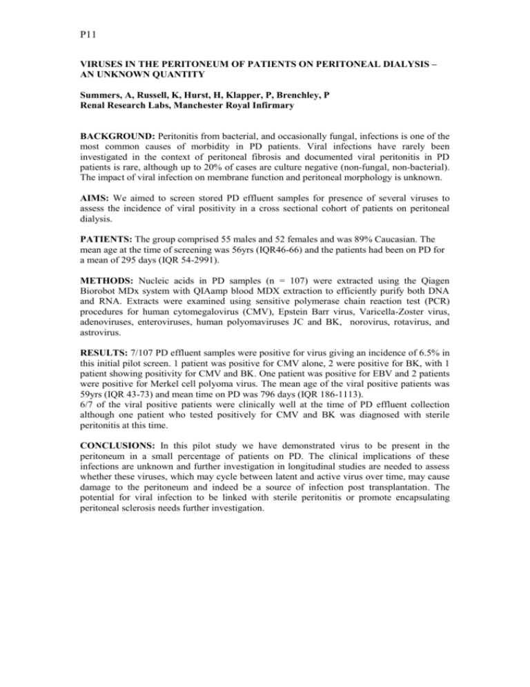
P11 VIRUSES IN THE PERITONEUM OF PATIENTS ON PERITONEAL DIALYSIS – AN UNKNOWN QUANTITY Summers, A, Russell, K, Hurst, H, Klapper, P, Brenchley, P Renal Research Labs, Manchester Royal Infirmary BACKGROUND: Peritonitis from bacterial, and occasionally fungal, infections is one of the most common causes of morbidity in PD patients. Viral infections have rarely been investigated in the context of peritoneal fibrosis and documented viral peritonitis in PD patients is rare, although up to 20% of cases are culture negative (non-fungal, non-bacterial). The impact of viral infection on membrane function and peritoneal morphology is unknown. AIMS: We aimed to screen stored PD effluent samples for presence of several viruses to assess the incidence of viral positivity in a cross sectional cohort of patients on peritoneal dialysis. PATIENTS: The group comprised 55 males and 52 females and was 89% Caucasian. The mean age at the time of screening was 56yrs (IQR46-66) and the patients had been on PD for a mean of 295 days (IQR 54-2991). METHODS: Nucleic acids in PD samples (n = 107) were extracted using the Qiagen Biorobot MDx system with QIAamp blood MDX extraction to efficiently purify both DNA and RNA. Extracts were examined using sensitive polymerase chain reaction test (PCR) procedures for human cytomegalovirus (CMV), Epstein Barr virus, Varicella-Zoster virus, adenoviruses, enteroviruses, human polyomaviruses JC and BK, norovirus, rotavirus, and astrovirus. RESULTS: 7/107 PD effluent samples were positive for virus giving an incidence of 6.5% in this initial pilot screen. 1 patient was positive for CMV alone, 2 were positive for BK, with 1 patient showing positivity for CMV and BK. One patient was positive for EBV and 2 patients were positive for Merkel cell polyoma virus. The mean age of the viral positive patients was 59yrs (IQR 43-73) and mean time on PD was 796 days (IQR 186-1113). 6/7 of the viral positive patients were clinically well at the time of PD effluent collection although one patient who tested positively for CMV and BK was diagnosed with sterile peritonitis at this time. CONCLUSIONS: In this pilot study we have demonstrated virus to be present in the peritoneum in a small percentage of patients on PD. The clinical implications of these infections are unknown and further investigation in longitudinal studies are needed to assess whether these viruses, which may cycle between latent and active virus over time, may cause damage to the peritoneum and indeed be a source of infection post transplantation. The potential for viral infection to be linked with sterile peritonitis or promote encapsulating peritoneal sclerosis needs further investigation.


![Info. Speech Packet [v6.0].cwk (DR)](http://s3.studylib.net/store/data/008110988_1-db39bdd1f22b58bf46d9a39ab146e2e3-300x300.png)
