Block 5 Dr. C.R. Houser BRAINSTEM I
advertisement
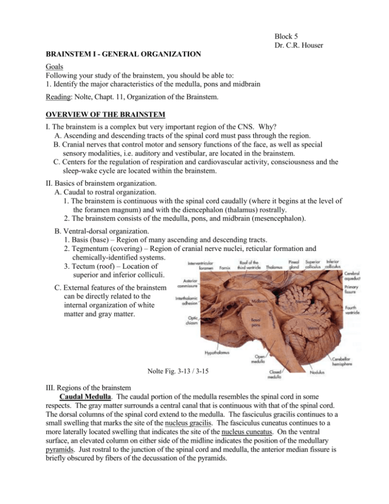
Block 5 Dr. C.R. Houser BRAINSTEM I - GENERAL ORGANIZATION Goals Following your study of the brainstem, you should be able to: 1. Identify the major characteristics of the medulla, pons and midbrain Reading: Nolte, Chapt. 11, Organization of the Brainstem. OVERVIEW OF THE BRAINSTEM I. The brainstem is a complex but very important region of the CNS. Why? A. Ascending and descending tracts of the spinal cord must pass through the region. B. Cranial nerves that control motor and sensory functions of the face, as well as special sensory modalities, i.e. auditory and vestibular, are located in the brainstem. C. Centers for the regulation of respiration and cardiovascular activity, consciousness and the sleep-wake cycle are located within the brainstem. II. Basics of brainstem organization. A. Caudal to rostral organization. 1. The brainstem is continuous with the spinal cord caudally (where it begins at the level of the foramen magnum) and with the diencephalon (thalamus) rostrally. 2. The brainstem consists of the medulla, pons, and midbrain (mesencephalon). B. Ventral-dorsal organization. 1. Basis (base) – Region of many ascending and descending tracts. 2. Tegmentum (covering) – Region of cranial nerve nuclei, reticular formation and chemically-identified systems. 3. Tectum (roof) – Location of superior and inferior colliculi. C. External features of the brainstem can be directly related to the internal organization of white matter and gray matter. Nolte Fig. 3-13 / 3-15 III. Regions of the brainstem Caudal Medulla. The caudal portion of the medulla resembles the spinal cord in some respects. The gray matter surrounds a central canal that is continuous with that of the spinal cord. The dorsal columns of the spinal cord extend to the medulla. The fasciculus gracilis continues to a small swelling that marks the site of the nucleus gracilis. The fasciculus cuneatus continues to a more laterally located swelling that indicates the site of the nucleus cuneatus. On the ventral surface, an elevated column on either side of the midline indicates the position of the medullary pyramids. Just rostral to the junction of the spinal cord and medulla, the anterior median fissure is briefly obscured by fibers of the decussation of the pyramids. Rostral Medulla. In the more rostral part of the medulla, the central canal expands into the fourth ventricle on the dorsal surface. The lower apex of the fourth ventricle, where it narrows into the central canal, is identified as the obex. The area postrema is located in the walls of the ventricle at approximately this location. This region lacks a normal blood brain barrier and is thought to monitor blood for the presence of toxins, and can then trigger vomiting if appropriate (thus the “vomiting center”). Looking down on the floor of the fourth ventricle, several bulges can be seen on either side of the midline. The most medial two elevations mark the sites of the hypoglossal nucleus and dorsal motor nucleus of the vagus. On the ventral surface, the pyramids are still evident and oval swellings are noted just lateral to each pyramid. These are the olives, and they indicate the location of the inferior olivary nuclei that provide a major input to the cerebellum. Dorsolaterally, two elevated columns extend toward the cerebellum; these are the inferior cerebellar peduncles. Pons. The pons is characterized by a large transversely oriented mass of fibers on its ventral surface. The region appears to be a "bridge" between the cerebellar hemispheres. However, the fibers do not actually interconnect the two cerebellar hemispheres but form connections between the pontine nuclei, located within the ventral pons (basis pontis), and the cerebellum. The large groups of fibers that enter the cerebellum are the brachium pontis or middle cerebellar peduncle. In the more rostral pons, much of the roof of the fourth ventricle is formed by the brachium conjunctivum or superior cerebellar peduncle. Midbrain. The fourth ventricle narrows and is continuous with the cerebral aqueduct (aqueduct of Sylvius) within the midbrain. The area of gray matter around the aqueduct is appropriately called the periaqueductal gray. The dorsal surface (roof or tectum) of the midbrain consists of four distinct elevations, the paired inferior colliculi (caudally) and superior colliculi (rostrally). The inferior colliculi are relay nuclei within the auditory path. In contrast, the superior colliculi are not located within the main visual path but instead receive visual input in parallel with the lateral geniculate nucleus of the thalamus (the main relay nucleus in the visual pathway) and participate in visuomotor control. The cerebral peduncles are evident on the ventral surface and contain large groups of fibers that are descending from the cortex to the brainstem and spinal cord (corticopontine and corticospinal fibers). A relatively deep fossa is found between the peduncles and is appropriately named the interpeduncular fossa. The oculomotor nerves emerge from this fossa. Immediately dorsal to the axons in the cerebral peduncle is the substantia nigra. This nuclear region is divided into two parts that have different anatomical connections and utilize different neurotransmitters. The substantia nigra, pars compacta, is located dorsally within the nucleus and contains dopaminergic neurons; the substantia nigra, pars reticulata, is located more ventrally and utilizes GABA as its neurotransmitter. Both regions are important components of the basal ganglia system, even though they are located in the midbrain. 2
