Exam questions
advertisement
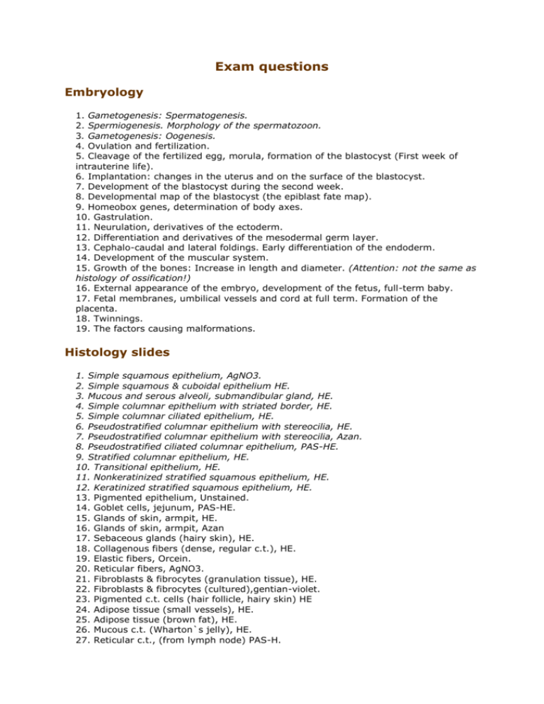
Exam questions Embryology 1. Gametogenesis: Spermatogenesis. 2. Spermiogenesis. Morphology of the spermatozoon. 3. Gametogenesis: Oogenesis. 4. Ovulation and fertilization. 5. Cleavage of the fertilized egg, morula, formation of the blastocyst (First week of intrauterine life). 6. Implantation: changes in the uterus and on the surface of the blastocyst. 7. Development of the blastocyst during the second week. 8. Developmental map of the blastocyst (the epiblast fate map). 9. Homeobox genes, determination of body axes. 10. Gastrulation. 11. Neurulation, derivatives of the ectoderm. 12. Differentiation and derivatives of the mesodermal germ layer. 13. Cephalo-caudal and lateral foldings. Early differentiation of the endoderm. 14. Development of the muscular system. 15. Growth of the bones: Increase in length and diameter. (Attention: not the same as histology of ossification!) 16. External appearance of the embryo, development of the fetus, full-term baby. 17. Fetal membranes, umbilical vessels and cord at full term. Formation of the placenta. 18. Twinnings. 19. The factors causing malformations. Histology slides 1. Simple squamous epithelium, AgNO3. 2. Simple squamous & cuboidal epithelium HE. 3. Mucous and serous alveoli, submandibular gland, HE. 4. Simple columnar epithelium with striated border, HE. 5. Simple columnar ciliated epithelium, HE. 6. Pseudostratified columnar epithelium with stereocilia, HE. 7. Pseudostratified columnar epithelium with stereocilia, Azan. 8. Pseudostratified ciliated columnar epithelium, PAS-HE. 9. Stratified columnar epithelium, HE. 10. Transitional epithelium, HE. 11. Nonkeratinized stratified squamous epithelium, HE. 12. Keratinized stratified squamous epithelium, HE. 13. Pigmented epithelium, Unstained. 14. Goblet cells, jejunum, PAS-HE. 15. Glands of skin, armpit, HE. 16. Glands of skin, armpit, Azan 17. Sebaceous glands (hairy skin), HE. 18. Collagenous fibers (dense, regular c.t.), HE. 19. Elastic fibers, Orcein. 20. Reticular fibers, AgNO3. 21. Fibroblasts & fibrocytes (granulation tissue), HE. 22. Fibroblasts & fibrocytes (cultured),gentian-violet. 23. Pigmented c.t. cells (hair follicle, hairy skin) HE 24. Adipose tissue (small vessels), HE. 25. Adipose tissue (brown fat), HE. 26. Mucous c.t. (Wharton`s jelly), HE. 27. Reticular c.t., (from lymph node) PAS-H. 28. 29. 30. 31. 32. 33. 34. 35. 36. 37. 38. 39. 40. 41. 42. 43. 44. 45. 46. 47. 48. 49. 50. 51. Fibrous cartilage, HE. Hyaline cartilage, HE. Elastic cartilage, Orcein-H. Ground compact bone, t.s. Fuchsin. Ground compact bone, l.s. Fuchsin. Intramembranous bone formation, HE. Intracartilaginous bone formation, HE. Intracartilaginous bone formation, Azan. Smooth muscle l.s. & t.s. HE. Skeletal muscle, HE. Skeletal muscle, Fe-H. Cardiac muscle l.s. HE. Cardiac muscle t.s. HE. Peripheral nerve, t.s.,HE. Peripheral nerve, t.s., Azan. Peripheral nerve, t.s.,OsO4 Sensory ganglion, HE. Sensory ggl., AgNO3. Autonomic ggl., AgNO3. Human blood smear, May-Grünwald-Giemsa. Red bone marrow, HE. Aorta HE. Medium sized artery & vein, HE. Medium sized artery & vein, Orcein. Exam questions Bones and joints. - Group I. 1. The types of connections between bones. The general structures and functions of the synovial joints. 2. Calvaria, fontanelles and emissary foramina of the skull. 3. The anterior and posterior cranial fossae. 4. The middle cranial fossa. 5. The external base of the skull. 6. The orbit. 7. The bony nasal cavity. 8. The pterygopalatine and infratemporal fossae. 9. The mandible. The bony palate. (Bony walls of the oral cavity.) 10. The temporomandibular joint. 11. The thoracic cage. Costovertebral and sternocostal connections, movements of the ribs. 12. The general features and types of vertebrae. Special characteristics of different types. 13. The intervertebral connections. Curvatures and movements of the vertebral column. 14. The atlantooccipital and atlantoaxial joints. Bones and joints. - Group II 1. 2. 3. 4. 5. The bones and joints of the shoulder girdle. The shoulder joint The elbow joint. The wrist joint. Bones and joints of the hand. 6. Components, position and diameters of the pelvis. 7. Bones and ligaments of the pelvis. 8. Connections between the bones of pelvis. Static characteristics. 9. The hip joint. 10. The knee joint. 11. The ankle joint. 12. The talotarsal joint. 13. Bones and joints of the foot (except the talotarsal joint). 14. Bony arches of the foot, and their supporting mechanisms. Regional Anatomy 1. Superficial veins of the limbs. The arteries for testing pulse. 2. Lymphatic drainage of the limbs, the thoracic wall, and the mammary gland. Medical importance of the primary lymph nodes, the axillary and the inguinal lymph nodes. 3. The infraclavicular and the axillary regions. 4. The nuchal region. 5. The dorsal scapular region. Muscles of the back. 6. The anterior and posterior brachial regions 7. The anterior cubital region. 8. The anterior antebrachial and carpal regions. 9. The posterior antebrachial region. 10. The palmar region. 11. The dorsal region of the hand and the posterior carpal region. 12. The subinguinal and anterior femoral regions. 13. The gluteal region. 14. The posterior femoral and popliteal regions. 15. The anterior genicular and crural regions. 16. The posterior crural region. 17. The medial and lateral malleolar regions. 18. The plantar region. 19. The dorsal region of the foot. 20. Nerves and the skin innervation of the upper limb. 21. Nerves and the skin innervation of the lower limb. 22. The arteries and the deep veins of the upper limb. 23. Arteries and deep veins of the lower limb 24. Regions and the structure of the anterior abdominal wall.
