Anatomy of the Eye Visual Tests & Experiments
advertisement
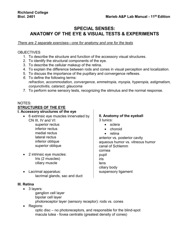
Richland College Biol. 2401 Marieb A&P Lab Manual - 11th Edition ACTIVITY 5: Audiometry SPECIAL SENSES: ANATOMY OF THE EYE & VISUAL TESTS & EXPERIMENTS There are 2 separate exercises—one for anatomy and one for the tests OBJECTIVES 1. To describe the structure and function of the accessory visual structures. 2. To identify the structural components of the eye. 3. To describe the cellular makeup of the retina. 4. To explain the difference between rods and cones in visual perception and localization. 5. To discuss the importance of the pupillary and convergence reflexes. 6. To define the following terms: refraction, accommodation, convergence, emmetropia, myopia, hyperopia, astigmatism, conjunctivitis, cataract, glaucoma 7. To perform some sensory tests, recognizing the stimulus and the normal response. NOTES: STRUCTURES OF THE EYE I. Accessory structures of the eye 6 extrinsic eye muscles innervated by CN III, IV and VI: superior rectus inferior rectus medial rectus lateral rectus inferior oblique superior oblique 2 intrinsic eye muscles: Iris (2 muscles) ciliary muscle Lacrimal apparatus: lacrimal glands, sac and duct II. Anatomy of the eyeball 3 tunics: sclera choroid retina anterior vs. posterior cavity aqueous humor vs. vitreous humor canal of Schlemm cornea pupil iris lens ciliary body suspensory ligament III. Retina 3 layers: ganglion cell layer bipolar cell layer photoreceptor layer (sensory receptor): rods vs. cones Regions: optic disc – no photoreceptors, and responsible for the blind-spot macula lutea - fovea centralis (greatest density of cones) ACTIVITY 1-2: IDENTIFYING EYE STRUCTURES EYE MODELS: 6 extrinsic eye muscles levator palpebrae muscle 3 nerves innervating the muscles (but NOT which nerve innervates which muscle) lacrimal apparatus: lacrimal gland, lacrimal sac, nasolacrimal duct 3 wall layers: sclera, choroid, retina ALL structures labeled on eye in lab book on diagram only (not on models): macula and fovea centralis Canal of Schlemm DISSECTION of cow eye (in lab book): optic nerve ciliary body cornea vitreous humor pupil aqueous humor iris optic disc lens tapedum lucidum 3 wall layers: sclera choroid retina ACTIVITY 3: MICROSCOPIC ANATOMY OF THE RETINA MICROSCOPY: section of the eye ID sclera, choroid, retina 3 layers of the retina: ganglion cells, bipolar cells, rods/cones layer ACTIVITY 4: VISUAL PATHWAYS cross over of fibers along the neural pathways optic chiasma optic nerve vs. optic tract visual fields of each eye VISUAL TEST PROCEDURES: ACTIVITY 1: BLIND SPOT ACTIVITY 2: NEAR-POINT OF ACCOMMODATION ACTIVITY 3: VISUAL ACUITY (Snellen eye chart) ACTIVITY 4: ASTIGMATISM ACTIVITY 5: COLOR BLINDNESS ACTIVITY 6: DEPTH PERCEPTION ACTIVITY 7: EYE REFLEXES Photopupillary Reflex Accommodation Pupillary Reflex Convergence Reflex FOR EACH SENSORY TEST: What is the stimulus? What is the normal response? What is the purpose of response? OPHTHALMOSCOPIC EXAMINATION NOTE: Histology Atlas at Martini website. Open MAP, click on STUDY AREA, which takes you to the Martini website. Vision.doc
