Appendicular Skeleton Lab
advertisement

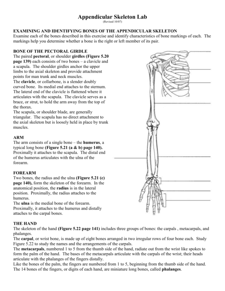
Appendicular Skeleton Lab (Revised 10/07) EXAMINING AND IDENTIFYING BONES OF THE APPENDICULAR SKELETON Examine each of the bones described in this exercise and identify characteristics of bone markings of each. The markings help you determine whether a bone is the right or left member of its pair. BONE OF THE PECTORAL GIRDLE The paired pectoral, or shoulder girdles (Figure 5.20 page 139) each consists of two bones – a clavicle and a scapula. The shoulder girdles anchor the upper limbs to the axial skeleton and provide attachment points for man trunk and neck muscles. The clavicle, or collarbone, is a slender doubly curved bone. Its medial end attaches to the sternum. The lateral end of the clavicle is flattened where it articulates with the scapula. The clavicle serves as a brace, or strut, to hold the arm away from the top of the thorax. The scapula, or shoulder blade, are generally triangular. The scapula has no direct attachment to the axial skeleton but is loosely held in place by trunk muscles. ARM The arm consists of a single bone – the humerus, a typical long bone (Figure 5.21 (a & b) page 140). Proximally it attaches to the scapula. The distal end of the humerus articulates with the ulna of the forearm. FOREARM Two bones, the radius and the ulna (Figure 5.21 (c) page 140), form the skeleton of the forearm. In the anatomical position, the radius is in the lateral position. Proximally, the radius attaches to the humerus. The ulna is the medial bone of the forearm. Proximally, it attaches to the humerus and distally attaches to the carpal bones. THE HAND The skeleton of the hand (Figure 5.22 page 141) includes three groups of bones: the carpals , metacarpals, and phalanges. The carpal, or wrist bone, is made up of eight bones arranged in two irregular rows of four bone each. Study Figure 5.22 to study the names and the arrangements of the carpals. The metacarpals, numbered 1 to 5 from the thumb side of the hand, radiate out from the wrist like spokes to form the palm of the hand. The bases of the metacarpals articulate with the carpals of the wrist; their heads articulate with the phalanges of the fingers distally. Like the bones of the palm, the fingers are numbered from 1 to 5, beginning from the thumb side of the hand. The 14 bones of the fingers, or digits of each hand, are miniature long bones, called phalanges. THE PELVIC GIRDLE The pelvic girdle or hip girdle (Figure 5.23 page 142), is formed by the two coxal bones. The bony pelvis is made up of the pelvic girdle together with the sacrum and the coccyx. Each coxal bone is a result of the fusion of three bones – the ilium, ischium, and pubis. The ilium, a large flaring bone, forms most of the coxal bone. It connects posteriorly with the sacrum. The ischium, forming the inferior portion of the coxal bone, is the “sit-down” bone. It receives majority of the weight of the body when we sit. The pubis is the most anterior part of the coxal bone. Two pubic bones meet anteriorly. The ilium, ischium, and pubis unite at the deep hemispherical socket called the acetabulum, which receives the head of the thigh bone. The female pelvis is modified for childbearing. Generally speaking, the female pelvis is wider, shallower, lighter, and rounder than that of the male. THE THIGH The femur, or thigh bone (Figure 5.24 (a & b), is the only bone of the thigh. It is the heaviest, strongest bone in the body. Its ball-like head articulates with the hop bone via the acetabulum. Distally, the femur articulates with the tibia below. The femur’s anterior surface attaches to the patella (kneecap). THE LEG Two bones, the tibia and the fibula (Figure 5.24 (c)), form the skeleton of the leg. The tibia, or shinbone, is the larger and more medial bone. At the proximal end, the tibia articulates with the distal portion of the femur. Distally, the tibia articulates with the tarsal bones that make up the ankle bones. The sticklike fibula, which lies parallel to the tibia, takes no part in forming the knee joint. Its proximal end articulates with the lateral head of the tibia. The fibula is not a weight-bearing bone. THE FOOT The bones of the foot include 7 tarsal bones forming the ankle, 5 metatarsals which forms the sole of the foot, and 14 phalanges which form toes (Figure 5.25 page145). IDENTIFY BONES: Go around the room, find the numbered bones, and identify the bones. 1. ____________________ 2. ____________________ 3. ____________________ 4. ____________________ 5. ____________________ 6. ____________________ 7. ____________________ 8. ____________________ 9. ____________________ 10. ____________________ 11. ____________________ 12. ____________________ 13. ____________________ 14. ____________________ 15. ____________________ 16. ____________________ 17. ____________________


