ECG & Heart Sounds Student Handout
advertisement
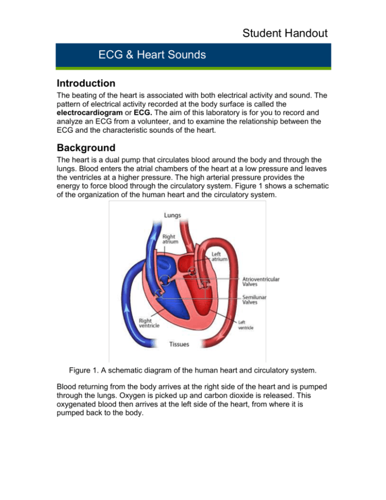
Student Handout ECG & Heart Sounds Introduction The beating of the heart is associated with both electrical activity and sound. The pattern of electrical activity recorded at the body surface is called the electrocardiogram or ECG. The aim of this laboratory is for you to record and analyze an ECG from a volunteer, and to examine the relationship between the ECG and the characteristic sounds of the heart. Background The heart is a dual pump that circulates blood around the body and through the lungs. Blood enters the atrial chambers of the heart at a low pressure and leaves the ventricles at a higher pressure. The high arterial pressure provides the energy to force blood through the circulatory system. Figure 1 shows a schematic of the organization of the human heart and the circulatory system. Figure 1. A schematic diagram of the human heart and circulatory system. Blood returning from the body arrives at the right side of the heart and is pumped through the lungs. Oxygen is picked up and carbon dioxide is released. This oxygenated blood then arrives at the left side of the heart, from where it is pumped back to the body. Student Handout ECG & Heart Sounds The electrical activity of the heart Cardiac contractions are not dependent upon a nerve supply. However, innervation by the parasympathetic (vagus) and sympathetic nerves does modify the basic cardiac rhythm. Thus the central nervous system can affect this rhythm. The best known example of this is so-called sinus arrhythmia where respiratory activity affects the heart rate. A group of specialized muscle cells, the sinoatrial, or sinuatrial (SA) node acts as the pacemaker for the heart (Figure 2). These cells rhythmically produce action potentials that spread through the muscle fibers of the atria. The resulting contraction pushes blood into the ventricles. The only electrical connection between the atria and the ventricles is via the atrioventricular (AV) node. The action potential spreads slowly through the AV node, thus allowing atrial contraction to contribute to ventricular filling, and then rapidly through the AV bundle and Purkinje fibers to excite both ventricles. Figure 2. Components of the human heart involved in conduction. The cardiac cycle involves a sequential contraction of the atria and the ventricles. The combined electrical activity of the different myocardial cells produces electrical currents that spread through the body fluids. These currents are large enough to be detected by recording electrodes placed on the skin (Figure 3). Student Handout ECG & Heart Sounds Figure 3. Standard method for connecting the electrodes to a volunteer. The regular pattern of peaks during one cardiac cycle is shown in Figure 4. Figure 4. One cardiac cycle showing the P wave, QRS complex and T wave. The action potentials recorded from atrial and ventricular fibers are different from those recorded from nerves and skeletal muscle. The cardiac action potential is composed of three phases: a rapid depolarization, a plateau depolarization (which is very obvious in ventricular fibers) and a repolarization back to resting membrane potential (Figure 5). Student Handout ECG & Heart Sounds Figure 5. A typical ventricular muscle action potential. The components of the ECG can be correlated with the electrical activity of the atrial and ventricular muscle: The P-wave is produced by atrial depolarization. The QRS complex is produced by ventricular depolarization; atrial repolarization also occurs during this time, but its contribution is insignificant. The T-wave is produced by ventricular repolarization. Heart valves and heart sounds Each side of the heart is provided with two valves, which convert the rhythmic contractions into a unidirectional pumping. The valves close automatically whenever there is a pressure difference across the valve that would cause backflow of blood. Closure gives rise to audible vibrations (heart sounds). Atrioventricular (AV) valves between the atrium and ventricle on each side of the heart prevent backflow from ventricle to atrium. Semilunar valves are located between the ventricle and the artery on each side of the heart, and prevent backflow of blood from the aorta and pulmonary artery into the respective ventricle. The closure of these valves is responsible for the characteristic sound produced by the heart, usually referred to as a ‘lub-dup’ sound. The lower-pitched ‘lub’ sound occurs during the early phase of ventricular contraction. This is produced by closing of the atrioventricular (mitral and tricuspid) valves. These valves prevent blood from flowing back into the atria. When the ventricles relax, the blood pressure drops below that in the artery, and the semilunar valves (aortic and pulmonary) close, producing the higher-pitched ‘dup’ sound. Malfunctions of these valves often produce an audible murmur, which can be detected with a stethoscope. Student Handout ECG & Heart Sounds The cardiac cycle The sequence of events in the heart during one cardiac cycle is summarized in Figure 6. During ventricular diastole blood is returning to the heart. Deoxygenated blood from the periphery enters the right atrium and flows into the right ventricle through its open AV valve. Oxygenated blood from the lungs enters the left atrium and flows into the left ventricle through its open AV valve. Filling of the ventricles is completed when the atria contract (atrial systole). In the resting state, atrial systole accounts for some 20% of atrial filling. Atrial contraction is followed by contraction of the ventricles (ventricular systole). Initially, as the ventricles begin to contract the pressure in them rises and exceeds that in the atria. This closes the AV valves. But, until the pressure in the left ventricle exceeds that in the aorta (and in the right ventricle exceeds that in the pulmonary artery), the volume of the ventricles can not change. This is the so-called isovolumic phase of ventricular contraction. Finally, when the pressure in the left ventricle exceeds that in the aorta (and the pressure in the right ventricle exceeds that in the pulmonary artery), the aortic and pulmonary valves open and blood is ejected into the aorta and pulmonary arteries. As the ventricular muscle relaxes, pressures in the ventricles fall below those in the aorta and pulmonary artery, and the aortic and pulmonary valves close. Ventricular pressure continues to fall and once it has fallen below that in the atria, the AV valves open and ventricular filling begins again. Figure 6. The cardiac cycle. Student Handout ECG & Heart Sounds Changes in a variety of parameters during one cardiac cycle are summarized in a figure introduced by Wiggers. A modified form of this is shown in Figure 7. The importance of this representation is that it allows you to see the temporal relationships between the different parameters. Figure 7. A Wiggers' diagram. Student Handout ECG & Heart Sounds What you will do in the laboratory 1. ECG in a resting volunteer. You will record the ECG, analyze the signal and observe the effects of slight movement on the signal. 2. ECG recorded from several other volunteers. You will identify and discuss similarities and differences in the ECGs of the different participants. 3. ECG and heart sounds. You will use a stethoscope to listen to the heart and an event marker to determine the relationship between what you are hearing and the ECG being recorded at the same time. 4. ECG and phonocardiography. You will also record the heart sounds (phonocardiogram) together with the ECG.


