AP Lab 7 – Fruit Fly Inheritence Project
advertisement
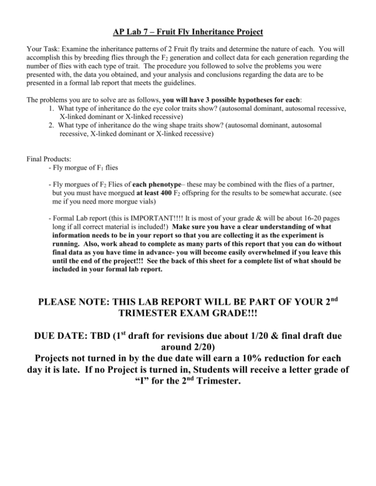
AP Lab 7 – Fruit Fly Inheritance Project Your Task: Examine the inheritance patterns of 2 Fruit fly traits and determine the nature of each. You will accomplish this by breeding flies through the F2 generation and collect data for each generation regarding the number of flies with each type of trait. The procedure you followed to solve the problems you were presented with, the data you obtained, and your analysis and conclusions regarding the data are to be presented in a formal lab report that meets the guidelines. The problems you are to solve are as follows, you will have 3 possible hypotheses for each: 1. What type of inheritance do the eye color traits show? (autosomal dominant, autosomal recessive, X-linked dominant or X-linked recessive) 2. What type of inheritance do the wing shape traits show? (autosomal dominant, autosomal recessive, X-linked dominant or X-linked recessive) Final Products: - Fly morgue of F1 flies - Fly morgues of F2 Flies of each phenotype– these may be combined with the flies of a partner, but you must have morgued at least 400 F2 offspring for the results to be somewhat accurate. (see me if you need more morgue vials) - Formal Lab report (this is IMPORTANT!!!! It is most of your grade & will be about 16-20 pages long if all correct material is included!) Make sure you have a clear understanding of what information needs to be in your report so that you are collecting it as the experiment is running. Also, work ahead to complete as many parts of this report that you can do without final data as you have time in advance- you will become easily overwhelmed if you leave this until the end of the project!!! See the back of this sheet for a complete list of what should be included in your formal lab report. PLEASE NOTE: THIS LAB REPORT WILL BE PART OF YOUR 2nd TRIMESTER EXAM GRADE!!! DUE DATE: TBD (1st draft for revisions due about 1/20 & final draft due around 2/20) Projects not turned in by the due date will earn a 10% reduction for each day it is late. If no Project is turned in, Students will receive a letter grade of “I” for the 2nd Trimester. Your Lab report should follow the information described in the packet “guidelines for Formal Lab Reports” that accompanies this assignment packet. All concepts addressed in those guidelines should be addressed in your lab report. This should include (but is not necessarily limited to) the following information in the appropriate section of the report: - Background information on why studying fly characteristics is useful in the study of genetics & on the various types of inheritance shown by your traits. (Background information) - Qualitative data tables on a weekly basis for each offspring generation (F1 & F2) that includes observations of: the vial (appearance of substrate, moisture level, condensation, temperature kept at, lighting conditions, any other environmental conditions the vial was subject to, etc.) larvae (emergence, size, behavior, coloration, when pupating/ pupae coloration, etc) fly behavior (active, inactive, at top/bottom, dead adults, & hatching times vs. sex of hatched individuals) - Raw data tables that include Drosophila record sheet showing data for eye color trait & Drosophila record sheet showing data for wing shape trait. - Condensed data table that shows the totals of the F1 & F2 generations as well as the Chi2 results for the F2 generation in a logical manner that helps clarify the results of the experiment. (results table) - A discussion of and application of the following concepts AS THEY RELATE TO YOUR DATA: The Law of Independent Assortment, The Law of Dominance, Homozygous, Heterozygous, Genotype, Phenotype, Segregation of alleles, Autosomal traits, X-linked traits, and any other major genetics concepts we have discussed throughout the course of the unit. (see analysis checklist on the back of the fly project grading rubric for more detail) - Show the calculations of the probabilities of each possible outcome to represent the F2 offspring of your crosses & provide expected ratios to accompany the explanations in your analysis. - Create a graph to compare the results of the F2 generation. - Complete Chi2 analysis calculations and use them to evaluate each of your 3 hypotheses for each trait and to reject 2 of the hypotheses & support the other (a sample of these should be in your calculations section too). - significant bonus points can be earned by showing Hardy-Weinberg calculations using your data & applying these concepts to your results in the analysis section. Fly Lab Scoring Rubric Total Score: / 85 points TITLE (3 points Total) ____ Conveys to the reader the purpose of the paper PART I - INTRODUCTION Background Information (2 points each) _____ Provides a basic understanding of concepts relating to the experiment the reader may need as context to understand the experiment and its results _____ Is clear & concise without overwhelming depth or extra non-essential info Statement of problem (1/2 point each) _____ Not a yes/no question _____ Independent & Dependent variables included _____ Problem is clearly testable _____ Response is written in a clear and concise manner Hypothesis (1/2 point each) _____ Statement predicts the outcome of the experiment & statement gives enough information to understand prediction(s) _____ Prediction includes both independent and dependent Variables _____ Hypothesis is testable in an experiment _____ A rationale is given for the hypothesis (draws on/includes background information and/or scientific concepts) Variables _____ Independent variable correctly identified (1/2 pt.) _____ Dependent variable correctly identified (1/2 pt.) _____ Constants/Controlled variables identified (1 pt.) _____ Method for keeping constants controlled is identified (1 pt.) Experimental Control (2 points) _____ Included a standard of comparison /control set-up PART II - METHODS Materials _____ All materials used are clearly listed separate from procedure, in detail (1 pt.) Procedure _____ Procedure is well organized in a logical fashion (1 pt.) _____ How to measure the dependent variable is specifically explained. (1 pt.) _____ Where to record data is correctly listed by table name and number (1pt.) _____ Enough information is given in detail so another could repeat procedure (1 pt.) _____ Diagram that clarifies understanding of instructions is included. (1 pt) _____ Repeated trials/criteria for additional trials (2 pts.) PART III - RESULTS Qualitative Observations From Experiment _____ Observations given that reflect multiple points during the Experiment (ex: beginning, middle & end) (1 pts.) _____ Adequate detail to understand in a clearly organized table (1pts.) Raw Data Table(s) _____ All raw data is given (2 pt.) _____ Table(s) labeled properly (1 pt.) Calculations ____ Shows a word equation and simple calculation for each value shown in the results table (3 pts.) Results Table _____ Results table with most important data included (2 pts.) Graphs _____ Data converted into appropriate type of graph (1/2 pt.) _____ Graph is titled & labeled appropriately (1/2 pt.) _____ x-axis addresses appropriate variable (1/2 pt.) _____ y-axis addresses appropriate variable (1/2 pt.) _____ Graph clarifies data visually to reader & is useful in analyzing results (2 pts.) Summary of Results (1 point each) ____ In paragraph form _____ Describe WHAT happened (not why it happened) _____ Describe the trend(s) in data. ____ Unusual data points pointed out PART IV - DISCUSSION Analysis of Results _____ What your results from the previous section mean & included major concepts the investigation focused on (5 pts.) _____ Explain cause and effect relationships (1 pt.) _____ Thorough detail is given to understand the data (2 pts) _____ Trends in data explained (1 pt) _____ Unusual data points explained (2 pts) _____ Response is clear and concise (2 pts) _____All statements are supported by the data (2 pts) Experimental Error (errors that actually took place) (1pt each = 3pts) _____ Explanation of errors is given (at least 2) _____ Important information about accuracy & precision is explained. _____ Effect errors had on data identified for each error & discussed in detail PART V - CONCLUSION Conclusion _____ Hypothesis is evaluated according to data (accept or reject) (1 pt.) _____ hypothesis is re-stated (1/2 pt.) _____ Reasons to accept/reject hypothesis given and linked to specific results that supports or refutes the hypothesis (2 pts.) _____ All statements are supported by the data (1 pt.) _____ The conclusion is scientifically correct (1/2 pt.) _____ Obvious link made between Scientific concepts and your conclusion (2 pts.) Recommendations for further experimentation (1 point each) _____ Suggestions for improving THIS experiment _____ Suggestions for future experiments given (new problem statement formulated with different variables) _____ Further predictions made based on results _____ Practical application(s) of experiment given _____ How can this be used in the real world? Citations _____ Information used in any part of this report that was not directly produced by this investigation was properly referenced and linked to a citation in the body of the report. (4 pts) _____ Proper citations given in acceptable format at the end of the report for all cited material (3 pts.) Format _____ Typed, spelling checked, double spaced, tables/section labels break in appropriate places (5 pts.) _____ Graphs, tables, diagrams all neat and clearly legible (2 pts.) _____ grammar, sentence flow, terminology convey competency and establish credibility of the author (3 pts.) Analysis of Results Evaluation ____ Explain any cause-and-effect relationships. ____ Are you sure that there was only one variable that could have caused that result? ____ How much variability is there in the results? ____ What are some possible causes of this variability? ____ How reliable is the data? ____ What general conclusions can be drawn? ____ What is the importance of the discoveries you have made? ____ Look at the mean & median and determine if any differences between them are significant. If so, what may be the cause of this difference? ____ Is there a large range in your data points, or do they all group together? A discussion of and application of the following concepts AS THEY RELATE TO YOUR DATA: ____ The Law of Independent Assortment ____ The Law of Dominance ____ Homozygous ____ Heterozygous ____ Genotype ____ Phenotype ____ Segregation of alleles ____ Autosomal traits/inheritance ____ X-linked traits/inheritance General Timeline for the experiment A culture containing several pairs of experimental flies will be given to you. The Male flies are all homozygous and genetically identical. Your female fruit flies were all virgins when placed in the vial and are all homozygous and genetically identical. These flies are the P1 generation. The females will already be laying eggs & when these hatch, they will represent the First Filial (F1) generation. First week: Before any F1 flies emerge, you must anesthetize your Parent flies, record their characteristics (be sure to separate the males & females when doing so) in your data tables and discard them (no need to morgue these). Second week: Begin collecting the F1 flies. Always collect the flies at the end of the school day. Most flies emerge in the morning, so this brief aging will allow coloration to develop and wings to take shape. This will also allow mutations to become more fully expressed making your phenotyping more accurate. As soon as possible, place five or six pairs of anesthetized F1 flies into a fresh culture bottle (this new vial will produce the F2 generation & should be labeled as such). Make sure you leave the vial on its side until the flies are fully active so they don’t get stuck in the culture medium at the bottom. For this mating, flies do not need to be virgin because all flies in the F1 generation will be genetically identical heterozygotes. Be sure to include these pairs of F1 flies in your characteristic tallies in your data tables. Once you have removed flies from the vial to parent the F2 generation, all other F1 flies should be collected once per day, counted by characteristic, recorded in your data tables & morgued in your morgue vial labeled F1. Third week: You should have about the same amount of time to collect F1 flies as it took to produce them from the time you got your parents from your teacher (about 2 weeks or so). Stop collecting flies before there is any chance of contaminating the results with new F2 flies produced. In general, once you see a new batch of larvae beginning to crawl up the side of the vial & pupate, you should stop collecting F1 offspring. Once you have OK’d it with your teacher, freeze the container to kill everything off and then clean out your vial with warm soapy water. Fourth week: Begin removing the F2 flies once per day. Determine the sex & phenotype of each & record this in your data tables. Place all mutated offspring into your second morgue (F2 mutated) and all your nonmutated offspring into your third morgue (F2 non-mutated). Stop removing F2 flies before there is any chance of contamination by F3 flies. Off spring should be in the 100 + or more range for a sample size large enough to yield reliable statistical data. F1 Parents (P1) to trash 10 F1 ♂ & 10 F1♀ From F1Offspring to a new culture vial to produce F2 offspring. After F2 larvae begin to climb up the sides to pupate, remove F1 parent flies & count them & add them to the F1 morgue. Extra F1 Offspring to F1 morgue after counting F2 All F2 Offspring to F2 morgues (1 per phenotype class) after counting Fruit fly lifecycle The length of the fruit fly life cycle is determined by a number of factors. Of these, temperature is more important. At normal room temperature (25C), the life cycle takes from 10-14 days. During warmer conditions, the lifecycle can be complete in as few as 7 days. The entire lifecycle is known as a complete lifecycle because fruit flies change form several times before they are adults. This includes 4 stages that progress from egg, to larvae, to pupae, to adult. Eggs – The eggs are small, oval and have 2 filaments at one end. They are laid on the surface of the nutrient medium and with practice can be seen. Eggs are often seen passing from the rear of anesthetized females. Eggs hatch into larvae in about 1 day. A typical female can lay as many as 400 eggs. Larva- the larva are worm like and white/clear in color. Their mouth parts are black and can be seen moving back & forth in the blue culture medium. Larvae channel through the culture media while eating; thus channels are a good indicator of successful larvae growth. The larva molt or shed their skin twice as they grow. In the last of the 3 larval stages, the cells of the salivary glands contain giant chromosomes, which can be seen under a microscope with proper staining. Pupa – When larvae are ready to pupate (become pupa) they usually climb up the side of the culture vial or onto netting in the vial. The last larval covering then becomes harder and darker, forming the pupal case. Through this case you can watch the metamorphosis into an adult fly. In the later stages, the eyes, wings, and legs become readily visible. Adult – When adults first emerge from the pupal case they are fragile and light in color and their wings are not expanded. In 2-3 hours their pigmentation develops, wings take on their final form, and their bodies round out into their final adult shape. They live a month or more & then die. A female doesn’t mate for about 10-12 hours after emerging from the pupa & is called a Virgin female until she does so. Females will mate several times & can store sperm from several males & fertilizes eggs as she lays them. Because of this, virgin females must be separated from the males before mating to ensure the cross you intend to create takes place. Adult males have an abdomen with a heavily pigmented tip and dark sex combs on their front forelegs. Adult females have an abdomen with several transverse stripes and no sex combs. Females also tend to have significantly lighter and larger abdomens than the males. Anesthetizing your flies Step 1: Transfer flies to Anesthetizing vial using “tap method” taught in class. Step 2: Dip the bristled end of the anesthetic wand into the fly nap & remove excess by running the wand along the lid of the bottle. Use one finger to push the plug in the vial aside slightly & quickly put wand in the vial so tip is just below the plug. Keep the vial upright & the wand in place as the anesthetic begins to take effect. Watch the flies closely & remove the wand when they are all anesthetized (if you don’t you will kill them). Step 3: Dump flies onto a note card for easy sorting under the dissecting scope. Use a paintbrush to gently move flies around. They will often stay asleep for 15-30 minutes or more depending on the number of flies and their age & size. If they start to wake up, just repeat this process. Step 4: Place unconscious flies into a morgue if they are to be killed, or into a new culture vial on its side until they are fully conscious if they are to be mated for new offspring. The Chi-square test of significance Chi-square is a statistical test commonly used to compare observed data with data we would expect to obtain according to a specific hypothesis. For example, if, according to Mendel's laws, you expected 10 of 20 offspring from a cross to be male and the actual observed number was 8 males, then you might want to know about the "goodness to fit" between the observed and expected. Were the deviations (differences between observed and expected) the result of chance, or were they due to other factors. How much deviation can occur before you, the investigator, must conclude that something other than chance is at work, causing the observed to differ from the expected. The chi-square test is always testing what scientists call the null hypothesis, which states that there is no significant difference between the expected and observed result. The formula for calculating chi-square (2) is: 2= (o-e)2/e That is, chi-square is the sum of the squared difference between observed (o) and the expected (e) data (or the deviation, d), divided by the expected data in all possible categories. For example, suppose that a cross between two pea plants yields a population of 880 plants, 639 with green seeds and 241 with yellow seeds. You are asked to propose the genotypes of the parents. Your hypothesis is that the allele for green is dominant to the allele for yellow and that the parent plants were both heterozygous for this trait. If your hypothesis is true, then the predicted ratio of offspring from this cross would be 3:1 (based on Mendel's laws) as predicted from the results of the Punnett square (Figure B.1). Figure B.1 - Punnett Square. Predicted offspring from cross between green and yellow-seeded plants. Green (G) is dominant (3/4 green; 1/4 yellow). Table B.1-Calculating Chi-Square Observed (o) Expected (e) Deviation (o – e) Deviation2 (o – e)2 (o – e)2 / e 2 Green Yellow 639 241 660 220 -21 21 441 441 0.668 2 2.668 To calculate 2 , first determine the number expected in each category. If the ratio is 3:1 and the total number of observed individuals is 880, then the expected numerical values should be 660 green and 220 yellow. Then calculate 2 using this formula, as shown in Table B.1. Note that we get a value of 2.668 for 2. But what does this number mean? Here's how to interpret the 2 value: 1. Determine degrees of freedom (df). Degrees of freedom can be calculated as the number of categories in the problem minus 1. In our example, there are two categories (green and yellow); therefore, there is I degree of freedom. 2. Determine a relative standard to serve as the basis for accepting or rejecting the hypothesis. The relative standard commonly used in biological research is p > 0.05. The p value is the probability that the deviation of the observed from that expected is due to chance alone (no other forces acting). In this case, using p > 0.05, you would expect any deviation to be due to chance alone 5% of the time or less. 3. Refer to a chi-square distribution table (Table B.2). Using the appropriate degrees of 'freedom, locate the value closest to your calculated chi-square in the table. Determine the closest p (probability) value associated with your chi-square and degrees of freedom. In this case (2=2.668), the p value is about 0.10, which means that there is a 10% probability that any deviation from expected results is due to chance only. Based on our standard p > 0.05, this is within the range of acceptable deviation. In terms of your hypothesis for this example, the observed chi-square is not significantly different from expected. The observed numbers are consistent with those expected under Mendel's law. Step-by-Step Procedure for Testing Your Hypothesis and Calculating Chi-Square 1. State the hypothesis being tested and the predicted results. Gather the data by conducting the proper experiment (or, if working genetics problems, use the data provided in the problem). 2. Determine the expected numbers for each observational class. Remember to use numbers, not percentages. Chi-square should not be calculated if the expected value in any category is less than 5. 3. Calculate 2 using the formula. Complete all calculations to three significant digits. Round off your answer to two significant digits. 4. Use the chi-square distribution table to determine significance of the value. a. b. c. Determine degrees of freedom and locate the value in the appropriate column. Locate the value closest to your calculated 2 on that degrees of freedom df row. Move up the column to determine the p value. 5. State your conclusion in terms of your hypothesis. a. b. If the p value for the calculated 2 is p > 0.05, accept your hypothesis. 'The deviation is small enough that chance alone accounts for it. A p value of 0.6, for example, means that there is a 60% probability that any deviation from expected is due to chance only. This is within the range of acceptable deviation. If the p value for the calculated 2 is p < 0.05, reject your hypothesis, and conclude that some factor other than chance is operating for the deviation to be so great. For example, a p value of 0.01 means that there is only a 1% chance that this deviation is due to chance alone. Therefore, other factors must be involved. Table B.2 Chi-Square Distribution Degrees of Freedom (df) Probability (p) 0.95 0.90 0.80 0.70 0.50 0.30 0.20 0.10 0.05 0.01 0.001 1 0.004 0.02 0.06 0.15 0.46 1.07 1.64 2.71 3.84 6.64 10.83 2 0.10 0.21 0.45 0.71 1.39 2.41 3.22 4.60 5.99 9.21 13.82 3 0.35 0.58 1.01 1.42 2.37 3.66 4.64 6.25 7.82 11.34 16.27 4 0.71 1.06 1.65 2.20 3.36 4.88 5.99 7.78 9.49 13.28 18.47 5 1.14 1.61 2.34 3.00 4.35 6.06 7.29 9.24 11.07 15.09 20.52 6 1.63 2.20 3.07 3.83 5.35 7.23 8.56 10.64 12.59 16.81 22.46 7 2.17 2.83 3.82 4.67 6.35 8.38 9.80 12.02 14.07 18.48 24.32 8 2.73 3.49 4.59 5.53 7.34 9.52 11.03 13.36 15.51 20.09 26.12 9 3.32 4.17 5.38 6.39 8.34 10.66 12.24 14.68 16.92 21.67 27.88 10 3.94 4.86 6.18 7.27 9.34 11.78 13.44 15.99 18.31 23.21 29.59 Nonsignificant Significant
