Gram Stain Lab Report: Identifying Bacteria Types
advertisement
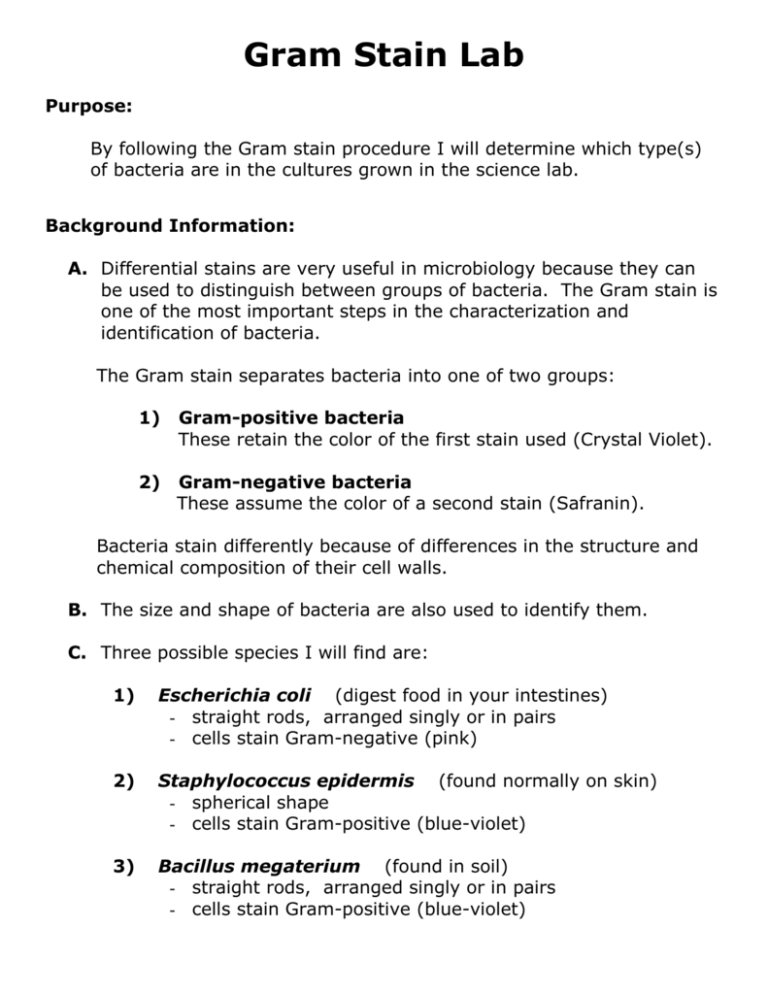
Gram Stain Lab Purpose: By following the Gram stain procedure I will determine which type(s) of bacteria are in the cultures grown in the science lab. Background Information: A. Differential stains are very useful in microbiology because they can be used to distinguish between groups of bacteria. The Gram stain is one of the most important steps in the characterization and identification of bacteria. The Gram stain separates bacteria into one of two groups: 1) Gram-positive bacteria These retain the color of the first stain used (Crystal Violet). 2) Gram-negative bacteria These assume the color of a second stain (Safranin). Bacteria stain differently because of differences in the structure and chemical composition of their cell walls. B. The size and shape of bacteria are also used to identify them. C. Three possible species I will find are: 1) Escherichia coli (digest food in your intestines) - straight rods, arranged singly or in pairs - cells stain Gram-negative (pink) 2) Staphylococcus epidermis (found normally on skin) - spherical shape - cells stain Gram-positive (blue-violet) 3) Bacillus megaterium (found in soil) - straight rods, arranged singly or in pairs - cells stain Gram-positive (blue-violet) Procedure: Part I -- Culture Transfer: A. Wash, rinse and dry a slide. B. Pass the end of the inoculating loop through the flame of a Bunsen burner until it is red hot. C. Let it cool. D. Put a drop of water on the slide. E. Use the inoculating loop to pick up a small amount of the bacteria culture from the petri dish. F. Add the bacteria on the loop into the water drop on the slide, mixing it thoroughly, and spreading it out into a large, thin layer. G. Air dry the slide on the warming tray until the water is gone. H. Holding the slide in a clothespin, heat fix the bacteria by passing the bottom of the slide through the flame of the Bunsen burner for 5 seconds. I. Let it cool. Leave the clothespin on it for the staining portion. Part II – Staining: Step 1 To be done over the sink!! Crystal Violet 60 seconds Cover the smear with an even layer of stain. Keep it horizontal. This is the primary stain that enters the bacterial cells making them visible. Step 2 Water Rinse brief Rinse slide with a gentle stream of water. NO RUBBING! Step 3 Gram’s Iodine 60 seconds Cover the smear with an even layer of stain. Keep it horizontal. This stain forms a chemical complex with the crystal violet that helps the dye attach to the bacteria’s cell wall, membrane, and cytoplasm. Step 4 Water Rinse thorough Rinse slide for several seconds with a gentle stream of water. NO RUBBING! Step 5 95% Ethanol 10 seconds Cover the smear with an even layer of stain. Keep it horizontal. This draws out the dye from the bacteria cells. Since the Gram negative bacteria have a much thinner wall, the dye is removed more rapidly from them. Step 6 Water Rinse thorough Immediately rinse slide for several seconds with a gentle stream of water. NO RUBBING! If the Ethanol remains for more than 10 seconds, it will remove all of the dye from each type of bacteria. If done correctly, the Gram positive cells will still be dark blue, while the Gram negative cells will be clear and difficult to see. Step 7 Safranin 60 seconds Cover the smear with an even layer of stain. Keep it horizontal. This stain acts as a counterstain to color the clear, Gram negative bacteria red. Step 8 Water Rinse thorough Immediately rinse slide for several seconds with a gentle stream of water. NO RUBBING! Step 9 Dry The Slide carefully Carefully blot the slide dry with some paper towel. NO RUBBING! Step 10 Remove the clothespin for the slide and place a cover slip over the area that you plan to view through the microscope. Step 11 When finished with the lab, wash the slide and cover slip carefully in the tub of soapy water provided. Dry, and put them away. Observations: 1. I took bacteria from dish __________. Get the smear in focus using the 4x objective lens. Then switch to the 10x objective lens and refocus. Now, draw and label what you observe. Bacteria @ 100x 2. Switch to the 40X objective lens. Draw and label what you observe. The color and shape of the bacteria should be exact in your drawings! Conclusion: Bacteria @ 400X Explain how it went, what you observed, what you learned. Be sure to include what type of bacteria you had, and how you identified them.

