a p p l i c at i o n N o t e
SPA Technology
Assay Development Guide to
Protein:Protein Studies
Introduction
Interactions between proteins are a key feature of many biochemical processes, for example cell signalling. In the absence
of any enzymatic activity, measurement of protein:protein interactions has presented problems. Scintillation Proximity Assay
(SPA) technology permits the direct measurement of binding of
one protein to another. In addition, in cases where the dissociation
rate of the interaction is high, SPA provides a means of equilibrium
counting in an homogeneous assay format. An SPA system for
the measurement of the direct binding of the GTP-ase activating
protein, Neurofibromin-1 (NF1) to an oncogenic Ras protein
(Leu-61 Ras) has been developed(1). The assay development
and optimization process is used to illustrate the key features
in designing a protein-protein interaction SPA.
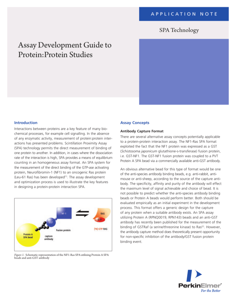
Figure 1. Schematic representation of the NF1-Ras SPA utilizing Protein A SPA
beads and anti-GST antibody
Assay Concepts
Antibody Capture Format
There are several alternative assay concepts potentially applicable
to a protein-protein interaction assay. The NF1-Ras SPA format
exploited the fact that the NF1 protein was expressed as a GST
(Schistosoma japonicum glutathione-s-transferase) fusion protein,
i.e. GST-NF1. The GST-NF1 fusion protein was coupled to a PVT
Protein A SPA bead via a commercially available anti-GST antibody.
An obvious alternative bead for this type of format would be one
of the anti-species antibody binding beads, e.g. anti-rabbit, antimouse or anti-sheep, according to the source of the capture antibody. The specificity, affinity and purity of the antibody will effect
the maximum level of signal achievable and choice of bead. It is
not possible to predict whether the anti-species antibody binding
beads or Protein A beads would perform better. Both should be
evaluated empirically as an initial experiment in the development
process. This format offers a generic design for the capture
of any protein when a suitable antibody exists. An SPA assay
utilizing Protein A (RPNQ0019, RPN143) beads and an anti-GST
antibody has recently been published for the measurement of the
binding of GSTRaf (a serine/threonine kinase) to Ras(2). However,
the antibody capture method does theoretically present opportunity
for non-specific inhibition of the antibody/GST fusion protein
binding event.
Streptavidin-biotin format
If one of the proteins can be biotinylated without loss of biological activity, then the use of the streptavidin bead becomes
an option. The avidity and stability of the biotin-streptavidin
bond can be an advantage. GST-fusion proteins have been
biotinylated for other assays without interfering with the functional activity of the protein (Figure 2). This format has been
used for an SPA system designed to measure the interaction
between Ras and Raf(4). In this assay, a GSTRaf fusion protein was
biotinylated using sulfosuccinimidyl-6-(biotinamido) hexanoate.
for loading Ras with radiolabelled GTPgS. The use of GTPgS
permits the use of wild type Ras proteins. For the NF1-Ras SPA,
an oncogenic Ras protein (Leu61 Ras) was used. This mutant
does not hydrolyze GTP even upon binding of a GTP-ase activating protein. This enabled the Leu61 Ras to be loaded with
[3H]GTP using an exchange reaction performed in the presence
of a GTP regenerating system (phosphoenol pyruvate and
pyruvate kinase) under optimized conditions(1). GDP and GTP
rapidly exchange from the protein into solution and any GDP
in solution will be converted into GTP.
Assay Optimization
Factors affecting the signal:noise ratio will be the relative amounts
of each reagent present, quality and affinity of the capture
antibody and the affinity of the two proteins participating in
the interaction.
Figure 2. Schematic diagram of potential Streptavidin-biotin capture format for the
NF1-Ras SPA
As has been mentioned earlier, to obtain a maximum signal it is
necessary that the capture protein (either biotinylated or coupled
to the antibody) be present in excess to the radiolabelled protein
and then that bead be present in excess to capture the complex.
This section will focus on the optimization process followed for
the NF1-Ras SPA.
Antibody
If biotinylation is considered, it is necessary to optimize the
ratio of biotinylation reagent to protein. This must be done
empirically for the system under study. There is currently no
method for the precise determination of the number of biotins
that become attached to a protein.
For all assays using an antibody capture format, the purity and
affinity of the antibody will greatly influence the amount of
antibody (and hence the amount of bead) required for maximal
signal. It will be essential that the antibody:protein interaction
does not interfere with the protein:protein interaction.
A suggested protocol would be to biotinylate the protein using
increasing molar ratios of biotinylation reagent to protein from
1:1 up to 50:1.
For the NF1-Ras assay, the initial experiment assessed the titre
of the anti-GST antibody required. The anti-GST antibody was
diluted in assay buffer and a constant volume of each dilution
added to each assay well. Maximal binding was achieved using
a dilution of 1:20.
Having biotinylated the protein and separated it from the biotinylation reagent, the biotinylated protein could be tested in a
simple binding experiment using 1mg of streptavidin bead and
a constant amount of the radiolabelled protein. Once an optimal
ratio of biotinylation reagent to protein is established, the ratio
of bead to biotinylated protein can be determined.
An important point to remember with all the formats described
in this section is that the bead or bead-antibody has to be present in excess with respect to the protein to maximize capture
by the beads.
Radiolabelling
For detection, radiolabelling one of the proteins is a requirement.
In order to achieve a good signal, a chemically pure protein
labelled to a high specific activity is desirable. The methods for
tritiation and iodination of proteins are described in the course
manual. Kahl et al(4) compared both iodine and tritium for
labelling Ras directly using conventional labelling methods and
2
Figure 3. The effect of anti-GST antibody dilution on the binding of HRasL61. GTP
to GST-NF1 in the presence of 1 mg Protein A bead in 50 mM Tris.HCl, pH7.5,
2mM dithiothreitol. p = SPA cpm bound in absence of GST-NF1, q = SPA cpm
bound in presence of 30pmol/well GST-NF1, n = specific SPA cpm bound.
SPA Bead
As shown in Figure 4, four different bead concentrations were
tested in the presence of constant amounts of anti-GST antibody,
GST-NF1 (Figure 3).
Figure 5. Effect of increasing GST-NF1 and H-RasL61.[3H]GTP concentration on
specific SPA cpm bound. Assay conditions: 3.75mg anti- GST antibody (Molecular
Probes), 0.5 mg Protein A bead in 50 mM Tris.HCl, pH7.5, 2 mM dithiothreitol.
n = 10p mol/well HRasL61.[ 3H]GTP, p= 20 pmol/well H-RasL61.[3H]GTP,
q = 30pmol/well H-RasL61.[3H]GTP, u = 40 pmol/well H-RasL61.[3H]GTP.
The effect of ionic strength of the buffer was also examined.
Figure 4. Effect of increasing Protein A bead concentration on the specific SPA cpm
Both sodium chloride (NaCl) and magnesium chloride (MgCl2)
bound (n) and percentage non-specific binding (NSB) (p). Assay conditions:
10 pmol/well H-RasL61.[3H]GTP, 3.75 μg/well GST-NF1 in 50 mM Tris.HCl,
were shown to elicit a significant dose dependent decrease
pH7.5, 2 mM dithiothreitol.
The maximal signal under the described conditions is achieved
with 1mg Protein A bead. However, using 0.5mg Protein A
bead, the signal is only slightly reduced but the reduction in
the percentage of nonspecific binding is in the region of 50%.
An important point demonstrated by this figure is the increase
in non-specific binding caused by increasing the concentration
of bead.
in signal (see figure 6). Increasing ionic strength is reported
to dramatically reduce the affinity of interaction between Ras
and Neurofibromin(3). Skinner et al demonstrated the increase
in NaCl or MgCl2 concentration effected only the interaction
between Ras and NF1 and not the interaction between the
anti-GST antibody and the GST fusion protein by the use of a
GST-Ras [3H]GTP fusion protein(1).
GST-NF1 fusion protein / Ras [3H]GTP
To optimize the concentration of the GST-NF1 fusion protein,
increasing concentrations of GST-NF1 were added to each
assay in the presence of 0.5mg Protein A SPA bead and a
constant amount of Ras [3H]GTP. The titration of GST-NF1 was
performed at four concentrations of Ras [3H]GTP (Figure 5).
The signal increases with the concentration of GST-NF1 until all
the available binding sites (i.e. anti-GST antibody bound to the
Protein A beads) are saturated. Further increases in the GST-NF1
concentration result in a reduction in signal as a competition
is established for anti-GST antibody by GST.NF1 and a complex
of GST.NF1 bound to Ras [3H]GTP. Improved signal was also
observed by increasing the concentration of Ras [3H]GTP.
Figure 6. Effect of ionic strength on the capture of Ras.[3H]GTP. n = NaCl,
p=MgCl2. Assay conditions as for figure 5 using 10pmol HRasL61.[ 3H]GTP and
40 pmole GST-NF1.
Assay buffer and interfering substances
For the NF1-Ras SPA, a simple Tris buffer was used. The presence of dithiothreitol was found to be essential to achieving
consistent assay results. It is probable that the dithiothreitol is
necessary for the GST fusion protein as a similar requirement
for dithiothreitol has been observed with other GST fusion proteins e.g. the SH2 fusion proteins and NF-kB.
3
Agent Concentration
% of control
binding
DMSO
0.25%
1.0%
2.5%
10%
100
96
92
97
Ethanol
1%
10%
87
80
Methanol
1%
10%
99
99
Acetone
1%
10%
93
65
HECAMEG
10%
98
SDS
10%
0
Tween 20
10%
88
CHAPS
10%
28
Glycerol
10%
72
Zwittergent
10%
90
Deoxycholate
10%
23
Triton X-100
10%
108
Polylysine
10%
96
K+(Cl)
10 mM
113
Ca2+(Cl)
10 mM
25
2+
Mg (Cl)
10 mM
93
2-
SO4 (Na)
10 mM
88
3-
PO4 (Na)
10 mM
65
CO2 (Na)
10 mM
62
3-
Table 1. Effect of potential interfering substances on the NF1-Ras SPA assay
Other potential interfering substances, e.g. detergents and
organic solvents were also examined. (Table 1). As expected,
acetone significantly reduces the signal due to its effect on
the integrity of the bead. Some detergents (SDS, CHAPS and
deoxycholate) significantly reduce the signal.
Order of Addition
The NF1 Ras SPA was designed as a T0 addition assay. This
means that each reagent was added at the start of the incubation
and allowed to equilibrate. This format is convenient for high
throughput screening purposes.
As has been briefly mentioned, it is possible that the
antibody:protein interaction could inhibit the protein:protein
interaction. This would occur if the epitope recognized by the
antibody was located close to, or within the binding site of the
second radiolabelled protein. To remove any potential interference, a delayed addition assay format can be utilized. In this
format, the two proteins will be permitted to equilibrate and
bind prior to the addition of the antibody and beads.
4
Equilibrium Counting
A common feature of all the potential formats discussed here
will be the need for establishing equilibrium counting conditions.
The signal will vary until all the interactions occurring in the
assay attain equilibrium. Due to the homogeneity of the SPA
technology, the time course of the assay can easily be followed
by repeat counting.
Assay Validation
The specificity of the NF1-Ras interaction was determined using
unlabelled GTP, ATP and GDP over a range of concentrations
in a SPA competition binding assay (Figure 7). GTP appeared to
have an inhibitory effect on the NF1-Ras interaction under normal assay conditions with IC50 values of 0.1 to 1.0 mM being
observed.
Table 2. Inhibition of H-RasL61.[3H]GTP to GST-NF1 by GTP (n), ATP (q) and
GDP(p). Assay conditions as for figure 5 using 10 pmol HRasL61.[3H]GTP and 40
pmole GST-NF1
Skinner et al(1) used a neutralizing anti-Ras monoclonal antibody Y13- 259 and a detergent n-dodecyl maltoside (a specific
inhibitor of NF1 catalytic activity) to demonstrate specific binding.
Both reagents abolished the signal from the NF1-Ras SPA but
neither effected the signal from a control SPA in which a [3H]
GTP. GST-Ras fusion protein was bound to Protein A beads.
The interaction between Ras and NF1 is known to show a high
degree of specificity for the GTP-bound form of Ras. This was
demonstrated using both Ras [3H]GTP and Ras [3H]GDP where
no binding of Ras [3H]GTP to GST.NF1 was detectable. The
NF1-Ras assay has been used to examine the reported inhibitory effect of mitogenic lipids such as arachidonic acid on the
GTP-ase activating activity of NF1. Sermon et al(3) showed that
Arachidonic acid could abolish the assay signal with 50% inhibition occurring at 5-10 mM arachidonic acid.
The kinetics of the NF1-Ras interaction and also the Ras-Raf
interaction have been studied(1,2,3). The homogeneous nature of
the SPA technology permits the kinetic analysis of such interactions despite the very fast dissociation rates (>1min-1). Skinner
et al(1) were able to calculate the affinity of the NF1-Ras interaction, giving a Kd of approximately 40 nM.
Conclusions
The measurement of protein:protein interactions in the absence
of enzymatic activity is feasible using SPA technology. The NF1Ras SPA format demonstrates that interactions of micromolar
affinity and fast dissociation rates can be measured using SPA,
permitting kinetic analysis and inhibitor screening.
References
1. SKINNER, R. H., PICARDO, M. and GANE, N. M. et
al. Direct measurement of the binding of RAS to
Neurofibromin using a Scintillation Proximity Assay. Anal.
Biochem., 223, 259-265 (1994).
2. GORMAN, C., SKINNER, R. H. and SKELLY, J. V., et al.
Equilibrium and kinetic measurements reveal rapidly
reversible binding of Ras to Raf. Journal of Biological
Chemistry, 271, 6713-6719 (1996).
3. SERMON, B. A., ECCLESTON, J. F. and SKINNER, R. H. et
al. Mechanism of inhibition of Arachidonic acid of the
catalytic activity of Ras GTP-ase-activating proteins. Journal
of Biological Chemistry, 271, 1566-1572 (1996).
Figure 7. Binding of H-RasL61.[3H]GTP (n) and H-RasL61.[3H]GDP (q) to
GST-NF1.
Typical Assay Conditions
4. KAHL, S. D., GYGI, T., EICHELBERGER, L. E. and
MANETTA, J. V. A multiple-approach Scintillation Proximity
Assay to measure the association between Ras and Raf.
Anal. Biochem., 243, 282-283 (1996).
The following assay conditions are those optimized for the
NF1-Ras SPA.
• 20 μl 50 mM Tris.HCl, pH7.5, 2 mM dithiothreitol
• 20 μl GST-NF1 (50 pmoles/assay)
• 50 μl Anti-GST antibody (3.75 μg/assay) Molecular Probes
• 10 μl Leu-61 Ras[3H]GTP (10 pmoles/assay)
• 50 μl Protein A SPA bead (0.5 mg/assay)
• 50 μl Sample/buffer
Total assay volume = 200 μl
Incubation conditions: One hour at 22°C
PerkinElmer, Inc.
940 Winter Street
Waltham, MA 02451 USA
P: (800) 762-4000 or
(+1) 203-925-4602
www.perkinelmer.com
For a complete listing of our global offices, visit www.perkinelmer.com/ContactUs
Copyright ©2009-2010, PerkinElmer, Inc. All rights reserved. PerkinElmer® is a registered trademark of PerkinElmer, Inc. All other trademarks are the property of their respective owners.
008959_01
Printed in USA
Oct. 2010


