Calculating Nucleic Acid or Protein Concentration
advertisement
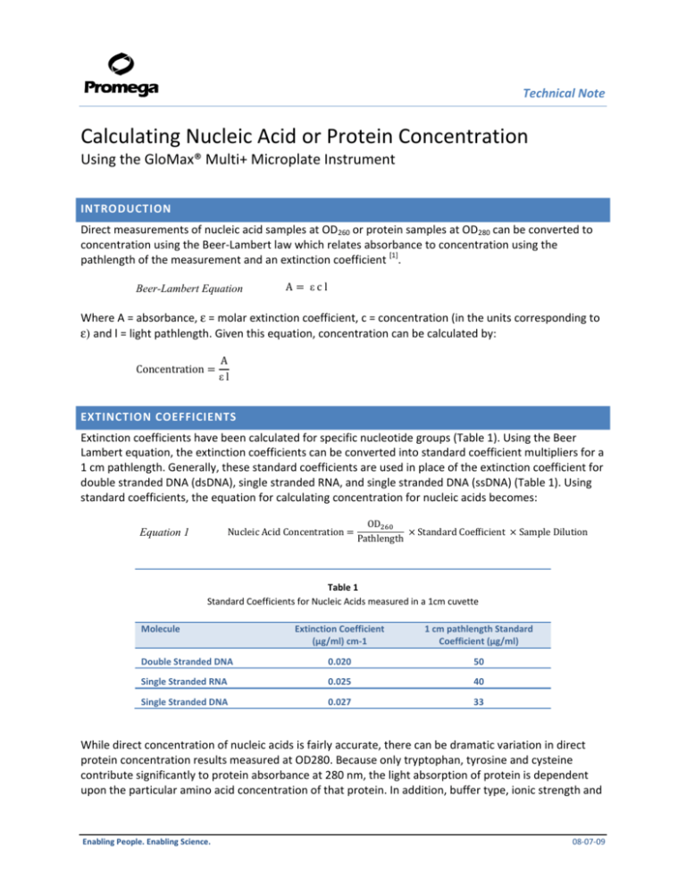
Technical Note Calculating Nucleic Acid or Protein Concentration Using the GloMax® Multi+ Microplate Instrument INTRODUCTION Direct measurements of nucleic acid samples at OD260 or protein samples at OD280 can be converted to concentration using the Beer-Lambert law which relates absorbance to concentration using the pathlength of the measurement and an extinction coefficient [1]. Beer-Lambert Equation A εcl Where A = absorbance, ε = molar extinction coefficient, c = concentration (in the units corresponding to ε) and l = light pathlength. Given this equation, concentration can be calculated by: A εl Concentration EXTINCTION COEFFICIENTS Extinction coefficients have been calculated for specific nucleotide groups (Table 1). Using the Beer Lambert equation, the extinction coefficients can be converted into standard coefficient multipliers for a 1 cm pathlength. Generally, these standard coefficients are used in place of the extinction coefficient for double stranded DNA (dsDNA), single stranded RNA, and single stranded DNA (ssDNA) (Table 1). Using standard coefficients, the equation for calculating concentration for nucleic acids becomes: Nucleic Acid Concentration Equation 1 OD Pathlength Standard Coefficient Sample Dilution Table 1 Standard Coefficients for Nucleic Acids measured in a 1cm cuvette Molecule Extinction Coefficient (μg/ml) cm-1 1 cm pathlength Standard Coefficient (μg/ml) Double Stranded DNA 0.020 50 Single Stranded RNA 0.025 40 Single Stranded DNA 0.027 33 While direct concentration of nucleic acids is fairly accurate, there can be dramatic variation in direct protein concentration results measured at OD280. Because only tryptophan, tyrosine and cysteine contribute significantly to protein absorbance at 280 nm, the light absorption of protein is dependent upon the particular amino acid concentration of that protein. In addition, buffer type, ionic strength and Enabling People. Enabling Science. 08-07-09 Technical Note pH affect absorptive values and even pure protein solutions may have different conformations and modifications. For OD280 based protein concentration calculations, the best approach is to empirically derive the extinction coefficient for the protein of interest. However, this may not be practical or necessary for most routine lab functions. A very rough protein concentration can be obtained by making the assumption that the protein sample has an extinction coefficient of 1, so 1 OD = 1 mg/ml protein. For better accuracy, some standard protein extinction coefficients have been published. See Table 2 for a few selected extinction coefficients or the Practical Handbook of Biochemistry and Molecular Biology for a more extensive table [2]. Finally, if the protein sequence of the protein to be measured is known, the theoretical extinction coefficient can be calculated using the equation ε = 5690(#Tryptophans) + 1280(#Tyrosines) + 60(#Cysteines) [3] or online tools such as ExPASy Protparam. Given a known or calculated extinction coefficient, protein concentration can be calculated using the Beer-Lambert equation. Equation 2 Protein Concentration OD Extinction coefficient Pathlength Sample Dilution Table 2 Calculated Extinction Coefficients for proteins measured in a 1cm cuvette Molecule Calculated Extinction Coefficient (mg/ml) cm-1 BSA .66 IgG 1.35 IgM 1.2 PATHLENGTH For single tube instruments, using a standard 10 x 10 cuvette, the light pathlength is fixed at 1 cm by the distance between the walls of the cuvette (Figure 1A). Absorbance measurements at 1 cm pathlength have been correlated with specific nucleic acid concentrations; for example an OD of 1.0 at 260 nm correlates to 50 μg/ml of dsDNA (Table 1). When using a 1 cm cuvette, the pathlength is 1 and equation 1 can be simplified to OD x Extinction Coefficient x sample dilution. For example, if an undiluted dsDNA sample measured in a 1 cm cuvette gives an OD 260 value of 0.9 OD, the dsDNA concentration would be calculated as: 0.9 OD 50 45 μg/ml DNA However, when using a microplate instrument, measurements are taken vertically so the distance light travels through a sample varies depending on the volume of liquid in the plate (Figure 1 B and C). Therefore, to calculate a nucleic acid concentration using equation 1, a pathlength correction value must be used to account for the different light pathlength corresponding to the sample volume. For example, if the same dsDNA sample was evaluated in a 96 well plate with a 200 μl sample volume, the OD value might be 0.50. Assuming a pathlength of 0.56 cm, the dsDNA concentration would be calculated as: 0.50 OD /0.56 50 45 μg/ml DNA By including sample pathlength information in the concentration calculation, both single tube measurements and microplate measurements provide comparable results. Pathlength can be calculated two ways: experimentally or mathematically. Enabling People. Enabling Science. 08-07-09 Technical Note A B Light in C Light out Light out Light in Light in Light out 1cm Figure 1: The light pathlength remains constant in the cuvette regardless of volume of liquid used but the pathlength varies in a microplate depending on how much volume is in a well. Light pathlength for A) standard 10x 10 cuvette B) 96 well microplate with 200 μl volume C) 96 well microplate with 100 μl volume. Experimentally derived Pathlength Experimentally derived pathlength’s are determined by using the absorbance properties of water at 900 nm and 977 nm wavelengths. While water does not typically absorb light, it does have a small absorbance peak at 977 nm. In a 1 cm cuvette, The OD of water a 977nm – 900 nm (900 nm is used as a blank) is approximately 0.18 OD at room temperature. Comparing this standard measurement with the OD values of water at 900 nm and 977 nm in a microplate allows calculation of the microplate sample pathlength using the following equation. Equation 3 OD . OD OD Sample Pathlength cm Using filters for 900 nm[*] and 980 nm and the sample pathlength equation, Modulus™ II Microplate pathlength values for 100 and 200 μl microplate volumes in a Corning 96 well UV compatible plate (# 3635) have been experimentally derived (Table 3). Table 3 Pathlength correction values calculated using 980 nm and 900 nm water measurements in Corning 96 well UV compatible plates (#3635) Sample volume 100 μl 200 μl Pathlength (cm) 0.29 0.56 Mathematically derived Pathlength Mathematically, pathlength values can be calculated using the sample volume and the diameter or height and width of the sample plate wells. Microplates have either circular (96 well plates) or square (384 well plates) wells. Using the formula’s in Figure 1, the height (pathlength) of the sample volume can be calculated. Because microplate wells have a slight taper, the mean diameter or width of a well can only be estimated. Further, this method does not account for the meniscus of the liquid. Enabling People. Enabling Science. 08-07-09 Technical Note A B h A. B. 4 V π d h V a b Calculation of pathlength (h) in plates with cylindrical wells. Where V = sample volume and d = mean diameter of the well Calculation of pathlength (h) in plates with square wells. Where V = sample volume, a = mean width of the well and b = mean depth of the well. Pathlength values for sample volumes ranging from 25 to 250 μl, depending on plate type, have been calculated for common microplates (Table 4). Because pathlength values are proportional to the volume of liquid used, a linear regression has been calculated and can be used to determine the pathlength of any volume between 50 and 250 μl where x = volume used and y = pathlength. Once the pathlength correction is determined, DNA concentration in a microplate is calculated using equation 1 above (Note: recommended sample volumes for 96 well plates are 100 μl to 250 μl). Table 4 Pathlength values and linear regression equation for different 96 well microplate well volumes. Pathlength (cm) UV compatible plates Part Number 353261 25 μl 50 μl 100 μl 200 μl 250 μl n/a n/a 0.28 0.56 0.70 y=0.0028x-3E-16 353262 0.19 0.39 0.77 n/a n/a y=0.0077x-4E-16 Corning 96 well UV plate 3635 n/a n/a 0.29 0.58 0.73 y=0.0029x+3E-16 Corning 384 well UV plate 3675 0.25 0.50 1.01 n/a n/a y=.0101x+4E-16 Corning 96 well half volume UV plate 3679 0.14 0.28 0.56 n/a n/a y=0.0056x 655801 n/a n/a 0.28 0.56 0.69 y=0.0028x 781801 0.20 0.41 0.82 n/a n/a y=0.0082x 675801 0.14 0.29 0.58 n/a n/a y=0.0058x BD Falcon 96 well UV plate BD Falcon 384 well UV plate Greiner 96 well UV Star (also Thermo Scientific/Nunc) Greiner 384 well UV Star (also Thermo Scientific/Nunc) Greiner 96 well half volume UV star (also Thermo Scientific/Nunc) Enabling People. Enabling Science. Linear Regression 08-07-09 Technical Note Comparing pathlengths for Corning 96 well UV plates (#3635) derived from the two methods shows that while the values for 200 μl pathlength are slightly different, the overall results are similar regardless of which method is used (Table 5). Table 5 Experimentally vs mathematically derived pathlength values in Corning 96 well UV compatible plates. Sample volume 100 μl Experimentally derived Pathlength (cm) 0.29 Concentration of dsDNA with OD of 0.9 155 μg/ml Mathematically derived Pathlength (cm) 0.29 Concentration of dsDNA with OD of 0.9 155 μg/ml 200 μl 0.56 80 μg/ml 0.58 78 μg/ml One caveat of using absorbance based measurements of nucleic acid samples is that proteins and reagents commonly used in the preparation of nucleic acids also absorb light at 260 nm and can lead to falsely elevated concentration results. Most reagents that can contaminate a sample also absorb light at 280 nm which provides a method of calculating DNA or RNA purity using the ratio of measurements at OD260/OD280. Generally an OD260/OD280 ratio ≥1.8 indicates “pure” DNA and an OD ratio of ~2.0 indicates “pure” RNA. A ratio below 1.8 indicates DNA or RNA that is contaminated by protein, phenol, or other aromatic compounds. The OD260/OD280 ratio does not necessarily indicate the absence of other nucleotides or single stranded nucleic acids. For protein concentration, the converse is true, if the sample is contaminated with nucleic acids, the OD260 value will be elevated so that a ratio of OD260/OD280 of <1.0 indicates “pure” protein where as a higher value indicates nucleic acid contamination. PROTOCOL FOR QUANTITATING NUCLEIC ACID USING THE GLOMAX MULTI+ MICROPLATE 1. Measure the DNA or RNA sample at both 260nm and 280nm wavelengths using the dual wavelength measurement feature on the GloMax Multi+ Microplate. 2. Calculate Nucleic Acid Concentration a. Divide the OD260 reading by the Table 3 pathlength value corresponding to the volume in the wells i. If a measurement volume not listed in Table 3 is used, calculate the pathlength correction using the linear regression equation shown in Table 3. b. Multiply this number by the DNA or RNA constant from Table 1 c. Multiply by the sample dilution factor 3. Estimate nucleic acid purity a. Subtract the blank value (well containing buffer) from the sample OD’s b. Divide the OD260/OD280 c. If the ratio of DNA OD260/OD280 is between 1.8 and 2.0, the DNA purity (free from protein contaminants) is ~90% or better. If the ratio of RNA OD260/OD280 is ~2.0, the RNA purity is ~90% or better. Enabling People. Enabling Science. 08-07-09 Technical Note PROTOCOL FOR QUANTITATING PROTEIN USING THE GLOMAX MULTI+ MICROPLATE 1. Measure the protein sample at both 280nm and 260nm wavelengths using the dual wavelength measurement feature on the GloMax Multi+ Microplate. 2. Calculate Protein Concentration a. Divide the OD280 reading by the Table 3 pathlength value corresponding to the volume in the wells multiplied by the extinction coefficient (Table 2, or calculated, or 1 if no value can be determined) i. If a measurement volume not listed in Table 3 is used, calculate the pathlength correction using the linear regression equation shown in Table 3. b. Multiply by the sample dilution factor 3. Estimate nucleic acid contamination a. Subtract the blank value (well containing buffer) from the sample OD’s b. Divide the OD260/OD280 c. If the ratio of DNA OD260/OD280 is < 1.0, the protein purity (free from nucleic acid contaminants) is ~95% or better. REFERENCES 1. Maniatis T, Fritsch EF, Sambrook J. Molecular cloning a laboratory manual. Cold Spring Harbor Laboratory, Cold Spring Harbor NY (1982). 2. Fasman G. Ed. Practical handbook of biochemistry and molecular biology. CRC press, Boston (1992) 3. Gill, SC and von Hippel, PH. Calculation of protein extinction coefficients from amino acid sequence data. (1989) Anal. Biochem 182:319-26. GloMax is a registered trademark of Promega Corporation. All other trademarks are the sole property of their respective owners. CONTACT INFORMATION Toll-Free: (800) 356-9526 Fax: (800) 356-1970 www.promega.com Email: custserv@promega.com Mailing Address: Promega Corporation 2800 Woods Hollow Rd. Madison, WI 53711 USA Enabling People. Enabling Science. 08-07-09
