Protein Measurements
advertisement
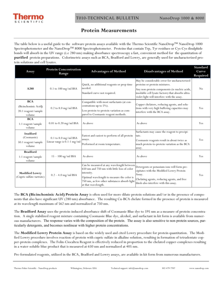
T010‐TECHNICAL BULLETIN NanoDrop 1000 & 8000 Protein Measurements The table below is a useful guide to the software protein assays available with the Thermo Scientific NanoDrop™ NanoDrop 1000 Spectrophotometer and the NanoDrop™ 8000 Spectrophotometer. Proteins that contain Trp, Tyr residues or Cys-Cys disulphide bonds will absorb in the UV range (i.e 280 nm) making absorbance spectroscopy a fast, convenient method for the quantitation of purified protein preparations. Colorimetric assays such as BCA, Bradford and Lowry, are generally used for uncharacterized protein solutions and cell lysates. Assay A280 BCA (Bicinchoninic Acid) 20:1 reagent/sample volume BCA 1:1 reagent/sample volume Bradford (Coomassie) 50:1 reagent/sample volume Bradford 1:1 reagent/sample volume Modified Lowry (Cupric sulfate-tartrate) Disadvantages of Method Standard Curve Required 0.1 to 100 mg/ml BSA Quick, no additional reagents or prep time needed. Standard curve not required. May be considerable error for uncharacterized proteins or protein mixtures. Any non-protein components (ie nucleic acids, insoluble cell lysate factors) that absorbs ultraviolet light will interfere with the assay. No 0.2 to 8.0 mg/ml BSA Compatible with most surfactants (at concentrations up to 5%). Less protein-to-protein variation as compared to Coomassie reagent methods. Copper chelators, reducing agents, and solutions with very high buffering capacities may interfere with the BCA assay. Yes As above As above Yes Protein Concentration Range 0.01 to 0.20 mg/ml BSA Advantages of Method Surfactants may cause the reagent to precipiFastest and easiest to perform of all protein tate. 0.1 to 8.0 mg/ml BSA assays. Coomassie reagents result in about twice as Linear range is 0.1-1 mg/ml Performed at room temperature. much protein-to-protein variation as the BCA assay. 15 – 100 ug/ml BSA As above As above Yes Yes C 0.2 – 4.0 mg/ml BSA Can be measured at any wavelength between 650 nm and 750 nm with little loss of color intensity. Optimal wavelength to measure the color is 750 nm, as few other substances absorb light at that wavelength. Detergents or potassium ions will form precipitates with the Modified Lowry Protein Assay. Chelating agents, reducing agents, and free thiols also interfere with this assay. Yes The BCA (Bicinchoninic Acid) Protein Assay is often used for more dilute protein solutions and/or in the presence of components that also have significant UV (280 nm) absorbance. The resulting Cu-BCA chelate formed in the presence of protein is measured at its wavelength maximum of 562 nm and normalized at 750 nm. The Bradford Assay uses the protein-induced absorbance shift of Coomassie Blue dye to 595 nm as a measure of protein concentration. A single stabilized reagent mixture containing Coomassie Blue dye, alcohol, and surfactant in kit form is available from numerous manufacturers. The response varies with the composition of the protein. The assay is also sensitive to non protein sources, particularly detergents, and becomes nonlinear with higher protein concentrations. The Modified Lowry Protein Assay is based on the widely used and cited Lowry procedure for protein quantitation. The Modified Lowry procedure involves reaction of protein with cupric sulfate in alkaline solution, resulting in formation of tetradentate copper-protein complexes. The Folin-Ciocalteu Reagent is effectively reduced in proportion to the chelated copper-complexes resulting in a water-soluble blue product that is measured at 650 nm and normalized at 405 nm. Pre-formulated reagents, utilized in the BCA, Bradford and Lowry assays, are available in kit form from numerous manufacturers. Thermo Fisher Scientific - NanoDrop products Wilmington, Delaware USA Technical support: info@nanodrop.com 302-479-7707 www.nanodrop.com 2 T010‐TECHNICAL BULLETIN NanoDrop 1000 & 8000 The NanoDrop 1000 and the NanoDrop 8000 A280 modules are used to determine the concentration of purified protein samples. The Beer-Lambert equation (A = E * b * c ) is used for all protein calculations to correlate absorbance with concentration. A is the absorbance value (A), E is the wavelength-dependent molar absorptivity coefficient (or extinction coefficient) with units of liter/mol-cm, b is the path length in centimeters, c is the analyte concentration in moles/liter or molarity (M) The software offers the following six options for selecting what extinction coefficient will be used in conjunction with Beer’s law to calculate sample concentration: A general reference setting based on a E 0.1% (1 mg/ml) protein solution producing an Absorbance of 1.0 at 280 nm (where the pathlength is 10 mm or 1 cm). This option is most commonly used when measuring an uncharacterized protein. Bovine Serum Albumin reference. With this option, BSA or unknown (sample) protein concentrations are calculated using the mass extinction coefficient of 6.7 at 280 nm for a 1% (10 mg/ml) BSA solution. IgG reference. With this option, IgG or unknown (sample) protein concentrations are calculated using the mass extinction coefficient of 13.7 at 280 nm for a 1% (10 mg/ml) IgG solution. Lysozyme reference. With this option, Lysozyme or unknown (sample) protein concentrations are calculated using the mass extinction coefficient of 26.4 at 280 nm for a 1% (10 mg/ml) Lysozyme solution. User-entered values for molar extinction coefficient (M-1 cm-1) and molecular weight (MW) in kilo Daltons for their respective protein reference. Maximum value for e is 999 X 1000 and maximum value for M.W. is 9999 X 1000. User-entered mass extinction coefficient (L gm-1cm-1) for a 10 mg/ml (1%) solution of the respective protein. E1% = (10* e) / MW Note- a general rule of thumb is to use 10 for the E1% if the true value is not known. This option is most commonly used when measuring purified and characterized proteins. C DETERGENTS While the most logical blanking solution is generally the buffer/solvent at the same pH and ionic strength as that of the unknown samples, it is important that the blanking solution have minimal absorbance at the wavelengths of interest. Although the operating software displays a flat baseline when a buffer blank is measured, the spectrometer still detects the absorbance of the buffer solution when measuring an unknown sample. Buffers, such as RIPA buffers containing NP-40 or its chemical equivalent Igepal CA-630, with very high A280 absorbance characteristics may result in reduced signal being available for the unknown sample at the wavelengths of interest. Some buffer components and reagents as well as detergents may cause the pedestal surfaces to become “unconditioned”. We have noted that routine use of the Bradford reagent may result in difficulty forming columns with 1 ul samples. Although the instrument was designed for 1 ul samples, using larger volumes (1.5-2.0 ul) will often overcome the inherent surface tension properties associated with some detergent based samples, allowing for proper column formation. Use an 8-channel pipettor when loading multiple samples on the NanoDrop 8000 to minimize evaporation due to delays in sample loading. It is recommended that spectrophotometric measurements be made immediately after pipetting samples onto the pedestals as delays can compromise accuracy. Use the instrument pedestal reconditioning kit (PR-1) as a rapid means of reconditioning the pedestals when the surface properties have been compromised and liquid columns break during measurement. For Technical Support contact us at 302-479-7707 or info@nanodrop.com. Rev 4/08 Thermo Fisher Scientific - NanoDrop products Wilmington, Delaware USA Technical support: info@nanodrop.com 302-479-7707 www.nanodrop.com
