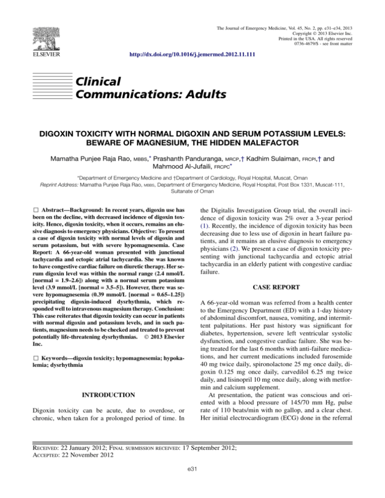
The Journal of Emergency Medicine, Vol. 45, No. 2, pp. e31–e34, 2013
Copyright Ó 2013 Elsevier Inc.
Printed in the USA. All rights reserved
0736-4679/$ - see front matter
http://dx.doi.org/10.1016/j.jemermed.2012.11.111
Clinical
Communications: Adults
DIGOXIN TOXICITY WITH NORMAL DIGOXIN AND SERUM POTASSIUM LEVELS:
BEWARE OF MAGNESIUM, THE HIDDEN MALEFACTOR
Mamatha Punjee Raja Rao, MBBS,* Prashanth Panduranga, MRCP,† Kadhim Sulaiman, FRCPI,† and
Mahmood Al-Jufaili, FRCPC*
*Department of Emergency Medicine and †Department of Cardiology, Royal Hospital, Muscat, Oman
Reprint Address: Mamatha Punjee Raja Rao, MBBS, Department of Emergency Medicine, Royal Hospital, Post Box 1331, Muscat-111,
Sultanate of Oman
, Abstract—Background: In recent years, digoxin use has
been on the decline, with decreased incidence of digoxin toxicity. Hence, digoxin toxicity, when it occurs, remains an elusive diagnosis to emergency physicians. Objective: To present
a case of digoxin toxicity with normal levels of digoxin and
serum potassium, but with severe hypomagnesemia. Case
Report: A 66-year-old woman presented with junctional
tachycardia and ectopic atrial tachycardia. She was known
to have congestive cardiac failure on diuretic therapy. Her serum digoxin level was within the normal range (2.4 nmol/L
[normal = 1.9–2.6]) along with a normal serum potassium
level (3.9 mmol/L [normal = 3.5–5]). However, there was severe hypomagnesemia (0.39 mmol/L [normal = 0.65–1.25])
precipitating digoxin-induced dysrhythmia, which responded well to intravenous magnesium therapy. Conclusion:
This case reiterates that digoxin toxicity can occur in patients
with normal digoxin and potassium levels, and in such patients, magnesium needs to be checked and treated to prevent
potentially life-threatening dysrhythmias. Ó 2013 Elsevier
Inc.
the Digitalis Investigation Group trial, the overall incidence of digoxin toxicity was 2% over a 3-year period
(1). Recently, the incidence of digoxin toxicity has been
decreasing due to less use of digoxin in heart failure patients, and it remains an elusive diagnosis to emergency
physicians (2). We present a case of digoxin toxicity presenting with junctional tachycardia and ectopic atrial
tachycardia in an elderly patient with congestive cardiac
failure.
CASE REPORT
A 66-year-old woman was referred from a health center
to the Emergency Department (ED) with a 1-day history
of abdominal discomfort, nausea, vomiting, and intermittent palpitations. Her past history was significant for
diabetes, hypertension, severe left ventricular systolic
dysfunction, and congestive cardiac failure. She was being treated for the last 6 months with anti-failure medications, and her current medications included furosemide
40 mg twice daily, spironolactone 25 mg once daily, digoxin 0.125 mg once daily, carvedilol 6.25 mg twice
daily, and lisinopril 10 mg once daily, along with metformin and calcium supplement.
At presentation, the patient was conscious and oriented with a blood pressure of 145/70 mm Hg, pulse
rate of 110 beats/min with no gallop, and a clear chest.
Her initial electrocardiogram (ECG) done in the referral
, Keywords—digoxin toxicity; hypomagnesemia; hypokalemia; dysrhythmia
INTRODUCTION
Digoxin toxicity can be acute, due to overdose, or
chronic, when taken for a prolonged period of time. In
RECEIVED: 22 January 2012; FINAL SUBMISSION RECEIVED: 17 September 2012;
ACCEPTED: 22 November 2012
e31
e32
M. P. Raja Rao et al.
Figure 1. Twelve-lead electrocardiogram demonstrating a narrow QRS tachycardia with a heart rate of 130 beats/min with inverted P waves falling on T waves (arrow), and upright P in aVR suggestive of a junctional tachycardia.
health center demonstrated narrow QRS tachycardia with
a heart rate of 130 beats/min with inverted P waves falling
on T waves and upright P in aVR (Figure 1, arrowheads),
suggestive of junctional tachycardia. An ECG done in our
ED demonstrated a narrow QRS tachycardia, peaked P
waves with prolonged PR interval at 110 beats/min heart
rate, and typical ECG signs of the ‘‘digoxin effect’’ in the
form of a scooped appearance of the asymmetric downsloping ST depression (‘‘reversed tick’’ sign) (Figure 2).
This ECG was suspicious for an ectopic atrial tachycardia
with 1:1 conduction. In view of the patient taking digoxin,
she was suspected to have digoxin toxicity, and her blood
was sent for routine blood tests along with digoxin levels.
All of the patient’s blood investigations were reported as
normal, including free digoxin level (2.4 nmol/L [normal
= 1.9–2.6] using Abbott AxSYM analyzer; Abbott Laboratories, Abbott Park, IL). Her troponin T was negative,
serum potassium was 3.9 mmol/L (normal = 3.5–5), creatinine was 91 mmol/L (normal = 45–90), calculated glomerular filtration rate (GFR) was 54 mL/min/1.73 m2,
and brain natriuretic peptide was 2017 pg/mL (normal
= 20–285). These results initially ruled out digoxin toxicity and suggested that the dysrhythmia may be due to underlying cardiomyopathy. However, in view of the ECG
changes being highly suspicious for digoxin toxicity,
her magnesium and calcium levels were requested. The
calcium level was normal, but the serum magnesium level
was low at 0.39 mmol/L (normal = 0.65–1.25). She was
diagnosed to have digoxin toxicity precipitated by hypomagnesemia.
Figure 2. Twelve-lead electrocardiogram (ECG) illustrating a narrow QRS tachycardia, peaked P waves with prolonged PR interval at 110 beats/min heart rate, and typical ECG signs of the ‘‘digoxin effect’’ in the form of a scooped appearance of the asymmetric down-sloping ST depression (‘‘reversed tick’’ sign; arrow). This ECG is suggestive of an ectopic atrial tachycardia with 1:1
conduction.
Magnesium and Digoxin Toxicity
The patient was admitted and was treated with intravenous magnesium sulfate 2 g in 100 mL saline over 60
min. The digoxin was discontinued. After 1 h of infusion,
her rhythm changed to sinus with a heart rate of 70 beats/
min and a prolonged PR interval (Figure 3). The P wave
morphology was different between the second ECG and
third ECG, suggesting that her second ECG showed ectopic atrial tachycardia. A further magnesium sulfate infusion of 5 g (approximately 40 mEq) in 500 mL saline
was given for 24 h. Repeat serum magnesium at 6 h
was 0.67 mmol/L. Her thyroid-stimulating hormone level
was normal. The patient was discharged after 72 h of observation, with no recurrence of any dysrhythmia. At discharge, digoxin level was 1.4 nmol/L and magnesium
level was 1.1 mmol/L.
DISCUSSION
In a relatively recent study, digoxin toxicity was identified in 0.04% of all admissions (2). The incidence rate
for digoxin toxicity-related admissions was 48 per
100,000 prescriptions, which corresponds to 1.94 admissions for toxicity per 1000 treatment-years. Women had
a 1.4-fold higher risk of intoxication than men. In patients
taking digoxin in recommended doses (0.125 to 0.25 mg
once daily), digoxin toxicity can occur when they are exposed to precipitating factors such as hypokalemia, hypomagnesemia, or hypothyroidism, even though the serum
digoxin level is within normal limits (3). In addition,
drug-to-drug interactions can increase digoxin level
(e.g., quinidine, verapamil, spironolactone, flecainide,
amiodarone, erythromycin, clarithromycin) and cause digoxin toxicity (3). Furthermore, elderly age, female gender, low muscle mass, and reduced GFR can predispose to
digoxin toxicity in patients taking a normal recommended dose (3,4).
e33
Hypokalemia or hypomagnesemia sensitize the myocardium to digoxin even when digoxin levels are within
the normal range (Lanoxin product information; Aspen
Pharma, St. Leonards, NSW, Australia). Digoxin directly
inhibits sodium-potassium ATPase pump in the membrane of cardiac myocyte, causing an increase in intracellular sodium and calcium (through a sodium and
calcium exchanger) with subsequent increase in myocardial contractility (5). Hypokalemia increases digoxin
cardiac sensitivity because potassium and digoxin compete for the same ATPase-binding site (5). This leads
to a decrease in atrioventricular (AV) node conduction,
and an increase in automaticity (increased intracellular
calcium) and ectopic pacemaker activity. In addition,
there is an increase in vagal tone (inhibits sodiumpotassium ATPase pump in vagal afferent fibers), causing bradycardia and AV blocks. Magnesium is a cofactor
of the sodium-potassium ATPase pump (6). Hypomagnesemia increases myocardial digoxin uptake, further inhibiting sodium-potassium ATPase pump activity (6).
Hypomagnesemia is associated with hypokalemia in
40–60% and hypocalcemia in 40% of toxicity cases
(7). However, hypomagnesemia can produce intracellular potassium depletion in the presence of normal serum
potassium levels (8). It is known that long-term digoxin
users often have hypokalemia or hypomagnesemia, presumably due to diuretic usage in patients with congestive
heart failure (5).
Even though this patient had several risk factors for digoxin toxicity, including elderly age, female gender, and
a moderately reduced GFR, it was the diuretic-induced
hypomagnesemia that precipitated digoxin-induced junctional and atrial tachycardia. This patient did not manifest
serum hypokalemia due to the co-administration of
a potassium-sparing diuretic and angiotensin-converting
enzyme inhibitor. However, it is possible that the
Figure 3. Twelve-lead ECG post-magnesium sulfate infusion for digoxin-induced dysrhythmia demonstrating sinus rhythm with
a heart rate of 70 beats/min and prolonged PR interval with ST depression suggestive of digoxin effect.
e34
hypomagnesemia was associated with intracellular potassium depletion, leading to digoxin-related dysrhythmias
(8). Enhanced automaticity and impaired conduction
are the hallmarks of digitalis toxicity (9). Digoxin can
cause any type of dysrhythmia, but especially the following: frequent premature ventricular complexes; atrial fibrillation with slow, regular ventricular rate or complete
AV block; non-paroxysmal junctional tachycardia; atrial
tachycardia with block; and bidirectional ventricular
tachycardia (9,10).
Normal levels of serum digoxin and the absence of hypokalemia (the most common precipitant of digoxininduced dysrhythmias) initially led to the conclusion
that the dysrhythmias may be due to underlying cardiomyopathy. However, magnesium was found to be very low
and magnesium replacement resulted in conversion to sinus rhythm without the need for any anti-dysrhythmic
agents. Management of digoxin toxicity involves withdrawal of the medication as well as administration of
digoxin-specific Fab antibodies for life-threatening cardiovascular compromise (9,10). In addition, it is
necessary to treat precipitating conditions such as
replacement of electrolyte deficiencies. In the patient
presented here, treatment of hypomagnesemia resulted
in conversion of atrial tachycardia to sinus rhythm
without the need for Fab antibodies. Magnesium therapy
requires monitoring of serum levels and is contraindicated in patients with bradycardia or AV block, or
severe renal failure.
M. P. Raja Rao et al.
CONCLUSION
This case reiterates that digoxin toxicity can occur in patients with normal digoxin and potassium levels, and in
such patients, magnesium must be monitored and treated
to prevent potentially life-threatening dysrhythmias.
REFERENCES
1. Rich MW, McSherry F, Williford WO, Yusuf S. Digitalis Investigation Group. Effect of age on mortality, hospitalizations and response
to digoxin in patients with heart failure: the DIG study. J Am Coll
Cardiol 2001;38:806–13.
2. Aarnoudse AL, Dieleman JP, Stricker BH. Age- and gender-specific
incidence of hospitalisation for digoxin intoxication. Drug Saf
2007;30:431–6.
3. Dec GW. Digoxin remains useful in the management of chronic
heart failure. Med Clin North Am 2003;87:317–37.
4. Ahmed A, Rich MW, Love TE, et al. Digoxin and reduction in mortality and hospitalization in heart failure: a comprehensive post hoc
analysis of the DIG trial. Eur Heart J 2006;27:178–86.
5. Chan KE, Lazarus JM, Hakim RM. Digoxin associates with mortality in ESRD. J Am Soc Nephrol 2010;21:1550–9.
6. Fawcett WJ, Haxby EJ, Male DA. Magnesium: physiology and
pharmacology. Br J Anaesth 1999;83:302–20.
7. Agus ZS. Hypomagnesemia. J Am Soc Nephrol 1999;10:1616–22.
8. Milionis HJ, Alexandrides GE, Liberopoulos EN, Bairaktari ET,
Goudevenos J, Elisaf MS. Hypomagnesemia and concurrent acidbase and electrolyte abnormalities in patients with congestive heart
failure. Eur J Heart Fail 2002;4:167–73.
9. Mercader MA, Bader Y. Internet diagnosis of digitalis toxicity. BMJ
Case Rep 2009;pii: bcr09.2008.0918, doi: 10.1136/bcr.09.2008.
0918. Epub 2009 Apr 7.
10. Ma G, Brady WJ, Pollack M, Chan TC. Electrocardiographic manifestations: digitalis toxicity. J Emerg Med 2001;20:145–52.

