The Effect of Glycation of CD59 on Complement
advertisement
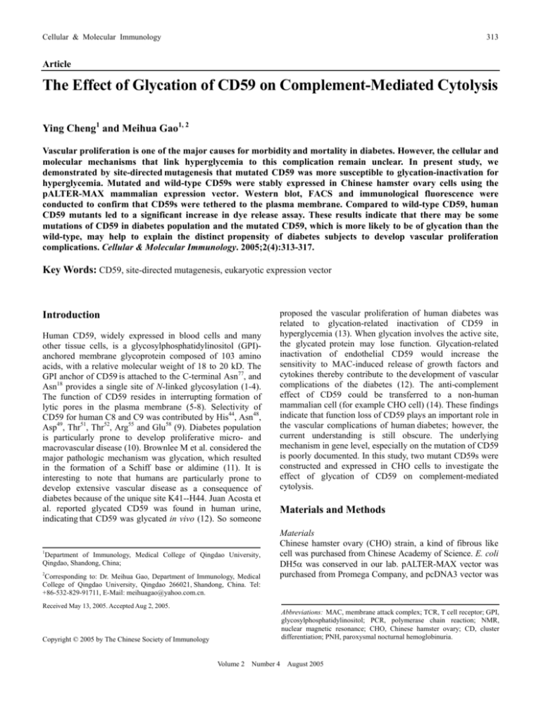
Cellular & Molecular Immunology 313 Article The Effect of Glycation of CD59 on Complement-Mediated Cytolysis Ying Cheng1 and Meihua Gao1, 2 Vascular proliferation is one of the major causes for morbidity and mortality in diabetes. However, the cellular and molecular mechanisms that link hyperglycemia to this complication remain unclear. In present study, we demonstrated by site-directed mutagenesis that mutated CD59 was more susceptible to glycation-inactivation for hyperglycemia. Mutated and wild-type CD59s were stably expressed in Chinese hamster ovary cells using the pALTER-MAX mammalian expression vector. Western blot, FACS and immunological fluorescence were conducted to confirm that CD59s were tethered to the plasma membrane. Compared to wild-type CD59, human CD59 mutants led to a significant increase in dye release assay. These results indicate that there may be some mutations of CD59 in diabetes population and the mutated CD59, which is more likely to be of glycation than the wild-type, may help to explain the distinct propensity of diabetes subjects to develop vascular proliferation complications. Cellular & Molecular Immunology. 2005;2(4):313-317. Key Words: CD59, site-directed mutagenesis, eukaryotic expression vector Introduction Human CD59, widely expressed in blood cells and many other tissue cells, is a glycosylphosphatidylinositol (GPI)anchored membrane glycoprotein composed of 103 amino acids, with a relative molecular weight of 18 to 20 kD. The GPI anchor of CD59 is attached to the C-terminal Asn77, and Asn18 provides a single site of N-linked glycosylation (1-4). The function of CD59 resides in interrupting formation of lytic pores in the plasma membrane (5-8). Selectivity of CD59 for human C8 and C9 was contributed by His44, Asn48, Asp49, Thr51, Thr52, Arg55 and Glu58 (9). Diabetes population is particularly prone to develop proliferative micro- and macrovascular disease (10). Brownlee M et al. considered the major pathologic mechanism was glycation, which resulted in the formation of a Schiff base or aldimine (11). It is interesting to note that humans are particularly prone to develop extensive vascular disease as a consequence of diabetes because of the unique site K41--H44. Juan Acosta et al. reported glycated CD59 was found in human urine, indicating that CD59 was glycated in vivo (12). So someone 1 Department of Immunology, Medical College of Qingdao University, Qingdao, Shandong, China; 2 Corresponding to: Dr. Meihua Gao, Department of Immunology, Medical College of Qingdao University, Qingdao 266021, Shandong, China. Tel: +86-532-829-91711, E-Mail: meihuagao@yahoo.com.cn. proposed the vascular proliferation of human diabetes was related to glycation-related inactivation of CD59 in hyperglycemia (13). When glycation involves the active site, the glycated protein may lose function. Glycation-related inactivation of endothelial CD59 would increase the sensitivity to MAC-induced release of growth factors and cytokines thereby contribute to the development of vascular complications of the diabetes (12). The anti-complement effect of CD59 could be transferred to a non-human mammalian cell (for example CHO cell) (14). These findings indicate that function loss of CD59 plays an important role in the vascular complications of human diabetes; however, the current understanding is still obscure. The underlying mechanism in gene level, especially on the mutation of CD59 is poorly documented. In this study, two mutant CD59s were constructed and expressed in CHO cells to investigate the effect of glycation of CD59 on complement-mediated cytolysis. Materials and Methods Materials Chinese hamster ovary (CHO) strain, a kind of fibrous like cell was purchased from Chinese Academy of Science. E. coli DH5α was conserved in our lab. pALTER-MAX vector was purchased from Promega Company, and pcDNA3 vector was Received May 13, 2005. Accepted Aug 2, 2005. Abbreviations: MAC, membrane attack complex; TCR, T cell receptor; GPI, glycosylphosphatidylinositol; PCR, polymerase chain reaction; NMR, nuclear magnetic resonance; CHO, Chinese hamster ovary; CD, cluster differentiation; PNH, paroxysmal nocturnal hemoglobinuria. Copyright © 2005 by The Chinese Society of Immunology Volume 2 Number 4 August 2005 314 Glycation Sensitizes CD59 to Complement-Mediated Cytolysis purchased from Invitrogen Company. Recombinant CD59 cDNA-pALTER was gifted by somebody from Harvard University. RPMI 1640 and F12 culture medium were purchased from Hyclone Company. Culture of CHO cells CHO cells were cultured in medium of F12 and RPMI 1640 containing 10% fetal calf serum, 100 U/ml penicillin and 100 μg/ml streptomycin. Oligonucleotides The sequences of the oligonucleotides used in this study are listed as follows: pT7, 5’-TAA TAC GAC TCA CTA TAG GC-3’; T3, 5’-ATT AAC CCT CAC TAA AGG GA-3’; HM 1F, 5’-AAG TGT TGG AAG CAT GAG CAG TGC AAT T-3’; HM 1R, 5’-ATT GCA CTG CTC ATG CTT CCA ACA CTT G-3’; HM 2F, 5’-GTG TTG GAA GTT TCA TCA GTG CAA TTT C-3’; HM 2R, 5’-GAA ATT GCA CTG ATG AAA CTT CCA ACA C-3’. Site-directed mutagenesis To obtain the mutant CD59, we employed the site-directed mutagenesis to replace residue 42(F) and 43(E), respectively. The mutagenic primers used were HM 1F and HM 1R for the 42-histidine substitution, and HM 2F and HM 2R for the 43-histidine substitution. Together with common primer (pT7 and T3) of CD59, tri-PCR was performed. A prominent band with an expected size of 0.6 kb was visible on 1% agarose gel. Point mutations, AAG to CAT (HM1) and AAC to CAC (HM2), were further verified by DNA sequencing determination. Construction of eukaryotic expression vector pALTER-MAXHMCD59 pALTER-MAX and pcDNA3, containing ampicillin resistance gene were amplified. Purified HM1, HM2 PCR product and eukaryotic expression vector pALTER-MAX are excised by EcoR I respectively, and gelatin electrophoresis was processed to test products. PCR product excised by EcoR I was about 500 bp. After HM1/2 PCR product and pALTER-MAX were ligased with T4 DNA ligase, recombinant eukaryotic expression vector was transformed into E. coli DH5α, screened by ampicillin resistance. Subsequently, recombinants were identified by PCR, EcoR I digestion and DNA sequencing. Transfection and expression of HMCD59 and wild-type CD59 in CHO cells Recombinant CD59-pALTER-MAX, together with plasmid pcDNA3 was transfected into CHO cells at the stage of log phase of growth. According to manufacture’s protocol, recombinant clones were screened by G418 resistance. Immunological fluorescence CHO cells stably transfected with HM1, HM2, empty pALTER-MAX plasmid, wild-type CD59 pALTER-MAX plasmid and normal CHO cells were washed twice with phosphate-buffered saline (PBS), then were fixed with 95% Volume 2 ethanol on glass slices, respectively. These cells were stained with FITC-labeled CD59 and observed under fluorescence microscope, with FITC-labeled IgG-2a as control. Flow cytometric analysis Cells were stained with FITC-labelled anti-CD59, followed at room temperature for 15-30 min. After washed with PBS twice, cells were filtered into net tube respectively and analyzed on a FACSCalibur utilizing CellQuest software (BD Biosciences). Western blot CHO cells were lysed in lysis buffer and proteins were denatured (3 min, 100oC). After electrophoresis, proteins were transferred to nitrocellulose film, and block was performed overnight. Subsequently, the goat-anti-human CD59 McAb and HRP-labelled rabbit-anti-goat IgG were added. At last, solution containing DAB and H2O2 was added to analyze the results. Dye release assay Anti-complement activities of HMCD59 proteins before and after glycation, as well as the functional difference between wild-type CD59 and HMCD59 were measured by dye release assay. In brief, CHO cells (2 × 10⁵/ml, transfected by wildtype CD59, HMCD59, empty pALTER-MAX plasmid) were seeded in 96-well plastic plate, and then 100 mM ribose was added to mimic the hyperglycemia. After 200 mM NaBH4, 2 μg/ml BCECF being added, rabbit-anti-CHO antibody and fresh serum with different dilution were used. Supernatant and fission product were collected respectively, and their fluorescence were detected at 525-535 nm (15). Statistical analysis Data are presented as the mean ± SD. p values of < 0.05 were considered statistically significant. Results Amplification and identification of target gene To gain mutant target gene, the 42 (F, HM1) and 43 (E, HM2) were replaced by histidine (H), respectively. One pair of common up-down stream primer (pT7 and T3) and two pairs of mutual oligonucleotide primer containing mutant sites (HM 1F/1R, HM 2F/2R) were synthesized. Using 3 pairs of primers mentioned above, tri-PCR was performed. A prominent band with an expected size of 0.6 kb was visible on 1% agarose gel. Point mutations were further verified by DNA sequence determination. Screening and identification of positive recombinant clones To directly assess whether human mutant MCD59 gene was transferred into vector and vector was transformed into E. coli DH5α, we conducted 1% gelatin electrophoresis to screen positive recombinants. The empty carrier of pALTER-MAX was 5,533 bp, the expected inverted segment 495 bp, so the recombinant pALTER-MAX-HMCD59 gene was about 6 kb. Number 4 August 2005 Cellular & Molecular Immunology bp 1 2 3 315 4 5 53.7% HM1 2000 1000 750 500 54.5% Figure 1. Identification of recombinant plasmid pALTERMAX-HMCD59 by EcoR I digestion. Purified HM1 [F (42) replaced by H, namely AAG replaced by CAT: TTT GAT GCG TGT CTC ATT ACC AAA GCT GGG TTA CAA GTG TAT AAC CAT TG], HM2 [E (43) replaced by H, namely AAC replaced by CAC: GAT TTT GAT GCG TGT CTC ATT ACC AAA GCT GGG TTA CAA GTG TAT CAC AGT GTT GGA AG] PCR product and eukaryotic expression vector pALTER-MAX are excised by EcoR I respectively. A prominent band with an expected size of 0.5 kb was visible in Lane 3 and Lane 5. Lane1, DL2000 DNA marker; Lane 2, pALTER-MAX-HM1 before enzyme digestion; Lane 3, pALTERMAX-HM1 after enzyme digestion; Lane 4, pALTER-MAX-HM2 before enzyme digestion; Lane 5, pALTER-MAX-HM2 after enzyme digestion. HM2 55.9% Wild-type CD59 Figure 2. Expression of recombinant human mutant CD59 in CHO cells. Cells were stained with FITC-labelled anti-CD59 at room temperature for 15-30 min. Cells were analyzed on the FACSCalibur utilizing CellQuest software (BD Biosciences). Groups with CD59 transfection (HM1 expression rate: 53.7%; HM2 expression rate: 54.5%; wild-type expression rate: 55.9%) were much higher than that of the control group. After excised by EcoR I, the product would be about 500 bp. With DL2000 DNA marker, a band with an expected size of 0.5 kb was visible, which was further verified by PCR and sequencing. The results indicated the recombinant plasmid PALTER-HMCD59 has been successfully constructed and transformed (Figure 1). Identification of expression on CHO cells To determine the human mutant CD59 (HM1/HM2) and wild-type expression on CHO cells, we conducted immunological fluorescence and flow cytometric assay with the known CD59 McAb. The expression of CD59 in transfected groups (HM1 expression rate: 53.7%; HM2 expression rate: 54.5%; wild-type expression rate: 55.9%) was significantly higher than those of the control group (Figure 2). Immunological fluorescence assay To further assess the protein expression on CHO cells, we conducted immunological fluorescence assay. CHO cells stably transfected by HM1, HM2, empty PALTER-MAX plasmid, wild-type CD59 and normal CHO cells were divided into different groups. With CD59 antibody labeled by FITC, results showed that fluorescence intensity of CHO transfected by HM1/HM2 and wild-type (Figure 3) was higher than that of the CHO transfected by empty PALTER plasmid, and the difference of them was quite significant. dye-release assay Before exposed to human complement, the target CHO cells were incubated with inactivated rabbit-anti-CHO antibody to measure its sensitivity to MAC-mediated cytolysis. As shown in Figure 5, before glycation, HMCD59 and wild-type CD59 have no obvious difference in anti-MAC activity; however, after glycation, function loss of HMCD59 is significant, indicating that HMCD59s are more sensitive to glycation than the wild-type. Identification of expression by Western blot After electrophoresis, proteins were transferred to nitrocellulose film. We found obvious band of 18-20 kD proteins, which was consistent with the molecular weight of HMCD59 (Figure 4). Expression of HMCD59 was successfully confirmed. Function detecting of HMCD59 and wild-type CD59 by Volume 2 Number 4 A B C D Figure 3. Immunological fluorescence assay. To further assess the protein expression on CHO cells, with CD59 antibody labeled by FITC, we conducted immunological fluorescence assay. Results showed CHO cells transfected with empty PALTER plasmid (A), HM1, HM2 plasmid (B, C), and wild-type CD59 (D) (100×). August 2005 316 Glycation Sensitizes CD59 to Complement-Mediated Cytolysis 2 1 3 4 Figure 4. Result of HM-CD59 protein by Western-blot test. CHO cells were lysed in lysis buffer and proteins were denatured (3 min, 100oC). After electrophoresis, we found obvious band of 18-20 kD proteins, which was consistent with the molecular weight of HMCD59. Lane 1, negtive control; Lane 2, postive control; Lane 3: HM1 group (18-20 kD); Lane 4, HM2 group (18-20 kD). Discussion Vascular complications cause substantial morbidity and mortality for patients with diabetes. Though the complement regulatory protein CD59 is proposed the most relevant to these diabetic complications, the underlying mechanism is not fully understood. The possibility that CD59 might be compromised in diabetics was suggested by the demonstration that human CD59 contained a glycation motif centred around amino acid residue K41 in close proximity to W40, a highly conserved amino acid that is known to be essential for CD59 function. Glycation of K41 may interfere with the function of CD59 and be responsible for the release of growth factors from endothelium cells, which contribute to the long-term vascular complications that characterize human diabetes (12). Here we clone and characterize for the first time the novel genes, namely human mutant CD59s, and establish their eukaryotic expression vectors. Furthermore, by dye-release assay we demonstrate that mutant CD59 has function loss and is more sensible to glycation-inactivation than the wild-type in hyperglycemia. 100 HM1 before glycation HM2 before glycation wild-type before glycation ** Presently, with the development of living standard and alteration of meal structure, there are at least 130 millions of diabetes subjects all over the world. In the USA, the treatment of chronic diabetic complications consumes nearly 15% of health care dollars. In developed country, the incidence is 5%-10%; however, in developing country, there exists an increasing incidence. Nephropathy, retinopathy, and accelerated atherosclerosis are the characteristic long-term proliferative complications of diabetes. CD59 has the function of signal transmit (16, 17) and relativity to paroxysmal nocturnal hemoglobinuria (PNH), replantation repulsion reaction, renal diseases and so on. Present research on construct and function of CD59 shows human CD59 has sites K41--H44, which contains a preferential glycation motif in or very close to its active site. H44 (histidine) may assistant glycation of lysine (K41), while K41 near to the active amino acid-W40 (tryptophan). The steric hindrance introduced by the glycation of K41 interferes with C8 and C9 binding on the surface of CD59 and may be responsible for the observed glycation-induced functional inhibition. We postulate that the previously unexplained vascular proliferation seen in diabetes could be complement-mediated as a consequence of glycation-inactivation of CD59. Thus, we design mutant CD59s of special sites in our study and believe they would better explain the glycation-inactivation mechanism than before. Gene mutagenesis is often conducted to research the relationship between construct and function (18). PCR site-directed mutagenesis (triplex PCR), is a kind of site-direct mutagenesis with high efficiency, making vitro site mutagenesis convenient and economical (19). Plasmid pALTER-MAX is a kind of efficient eukaryotic expression carrier controlled by promoter of cytom egalovirus (CMV), containing CMV early enhancer, multiple clone site and Poly(A) signal, and increasing expression of the inverted foreign gene. Cation liposome reagent, may provide a kind of HM1 after glycation HM2 after glycation wild-type after glycation ** ** Release rate (%) 75 ** 50 * ** ** ** * 25 0 1:8 1:4 1:2 1:1 Dilution of Complement Volume 2 Number 4 August 2005 Figure 5. Function difference between human mutant CD59 and wild-type CD59 before and after glycation. CHO cells (2 × 10⁵/ml, transfected by wild-type CD59, HMCD59, empty pALTER-MAX plasmid) were seeded in 96-well plastic plate, then 100 mM ribose was added to mimic the hyperglycemia. We added 200 mM NaBH4, 2 μg/ml BCECF, rabbit-anti-CHO antibody and fresh serum, then we detected the supernatant and fission product at 525-535 nm. **p < 0.01, *p < 0.05, compared with before glycation by t-test. Cellular & Molecular Immunology 317 efficient method to establish stable transfected strain (19). According to these, it is tempting to propose that gene mutation sensitive CD59 to glycation-inactivation. The mechanism involved may be that, after mutation, the site of histidine (H44) was closer to lysine, which would improve the glycation of lysine (K41). Glycated lysine (K41) would cause inactivation of W40, the active site, so it causes function loss of restriction (15) homogeneity deposition of MAC. In summary, this study has constructed eukaryotic expression system on mutant and wild-type CD59 genes and proposed mutant CD59 was more sensible to glycationinactivation in hyperglycemia. Thus, mutation of CD59 may also contribute to development of proliferative complications of chronic hyperglycemia and we tempt to postulate there was some gene mutation of CD59 in diabetes population. Acknowledgements This work was supported by the National Natural Science Foundation of China (No. 30170893). We would like to thank Professor Chunting Zhao for his technical help and Qiubo Wang for her valuable efforts in this research. References 1. Bodian DL, Davis SJ, Morgan BP, Rushmere NK. Mutational analysis of the active site and antibody epitopes of the complement-inhibitory glycoprotein, CD59. J Exp Med. 1997; 185:507-516. 2. Kieffer B, Driscoll PC, Campbell ID, Willis AC, van der Merwe PA, Davis SJ. Three-dimensional solution structure of the extracellular region of the complement regulatory protein CD59, a new cell-surface protein domain related to snake venom neurotoxins. Biochemistry. 1994;33:4471-4482. 3. Fletcher CM, Harrison RA, Lachmann PJ, Neuhaus D. Sequence-specific 1H-NMR assignments and folding topology of human CD59. Protein Sci. 1993;2:2015-2027. 4. Ninomiya H, Stewart BH, Rollins SA, Zhao J, Bothwell AL, Sims PJ. Contribution of the N-linked carbohydrate of erythrocyte antigen CD59 to its complement-inhibitory activity. J Biol Chem. 1992;267:8404-8410. 5. Lockert DH, Kaufman KM, Chang CP, Hüsler T, Sodetz JM, Sims PJ. Identity of the segment of human complement C8 recognized by complement regulatory protein CD59. J Biol Chem. 1995;270:19723-19728. 6. Rollins SA, Zhao J, Ninomiya H, Sims PJ. Inhibition of homologous complement by CD59 is mediated by a speciesselective recognition conferred through binding to C8 within Volume 2 C5b-8 or C9 within C5b-9. J Immunol. 1991;146:2345-2351. 7. Hüsler T, Lockert DH, Kaufman KM, Sodetz JM, Sims PJ. Chimeras of human complement C9 reveal the site recognized by complement regulatory protein CD59. J Biol Chem. 1995; 270:3483-3486. 8. Chang CP, Husler T, Zhao J, Wiedmer T, Sims PJ. Identity of a peptide domain of human C9 that is bound by the cell-surface complement inhibitor, CD59. J Biol Chem. 1994;269:2642426430. 9. Zhao XJ, Zhao J, Zhou Q, Sims PJ. Identity of the residues responsible for the species-restricted complement inhibitory function of human CD59. J Biol Chem. 1998;273:1066510671. 10. Duhault J, Koenig-Berard E. Diabetes mellitus and its animal models. Therapie. 1997;52:375-384. 11. Brownlee M, Vlassara H, Cerami A. Nonenzymatic glycosylation and the pathogenesis of diabetic complications. Ann Intern Med. 1984;101:527-537. 12. Acosta J, Hettinga J, Flückiger R, et al. Molecular basis for a link between complement and the vascular complications of diabetes. Proc Natl Acad Sci U S A. 2000;97:5450-5455. 13. Brooimans RA, van Wieringen PA, van Es LA, Daha MR. Relative roles of decay-accelerating factor, membrane cofactor prtotein, and CD59 in the protection of human endothelial cells against complement-mediated lysis. Eur J Immunol. 1992;22: 3135-3140. 14. Zhao J, Rollins SA, Maher SE, Bothwell AL, Sims PJ. Amplified gene expression in CD59-transfected Chinese hamster ovary cells confers protection against the membrane attack complex of human complement. J Biol Chem. 1991;266: 13418-13422. 15. Acossta J, Halperin JA. Inhibition of the complement regulatory protein CD59 by non-enzymatic glycation: its potential pathogenic role in the proliferative complications of diabetes mellitus. J Investig Med. 1996b;44:199A. 16. Halperin JA, Taratuska A, Rynkiewicz M, Nicholson-Weller A. Transient changes in erythrocyte membrane permeability are induced by sublytic amounts of the complement membrane attack complex (C5b-9). Blood. 1993;81:200-205. 17. Kilgore KS, Schmid E, Shanley TP, et al. Sublytic concentrations of the membrane attack complex of complement induce endothelial interleukin-8 and monocyte chemoattractant protein-1 through nuclear factor-κB activation. Am J Pathol. 1997;150: 2019-2031. 18. Ferriani VPL, Harrison RA, Lachman PJL. C5b-9 binds to N-terminal portion of CD59. Mol Immunol. 1993;30(Suppl 1): 10. 19. Sims PJ, Faioni EM, Wiedmer T, Shattil SJ. Complement proteins C5b-9 cause release of membrane vesicles from the platelet surface that are enriched in the membrane receptor for coagulation factor Va and express prothrombinase activity. J Biol Chem. 1988;263:18205-18212. Number 4 August 2005
