LENSES
advertisement

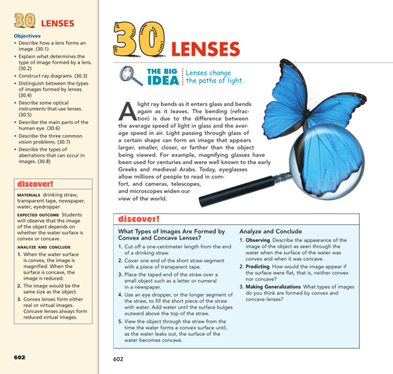
Objectives • Describe how a lens forms an image. (30.1) • Explain what determines the type of image formed by a lens. (30.2) 30 LENSES ........ LENSES THE BIG • Construct ray diagrams. (30.3) IDEA • Distinguish between the types of images formed by lenses. (30.4) • Describe some optical instruments that use lenses. (30.5) • Describe the main parts of the human eye. (30.6) • Describe the three common vision problems. (30.7) • Describe the types of aberrations that can occur in images. (30.8) discover! drinking straw, transparent tape, newspaper, water, eyedropper MATERIALS EXPECTED OUTCOME Students will observe that the image of the object depends on whether the water surface is convex or concave. ANALYZE AND CONCLUDE 1. When the water surface is convex, the image is magnified. When the surface is concave, the image is reduced. 2. The image would be the same size as the object. 3. Convex lenses form either real or virtual images. Concave lenses always form reduced virtual images. 602 Lenses change the paths of light. A light ray bends as it enters glass and bends again as it leaves. The bending (refraction) is due to the difference between the average speed of light in glass and the average speed in air. Light passing through glass of a certain shape can form an image that appears larger, smaller, closer, or farther than the object being viewed. For example, magnifying glasses have been used for centuries and were well known to the early Greeks and medieval Arabs. Today, eyeglasses allow millions of people to read in comfort, and cameras, telescopes, and microscopes widen our view of the world. discover! What Types of Images Are Formed by Convex and Concave Lenses? 1. Cut off a one-centimeter length from the end of a drinking straw. 2. Cover one end of the short straw segment with a piece of transparent tape. 3. Place the taped end of the straw over a small object such as a letter or numeral in a newspaper. 4. Use an eye dropper, or the longer segment of the straw, to fill the short piece of the straw with water. Add water until the surface bulges outward above the top of the straw. 5. View the object through the straw from the time the water forms a convex surface until, as the water leaks out, the surface of the water becomes concave. 602 Analyze and Conclude 1. Observing Describe the appearance of the image of the object as seen through the water when the surface of the water was convex and when it was concave. 2. Predicting How would the image appear if the surface were flat, that is, neither convex nor concave? 3. Making Generalizations What types of images do you think are formed by convex and concave lenses? 30.1 Converging and Diverging Lenses A lens is a piece of transparent material, such as glass, that refracts light. A lens forms an image by bending rays of light that pass through it. Learning about lenses is a hands-on activity. Not manipulating lenses while learning about them is like taking swimming lessons out of water. Shapes of Lenses The shape of a lens can be understood by considering a lens to be a large number of portions of prisms, as shown in Figure 30.1. When arranged in certain positions, the prisms bend incoming parallel rays so they converge to (or diverge from) a single point. The arrangement shown in Figure 30.1a is thicker in the middle; it converges the light. The arrangement in Figure 30.1b is thinner in the middle; it diverges the light. 30.1 Converging and Diverging Lenses FIGURE 30.1 A lens may be thought of as a set of prisms. a. Incoming parallel rays converge to a single point. b. Incoming rays seem to diverge from a single point. In both arrangements, the most net bending of rays occurs at the outermost prisms, for they have the greatest angle between the two refracting surfaces. No net bending occurs in the middle “prism,” for its glass faces are parallel and rays emerge in their original direction. Real lenses are made not of prisms, of course, but of solid pieces of glass or plastic with surfaces that are usually ground to a spherical shape. Figure 30.2 shows how smooth lenses refract rays of light and form wave fronts. The lens in Figure 30.2a is a converging lens. A converging lens, also known as a convex lens, is thicker in the middle, causing rays of light that are initially parallel (straight wave fronts) to meet at a single point called the focal point. The lens in Figure 30.2b is a diverging lens. A diverging lens, also known as a concave lens, is thinner in the middle, causing the rays of light to appear to originate from a single point. This chapter is an extension of the previous chapter. It applies refraction to lenses, and introduces the ideas of ray optics. It may be omitted without consequence to the chapters that follow. Key Terms lens, converging lens, convex lens, diverging lens, concave lens, principal axis, focal point, focal plane, focal length Teaching Tip Distribute diverging and converging lenses to your students. Let them play with the lenses for a few minutes. Ask them to describe what they see. Point out the need to define terms and then do so. Demonstration Show, with an actual prism, how a prism refracts a ray of light. Then introduce ray tracing on the board. FIGURE 30.2 Wave fronts travel more slowly in glass than in air. a. In the converging lens, the wave fronts are retarded more through the center of the lens, and the light converges. b. In the diverging lens, the waves are retarded more at the edges, and the light diverges. CHAPTER 30 LENSES 603 Teaching Tip Contrast the deviation of light in a prism with the absence of deviation in a pane of glass. Show that a pane only displaces light rays, and show how thicker panes produce greater displacements. 603 Teaching Tip Discuss the key features of a converging lens (Figure 30.3). Sketch a ray diagram for parallel light along the principal axis to define the focal point. ...... Key Features of Lenses Figure 30.3 illustrates some important features of a lens. The principal axis of a lens is the line joining the centers of curvature of its surfaces. The focal point for a converging lens is the point at which a beam of light parallel to the principal axis converges. The focal plane is a plane perpendicular to the principal axis that passes through either focal point of a lens. For a converging lens, any incident parallel beam converges to a point on the focal plane. A lens affects light coming from the right in the same way as light coming from the left (or has the same effect on light incident from either side). Therefore, a lens has two focal points and two focal planes. When the lens of a camera is set for distant objects, the film is in the focal plane behind the lens in the camera. A lens forms an image by bending rays of light that pass through it. CONCEPT CHECK Teaching Resources • Reading and Study Workbook • Laboratory Manual 81, 82 • PresentationEXPRESS FIGURE 30.3 • Interactive Textbook The key features of a converging lens include the principal axis, focal point, and focal plane. 30.2 Image Formation by a Lens Key Term real image For a diverging lens, an incident beam of light parallel to the principal axis is not converged to a point, but is diverged so that the light appears to originate from a single point. The focal length of a lens, whether converging or diverging, is the distance between the center of the lens and its focal point. When the lens is thin, the focal lengths on either side are equal, even when the curvatures on the two sides are not. ...... Teaching Tip Allow students to cast images of the windows on the wall, or ceiling lights on their desks, using converging lenses. Have them cast images of closer brightly illuminated objects (bright enough to cast a clearly seen image). CONCEPT CHECK Ask is there a relationship between the image distance (distance from the lens to the place where a sharp image appears) and the object distance (from object to lens)? Yes, the farther the object, the nearer the image. 30.2 Image Formation by a Lens For: Links on lenses Visit: www.SciLinks.org Web Code: csn – 3002 604 How does a lens form an image? 604 With unaided vision, an object far away is seen through a relatively small angle of view, as shown in Figure 30.4a. When you are closer, the same object is seen through a larger angle of view, as illustrated in Figure 30.4b. This wider angle allows the perception of more detail. Magnification occurs when the use of a lens allows an image to be observed through a wider angle than would be observed without the lens, and so more detail can be seen. A magnifying glass is simply a converging lens that increases the angle of view and allows more detail to be seen. The type of image formed by a lens depends on the shape of the lens and the position of the object. Teaching Tip Pass around the class a box with a pinhole and a translucent viewing screen on opposite ends. Using a simple ray diagram, describe how real images are formed, and why they are inverted. Discuss Activity 43 on page 621. Note that the size of the hole compared to the distance from the viewing screen affects the amount of light that makes the image and the sharpness of the image. A larger hole will admit more light, but overlapping images through different parts of the hole reduce sharpness. A large hole covered by a lens solves both problems. FIGURE 30.4 With unaided vision, the size of an object appears to change depending on your distance to the object. a. A distant object is viewed through a narrow angle. b. When the same object is viewed from a closer distance and thus through a wider angle, more detail is seen. Images Formed by Converging Lenses When you use a magnifying glass, you hold it close to the object you wish to see magnified. This is because a converging lens will magnify only when the object is between the focal point and the lens, as shown in Figure 30.5. The magnified image will be farther from the lens than the object and right-side up (erect). If a screen were placed at the image distance, no image would appear on the screen because no light is actually directed to the image position. The rays that reach your eye, however, behave as if they came from the image position, so the image is a virtual image. Recall from Chapter 29 that a virtual image originates from a location where light does not actually reach. FIGURE 30.5 A converging lens can be used as a magnifying glass to produce a virtual image of a nearby object. FIGURE 30.6 A converging lens forms a real, upside-down image of a more distant object. When the object is far enough away to be beyond the focal point of a converging lens, light originating from the object and passing through the lens converges and can be focused on a screen, as illustrated in Figure 30.6. An image formed by converging light is called a real image. A real image formed by a single converging lens is upside down (inverted). Converging lenses are used for projecting motion pictures onto a screen. CHAPTER 30 LENSES 605 Teaching Tip Choose a bright scene with noticeable depth, so perspective plays a role when the scene is viewed from close positions (for example, from each eye). Due to perspective, the images viewed through far-apart holes are noticeably different. When these different images are combined by a lens, parts of the composite image are fuzzy. A composite image viewed through closely spaced holes is sharp. This is why photographers take pictures through a small aperture for “depth of field” in their photographs. When a fuzzy background is desirable, the aperture of the camera is opened wide. The focal plane is set for sharpness of the object, and things closer or farther away appear fuzzy. Ask When the aperture setting of a camera is small, should the exposure time be correspondingly longer or shorter? The smaller aperture means less light enters, so the film should be exposed to light for a longer time. 605 ...... The type of image formed by a lens depends on the shape of the lens and the position of the object. CONCEPT CHECK FIGURE 30.7 A diverging lens always produces a virtual image. Teaching Resources • Reading and Study Workbook • PresentationEXPRESS • Interactive Textbook • Next-Time Question 30-1 Images Through Ray Diagrams Key Term ray diagram Learning takes place not in seeing diagrams made, but in making them. It is fruitless to show the techniques of ray diagram construction without students constructing their own. Your students should have a lens in hand, and an object and image to observe when they make their constructions. Let your discussion of this topic be a class activity. think! Why is the greater part of the photograph in Figure 30.7 out of focus? Answer: 30.2 Images Formed by Diverging Lenses When a diverging lens is used alone, the image is always virtual, right-side up, and smaller than the object, as can be seen in Figure 30.7. It makes no difference how far or how near the object is. A diverging lens is often used for the viewfinder on a camera. When you look at the object to be photographed through the viewfinder, you see a right-side up virtual image that approximates the same proportions as the photograph to be taken. CONCEPT What determines the type of image ...... 30.3 Constructing CHECK formed by a lens? 30.3 Constructing Images Through Ray Diagrams Teaching Tip Reproduce Figure 30.8 on the board using a different color for each ray. Stress that the three rays are samples of an immense number of rays leaving every part of the object. If the top part of the lens were covered, for example, the entire image would still appear, but it would be dimmer. FIGURE 30.8 606 606 In a ray diagram, the three useful rays from the object converge on the image. Ray diagrams show the principal rays that can be used to determine the size and location of an image. An example of a ray diagram is shown in Figure 30.8. The size and location of the object, its distance from the center of the lens, and the focal length of the lens are used to construct a ray diagram.30.3 An arrow is used to represent the object (which may be anything from a microbe viewed in a microscope to a galaxy viewed through a telescope). For simplicity, one end of the object is placed right on the principal axis. The Three Principal Rays To locate the position of the image, you only have to know the paths of two rays from a point on the object, represented by the vertical arrow. Any point except for the point on the principal axis will work, but it is customary to choose a point at the tip of the arrow. The path of one refracted ray is known from the definition of the focal point. A ray parallel to the principal axis will be refracted by the lens to the focal point, as shown in Figure 30.8. Another path is known: through the center of the lens where the faces are parallel to each other. A ray of light will pass through the center with no appreciable change in direction. Therefore, a ray from the tip of the arrowhead proceeds in a straight line through the center of the lens. A third path is known: A ray of light that passes through the focal point in front of the lens emerges from the lens and proceeds parallel to the principal axis. All three paths are shown in Figure 30.8, which is a typical ray diagram. The image is located where the three rays intersect. Any two of these three rays is sufficient to locate the relative size and location of the image. We use these particular rays only because their paths through the lens are easy to predict. You should know that all light passing through a lens contributes to image formation. FIGURE 30.9 The ray diagram for a magnifying glass shows that when the object is less than one focal length from the lens the image is virtual, rightside up, and magnified. The ray diagram for a converging lens used as a magnifying glass is shown in Figure 30.9. In this case, where the distance from the lens to the object is less than the focal length, the rays diverge as they leave the lens. The rays of light appear to come from a point in front of the lens (same side of the lens as the object). The location of the image is found by extending the rays backward to the point where they converge. The virtual image that is formed is magnified and right-side up. CHAPTER 30 LENSES 607 Teaching Tip Show how changing the position of the object changes the position of the image. Demonstration Show students how they can calculate the diameter of the sun by measuring the image of the sun formed by a pinhole. On a sunny day, go outside with your class and call attention to the round spots of light that are found beneath trees. Small spaces between leaves in the trees act as pinholes and cast images of the sun on the ground. If the sun is overhead, the images will be circles. If it is low in the sky, they will be ellipses. Hold a piece of cardboard with a small hole in it in the sunlight and note the circle of light on the ground. Place a coin over the image and adjust the position of the pinhole until the image is the same size as the coin. (If the image is an ellipse, the short diameter of the ellipse should equal the diameter of the coin.) Measure the distance from the pinhole to the image. The ratio of the diameter of the image (i.e., diameter of the coin) to the image’s distance from the pinhole is equal to the ratio of the sun’s diameter to its distance from the pinhole (150,000,000 km). Letting x represent the diameter of the sun, use the proportion “diameter of coin/image distance 5 x/150,000,000 km” to calculate x. Students’ answers should be approximately 1,400,000 km, which agrees with the accepted value. 607 It is quite profound that average students can calculate the diameter of the sun armed only with a meterstick and a coin! This is a striking example of how the richness in life is found by not only looking at the world with wide-open eyes, but knowing what to look for. You are the guide to that richness, for you are their physics teacher. How nice that your students can go home after school and tell their families that they measured the sun’s diameter with a meterstick! The most profound concepts in physics are sometimes the simplest. FIGURE 30.10 The ray diagrams illustrate the formation of an image when the object is at various positions in relation to a converging lens of focal length f. Object position: distance f from lens (at the focal point) Image position: at infinity Object position: between f and 2f from lens Image position: beyond 2f from lens Image size: magnified Demonstration Poke an extra hole in your piece of cardboard and hold it between a bright region (window or overhead light) and a dark viewing area. Two holes produce two images; three holes produce three images; many holes produce many images—one for each hole. Place a convex lens behind (or in front of) the holes and show that all the images are focused in one place. At the proper distance they neatly overlap to produce a clear and brighter image. An appropriately placed lens simply directs a multitude of images atop one another! Object position: distance 2f from lens Image position: distance 2f from lens Image size: same as object Object position: beyond 2f from lens Image position: between f and 2f from lens Image size: smaller Teaching Tip Discuss the rules for ray diagram construction. The summary of ray diagram construction is well worth class time. For: Mirrors and Lenses Visit: PHSchool.com Web Code: csp – 3003 608 608 Object position: at infinity Image position: distance f from lens (at the focal point) Demonstration The three rays useful for the construction of a ray diagram are summarized: 1. A ray parallel to the principal axis that passes through the focal point on the opposite side. 2. A ray passing through the center of the lens that is undeflected. 3. A ray through the focal point in front of the lens that emerges parallel to the principal axis after refraction by the lens. Interestingly, when half a lens is covered, half as much light forms the image. This does not mean half the image is formed! Even a piece of broken lens can form a complete image on a screen. Try it and see. Demonstrate the ray diagrams in Figure 30.10. Darken the room and use a candle or an unfrosted bulb on an optical bench with length markings. A converging lens with a focal length of 20–25 cm works nicely. Teaching Tip Draw Figure 30.11 on the board using a different color for each ray. Differentiate among the lens, the principal axis, and the rays. Ray Diagrams for Converging and Diverging Lenses The ray diagrams in Figure 30.10 show image formation by a converging lens as an object initially at the focal point is moved away from the lens along the principal axis. Since the object is not located between the focal point and the lens, all the images that are formed are real and inverted. As illustrated in Figure 30.11, the method of drawing ray diagrams applies also to diverging lenses. A ray parallel to the principal axis from the tip of the arrow will be bent by the lens as if it had come from the focal point. A ray through the center goes straight through. A ray heading for the focal point on the far side of the lens is bent so that it emerges parallel to the axis of the lens. Teaching Tip The focal point of a lens depends on the curvature and material of the lens, not on the location of any object. Teaching Tip Although ray diagrams usually show three rays, there are an infinite number of rays that can be drawn between the object and its image. FIGURE 30.11 ...... The ray diagram shows how a virtual image is formed by a diverging lens. The size and location of the object, its distance from the center of the lens, and the focal length of the lens are used to construct a ray diagram. CONCEPT CHECK Teaching Resources • Reading and Study Workbook ...... On emerging from the lens, the three rays appear to come from a point on the same side of the lens as the object, which defines the position of the virtual image. The image is nearer to the lens than the object, smaller than the object, and right-side up. The image formed by a diverging lens is always virtual, reduced, and right-side up. CONCEPT CHECK • Concept-Development Practice Book 30-1 • Laboratory Manual 83, 84, 85, 86 • Transparencies 72, 73 What information is used to construct a ray diagram? • PresentationEXPRESS • Interactive Textbook • Next-Time Question 30-2 CHAPTER 30 LENSES 609 609 30.4 Image Formation Summarized ...... Teaching Tip Summarize converging and diverging lenses, real images and virtual images, and image position and object position. A converging lens forms either a real or a virtual image. A diverging lens always forms a virtual image. CONCEPT CHECK 30.4 Image Formation Summarized think! Where must an object be located so that the image formed by a converging lens will be (a) at infinity? (b) as near the object as possible? (c) right-side up? (d) the same size? (e) inverted and enlarged? Answer: 30.4 Teaching Resources • Reading and Study Workbook • PresentationEXPRESS ...... • Interactive Textbook A converging lens forms either a real or a virtual image. A diverging lens always forms a virtual image. A converging lens is a simple magnifying glass when the object is within one focal length of the lens. The image is then virtual, magnified, and right-side up. When the object is beyond one focal length, a converging lens produces a real, inverted image. The location of the image depends on how close the object is to the focal point. If it is close to (but slightly beyond) the focal point, the image is far away (as with a slide projector or movie projector). If the object is far from the focal point, the image is nearer (as with a camera). In all cases where a real image is formed, the object and the image are on opposite sides of the lens. When the object is viewed with a diverging lens, the image is virtual, reduced, and right-side up. This is true for all locations of the object. In all cases where a virtual image is formed, the object and the image are on the same side of the lens. CONCEPT CHECK What types of images are produced by lenses? 30.5 Some Common 30.5 Some Common Optical Instruments Optical Instruments Key Terms eyepiece, objective lens Teaching Tip If possible, show disassembled samples of the variety of optical instruments discussed in the text. Note the simplicity of the diagrams of these instruments in the text, compared to their actual construction. FIGURE 30.12 In a simple camera, the lens forms a real, inverted image on the film. A valuable lesson is learned looking for the simplicity that underlies the seemingly complex. Teaching Tip Show students a camera and its lens. Point out the diaphragm and the focusing mechanism. 610 610 The advent of eyeglasses probably occurred in Italy in the late 1200s. If anybody at the time or before viewed objects through a pair of lenses held far apart, one in front of the other, there is no record of it, for curiously enough, the telescope wasn’t invented until some 300 years later. Today, lenses are used in many optical instruments. Optical instruments that use lenses include the camera, the telescope (and binoculars), and the compound microscope. Camera A camera consists of a lens and sensitive film (or lightdetecting chip) mounted in a light-tight box. In many cameras, the lens is mounted so that it can be moved back and forth to adjust the distance between the lens and film. The lens forms a real, inverted image on the film or chip. Figure 30.12 shows a camera with a single simple lens. In practice, most cameras make use of compound lenses to minimize distortions called aberrations. The amount of light that gets to the film is regulated by a shutter and a diaphragm. The shutter controls the length of time that the film is exposed to light. The diaphragm controls the opening that light passes through to reach the film. Varying the size of the opening (aperture) varies the amount of light that reaches the film at any instant. Telescope A simple telescope uses a lens to form a real image of a distant object. The real image is not caught on film but is projected in space to be examined by another lens used as a magnifying glass. The second lens, called the eyepiece, is positioned so that the image produced by the first lens is within one focal length of the eyepiece. The eyepiece forms an enlarged virtual image of the real image. When you look through a telescope, you are looking at an image of an image. Figure 30.13 shows the lens arrangement for an astronomical telescope. The image is inverted, and thus the image of the man on the moon would appear upside down. Teaching Tip Tell students that the telescope was invented in 1608 by Dutch optician Hans Lippershey. FIGURE 30.13 An astronomical telescope forms an inverted image. (For simplification, the image is shown close here; it is actually located at infinity.) Teaching Tip If a refracting astronomical telescope is available, show it to the students. In better telescopes, the primary lens is combined with a concave lens. Teaching Tip Show students a terrestrial telescope and a pair of binoculars. Teaching Tip Borrow a compound microscope from your school’s biology department. Use it to show the class how the objective lens forms a real image of the object being viewed and how the eyepiece forms a virtual image of the real image. A third lens or a pair of reflecting prisms is used in the terrestrial telescope, which produces an image that is right-side up. A pair of these telescopes side by side, each with a pair of prisms, makes up a pair of binoculars like those shown in Figure 30.14. Since no lens transmits 100% of the light incident upon it, astronomers prefer the brighter, inverted images of a two-lens telescope to the less bright, right-side-up images that a third lens or prisms would provide. For non-astronomical uses, such as viewing distant landscapes or sporting events, right-side-up images are more important to the viewer than brightness, so the additional lens or prisms are used. Teaching Tidbit A real image is formed where light rays intersect at a single point. A virtual image is formed by light rays that only appear to intersect. FIGURE 30.14 Each side of a pair of binoculars uses a pair of prisms that flip the image right-side up. Link to TECHNOLOGY The Digital Camera Rather than focusing an image onto film, the lens in a digital camera focuses light onto an array of millions of tiny light-sensitive semi-conductor photocells. Each cell produces an electrical signal in proportion to the amount of light hitting it. Usually, red, green, and blue filters on the different photocells make them sensitive to a particular color of light. All of the intensity and color information is relayed from the photocell array to a computer chip. Software processes the raw information to produce an image. CHAPTER 30 LENSES 611 611 ...... Optical instruments that use lenses include the camera, the telescope (and binoculars), and the compound microscope. CONCEPT CHECK FIGURE 30.15 A compound microscope uses two converging lenses. Teaching Resources • Reading and Study Workbook • Problem-Solving Exercises in Physics 15-1 Compound Microscope A compound microscope uses two converging lenses of short focal length, arranged as shown in Figure 30.15. The first lens, called the objective lens, produces a real image of a close object. Since the image is farther from the lens than the object, it is enlarged. A second lens, the eyepiece, forms a virtual image of the first image, further enlarged. The instrument is called a compound microscope because it enlarges an already enlarged image. • PresentationEXPRESS • Interactive Textbook 30.6 The Eye Teaching Tip Borrow a model of an eye from the biology department. Use it to show students the parts of the eye mentioned in the text. Teaching Tip Discuss the function of the rods and cones in the retina of the eye. Explain that color cannot be perceived in dim light, and that colored stars appear white to us, whereas they show up clearly colored in camera time exposures. ...... Key Terms cornea, iris, pupil, retina CONCEPT CHECK The giant squid has the largest eyes in the world. I show a colored photo that I took of the stars and discuss the curved lines encircling the north star. Then I get into a discussion of how long the camera shutter was held open. Name some optical instruments that use lenses. 30.6 The Eye In many respects, the human eye is similar to the camera. A diagram of the eye is shown in Figure 30.16. The main parts of the eye are the cornea, the iris, the lens, and the retina. Light enters through the transparent covering called the cornea. The amount of light that enters is regulated by the iris, the colored part of the eye that surrounds the pupil. The pupil is the opening in the iris through which light passes.30.6 Light passes through the pupil and lens and is focused on a layer of tissue at the back of the eye—the retina —that is extremely sensitive to light. Different parts of the retina receive light from different directions. FIGURE 30.16 Light enters the human eye through the cornea, passes through the pupil and lens, and is focused on the retina. The retina is not of uniform sensitivity. There is a small region in the center of our field of view where vision is most distinct. This spot is called the fovea. Much greater detail can be seen at the fovea than off to the side. 612 612 Demonstration The Blind Spot There is also a spot in the retina where the nerves carrying all the information leave the eye in a narrow bundle. This is the blind spot. You can demonstrate that you have a blind spot in each eye if you hold this book at arm’s length, close your left eye, and look at the round dot in Figure 30.17 with only your right eye. You can see both the round dot and the X at this distance. If you now move the book slowly toward your face, with your right eye still fixed upon the dot, you’ll reach a position about 20 to 25 cm from your eye where the X disappears. To establish the blind spot in your left eye, close your right eye and similarly look at the X with your left eye so that the dot disappears. With both eyes opened, you’ll find no position where either the X or the dot disappears because one eye “fills in” the part of the object to which the other eye is blind. Repeat the exercise of Figure 30.17 with small objects on various backgrounds. For: Links on the eye Visit: www.SciLinks.org Web Code: csn – 3006 FIGURE 30.17 Try to find your blind spot in each eye. ...... ...... CHECK Teaching Tip Although the pupil of the eye looks black, flashbulb photos often show it to be red. This is largely because the light from the flash reflects directly back from the assemblage of blood vessels on the retina. Teaching Tip Tell students that the particularly bright reflection from the eye pupils of many animals when illuminated at nighttime can cause them to look luminous. The reflection from a thin membrane called the tapetum, located behind the rods in the animal’s eyes, provides a “second chance” for the animal to perceive light that initially misses a photoreceptor. This arrangement, common in owls, cats, and other night predators, results in excellent night vision. The Camera and the Eye In both the camera and the eye, the image is upside down, and this is compensated for in both cases. You simply turn the camera film around to look at it. Your brain has learned to turn around images it receives from your retina! A principal difference between a camera and the human eye has to do with focusing. In a camera, focusing is accomplished by altering the distance between the lens and the film or chip. In the human eye, most of the focusing is done by the cornea, the transparent membrane at the outside of the eye. Adjustments in focusing of the image on the retina are made by changing the thickness and shape of the lens to regulate its focal length, as shown in Figure 30.18. This is called accommodation and is brought about by the action of the ciliary muscle, which surrounds the lens. CONCEPT Stand at a corner of the room and shake brightly colored cards, first turned backward so the color is hidden and students can adjust the position of their heads (looking toward the other corner of the room). When they can just barely see the moving cards at the corners of their eyes, turn them over and display the color. Try this with different colors. Your students will see the cards as they move, but not their colors. The main parts of the eye are the cornea, the iris, the lens, and the retina. CONCEPT CHECK Name the main parts of the human eye. Teaching Resources • Reading and Study Workbook FIGURE 30.18 The shape of the lens changes to focus light on the retina. CHAPTER 30 LENSES • PresentationEXPRESS • Interactive Textbook 613 613 30.7 Some Defects in Vision Physics on the Job Key Terms farsighted, nearsighted, astigmatism Photographer Photography blends art with physics. A photographer’s ideas are based on artistry, but the execution relies on a savvy use of physics and, for most photographers now, computer technology. A photographer knows that a camera seldom records just what the eye sees. Our eyes can discern detail simultaneously both in dark shadows and in light that is millions of times brighter; neither film nor digital image sensors can do this. Hence, the photographer experiments with brightness and contrast. The photographer who has knowledge of the physics of light and optics is better able to use available software to turn a digital image into a work of art. Teaching Tip In Figure 30.19, it appears that the outside of the lens is convex and the inside is concave. This is true, but the lens is still thicker in the center than at the edges so it is a converging lens. Similarly, the lens in Figure 30.20 is a diverging lens. It is thinner in the center than at the edges. Demonstration Simulate the human eye with a spherical flask filled with a bit of fluorescent dye. Paint an “iris” on the flask and position appropriate lenses behind the iris for normal, farsighted and nearsighted vision. Then show how corrective lenses placed in front of the eye focus the light on the retina. 30.7 Some Defects in Vision If you have normal vision, your eye can accommodate to clearly see objects from infinity (the far point) down to 25 cm (the near point, which normally recedes for all people with advancing age). Unfortunately, not everyone has normal vision. Three common vision problems are farsightedness, nearsightedness, and astigmatism. Farsightedness A farsighted person has trouble focusing on nearby objects because the eyeball is too short or the cornea is too flat so that images form behind the retina. As Figure 30.19 illustrates, farsighted people have to hold things more than 25 cm away to be able to focus them. The remedy is to increase the converging effect of the eye. This is done by wearing eyeglasses or contact lenses with converging lenses. Converging lenses will converge the rays that enter the eye sufficiently to focus them on the retina instead of behind it. FIGURE 30.19 The eyeball of the farsighted eye is too short. A converging lens moves the image closer and onto the retina. 614 614 ...... Three common vision problems are farsightedness, nearsightedness, and astigmatism. CONCEPT CHECK FIGURE 30.20 The eyeball of the nearsighted eye is too long. A diverging lens moves the image farther away and onto the retina. Teaching Resources • Reading and Study Workbook • Concept-Development Practice Book 30-2 Nearsightedness A nearsighted person can see nearby objects clearly, but does not see distant objects clearly because they are focused too near the lens, in front of the retina. As depicted in Figure 30.20, the eyeball is too long or the surface of the cornea is too curved. A remedy is to wear corrective lenses that diverge the rays from distant objects so that they focus on the retina instead of in front of it. • Problem-Solving Exercises in Physics 15-2 • PresentationEXPRESS • Interactive Textbook • Next-Time Question 30-3 30.8 Some Defects ...... Astigmatism Astigmatism of the eye is a defect that results when the cornea is curved more in one direction than the other, somewhat like the side of a barrel. Because of this defect, the eye does not form sharp images. The remedy is cylindrical corrective lenses that have more curvature in one direction than in another. CONCEPT CHECK of Lenses Key Term aberration Name and describe the three common vision problems. discover! MATERIALS 30.8 Some Defects of Lenses No lens gives a perfect image. The distortions in an image are called aberrations. By combining lenses in certain ways, aberrations can be minimized. For this reason, most optical instruments use compound lenses, each consisting of several simple lenses, instead of single lenses. Two types of aberration are spherical aberration and chromatic aberration. For: Links on abnormalities of the eye Visit: www.SciLinks.org Web Code: csn – 3008 discover! paper or cardboard EXPECTED OUTCOME When the page is viewed through the pinhole, the print will be seen more clearly. When the light is bright, the pupil (i.e., the aperture) is smaller. Someone who can’t quite read the small print in a telephone book without his or her glasses should squint to reduce the size of the “aperture” in the eyes. THINK Can a Pinhole Improve Your Vision? 1. Poke a tiny hole in a piece of paper or cardboard. 2. Hold the pinhole in front of your eye and close to this page. Can you see the print clearly? 3. Why is bright light needed? 4. Think What advice do you have for someone who wears glasses and misplaces them, and can’t see the small print in a telephone book? CHAPTER 30 LENSES 615 615 Demonstration Send a wide beam of white light through a converging lens. Make sure that the beam goes through the edges as well as the center of the lens. At the focal point, a circle of unfocused light is formed due to the spherical aberration. Using a diaphragm, show how the focus is improved by limiting the aperture. (If a diaphragm is not available, use paper or cardboard with holes of different sizes.) If you look carefully near the focus, you’ll note that different colors focus at different points. Aberrations Spherical aberration results when light passes through the edges of a lens, as in Figure 30.21a, and focuses at a slightly different place from light passing through the center of the lens. This can be remedied by covering the edges of a lens, as with a diaphragm in a camera. Spherical aberration is corrected in good optical instruments by a combination of lenses. Chromatic aberration is the result of the different speeds of light of various colors and hence the different refractions they undergo, as shown in Figure 30.21b. In a simple lens red light and blue light bend by different amounts (as in a prism), so they do not come to focus in the same place. Achromatic lenses, which combine simple lenses of different kinds of glass, correct this defect. FIGURE 30.21 a. Spherical aberration occurs when light passes through the edges of a lens. b. Chromatic aberration occurs because different colors of light travel at different speeds. In the eye, vision is sharpest when the pupil is smallest because light then passes through only the center of the eye’s lens, where spherical and chromatic aberrations are minimal. Also, light bends the least through the center of a lens, so minimal focusing is required for a sharp image. You see better in bright light because your pupils are smaller. ...... Methods for Correcting Vision An alternative to wearing eyeglasses for correcting vision is contact lenses. A more recent option is LASIK (acronym for laser-assisted in-situ keratomileusis), the procedure of reshaping the cornea using pulses from a laser. Another recent procedure is PRK (photorefractive keratectomy). Still another is IntraLase, where intraocular lenses are implanted in the eye like a contact lens, a procedure of choice for extremely nearsighted or farsighted people and for those who can’t have laser surgery. The wearing of eyeglasses and contact lenses may soon be a thing of the past. Two types of CHECK aberration are spherical aberration and chromatic aberration. CONCEPT ...... CONCEPT Name the types of aberrations that can occur in CHECK images formed by lenses. Teaching Resources think! • Reading and Study Workbook Why is chromatic aberration a problem for lenses but not for mirrors? Answer: 30.8 • PresentationEXPRESS • Interactive Textbook 616 616 REVIEW 30 REVIEW Concept Summary • • • • • • • • •••••• A lens forms an image by bending rays of light that pass through it. The type of image formed by a lens depends on the shape of the lens and the position of the object. The size and location of the object, its distance from the center of the lens, and the focal length of the lens are used to construct a ray diagram. A converging lens forms either a real or a virtual image. A diverging lens always forms a virtual image. Optical instruments that use lenses include the camera, the telescope (and binoculars), and the compound microscope. The main parts of the eye are the cornea, the iris, the lens, and the retina. Three common vision problems are farsightedness, nearsightedness, and astigmatism. Two types of aberrations are spherical aberration and chromatic aberration. Key Terms For: Self-Assessment Visit: PHSchool.com Web Code: csa – 3000 Teaching Resources • TeacherEXPRESS • Virtual Physics Lab 27 think! Answers 30.2 Both Jamie and his cat and the virtual image of Jamie and his cat are “objects” for the lens of the camera that took this photograph. Since the objects are at different distances from the camera lens, their respective images are at different distances with respect to the film in the camera. So only one can be brought into focus. 30.4 The object should be (a) at one focal length from the lens (at the focal point) (see Figure 30.10); (b) and (c) within one focal length of the lens (see Figure 30.9); (d) at two focal lengths from the lens (see Figure 30.10); (e) between one and two focal lengths from the lens (see Figure 30.10). 30.8 Different frequencies travel at different speeds in a transparent medium, and therefore refract at different angles. This produces chromatic aberration. Light reflection, on the other hand, has nothing to do with the frequency of light. One color reflects the same as any other. •••••• lens (p. 603) converging lens (p. 603) convex lens (p. 603) diverging lens (p. 603) concave lens (p. 603) principal axis (p. 604) focal point (p. 604) focal plane (p. 604) focal length (p. 604) real image (p. 605) ray diagram (p. 606) eyepiece (p. 611) objective lens (p. 612) cornea (p. 612) iris (p. 612) pupil (p. 612) retina (p. 612) farsighted (p. 614) nearsighted (p. 615) astigmatism (p. 615) aberration (p. 615) CHAPTER 30 30 CHAPTER LENSES LENSES 617 617 ASSESS Check Concepts 1. A converging lens is thickest in the middle; a diverging lens is thinnest in the middle. 2. The focal point is a single point on the principal axis; the focal plane is an area perpendicular to the principal axis. 30 ASSESS Check Concepts (continued) •••••• Section 30.1 3. Only a real image can be projected onto a screen. 1. Distinguish between a converging lens and a diverging lens. 4. Parallel to the principal axis; through the focal point; through center of the lens 2. Distinguish between the focal point and focal plane of a lens. 5. Any two 6. To both 7. Converging lenses form either real or virtual images, depending on where the object is with respect to the focal length of the lens. Section 30.2 3. Distinguish between a virtual image and a real image. 8. What types of images are formed by diverging lenses? Section 30.5 9. Explain what is meant by saying that in a telescope one looks at the image of an image. 10. What is the function of the eyepiece in an astronomical telecsope? 11. What type of lenses are used in a compound microscope? Section 30.6 8. Diverging lenses always form virtual images that are reduced and right-side up. 12. Which instrument—a telescope, a compound microscope, or a camera—is most similar to the eye? 9. The eyepiece forms an image of the image formed by the objective lens. 13. Why do you not normally notice a blind spot when you look at your surroundings? 10. For the viewer to look at the real image of the objective lens 11. Converging lenses 12. A camera 13. One eye fills in the blind spot of the other. 14. Farsighted—image falls behind the retina; nearsighted—image falls in front of the retina 15. The cornea is curved like a barrel; vision can be corrected with a cylindrical lens. 16. Spherical—light that passes through the lens’s edge focuses poorly; combine lenses; chromatic—different colors focus at different places; combine lenses of different kinds of glass 618 Section 30.3 4. There are three convenient rays commonly used in ray diagrams to estimate the position of an image. Describe these three rays in terms of their orientation with respect to the principal axis and focal points. Section 30.7 14. Distinguish between farsighted and nearsighted vision. 5. How many of the rays in Question 4 are necessary for estimating the position of an image? 6. Do ray diagrams apply only to converging lenses, or to diverging lenses also? Section 30.4 7. What types of images are formed by converging lenses? 618 15. What is astigmatism, and how can it be corrected? Section 30.8 16. Distinguish between spherical aberration and chromatic aberration, and cite a remedy for each. Think and Rank 17. B, C, D, A 18. C = B, A 19. C, B, A Think and Explain 20. It will only be dimmer! The full image is still present. Think and Rank •••••• Rank each of the following sets of scenarios in order of the quantity or property involved. List them from left to right. If scenarios have equal rankings, then separate them with an equal sign. (e.g., A ⫽ B) 19. Here Percy is in front of a diverging lens. Virtual images are seen by viewing from the right side of the lens. For object positions A, B, and C, rank the sizes of the virtual images from largest to smallest. 24. It can focus sunlight energy onto a flammable material and start a fire. Think and Explain •••••• 20. When you cover the top half of a camera lens, what effect does this have on the pictures taken? 18. Depending on the location of Percy relative to the focal point of the converging lens (only one of the symmetrical focal points is shown here), the image may be real or virtual. The size of the image varies for object positions A, B, and C. Rank the image sizes from largest to smallest. (You may want to draw ray diagrams on a separate piece of paper.) 22. a. The object must be between the lens and its focal point. b. A diverging lens cannot produce a real image. 23. If you can project an image onto a screen, it is a real image. 17. Percy stands at different distances from a lens with a focal length of 30 cm. Rank the image distance from farthest to Percy to closest to Percy. (A) Object is at 10 cm. (B) Object is at 35 cm. (C) Object is at 60 cm. (D)Object is at 90 cm. 21. It will only be dimmer! The full image is still present. 25. It is located one focal length from the film or chip. 26. No, because the image is behind the mirror. 21. What will happen to the image projected onto a screen by a lens when you cover most of the lens? 22. a. What condition must exist for a converging lens to produce a virtual image? b. What condition must exist for a diverging lens to produce a real image? 23. How could you demonstrate that an image was indeed a real image? 24. Why do you suppose that a magnifying glass has often been called a “burning glass”? 25. In terms of focal length, how far is the camera lens from the film or chip when very distant objects are being photographed? 26. Can you photograph yourself in a mirror and focus the camera on both your image and the mirror frame? Explain. CHAPTER 30 CHAPTER 30 LENSES LENSES 619 619 619 27. You should set your focus for 4 m, since your image is as far behind the mirror as you are in front. 28. Check students’ diagrams. 29. Moon maps are upside-down views of the moon to match with the upside-down image that moon watchers see in a telescope. 30. Yes; your brain re-inverts the images. 31. Greater change in speed when light travels from air to glass 32. Greater amount of refraction when light travels from air to glass 30 ASSESS (continued) 27. If you take a photograph of your image in a plane mirror, how many meters away should you set your focus if you are 2 m in front of the mirror? 28. Copy the three drawings in the figure. Then use ray diagrams to find the image of each arrow. 34. A whale’s eye uses hydraulics to move the lens of its eye closer or farther from the retina. When a whale dives after having had a look around in the air, in which direction should the lens be moved? 34. Farther from the retina; refraction is less, so the distance must be greater. 35. Focal-length variations in birds and reptiles are accomplished by eye muscles that change lens thickness and curvature. For near vision, should the lens be made thicker and more curved or thinner and less curved? 35. Thicker and more curved so that the focal length is less than the distance to the retina. 36. Would telescopes and microscopes magnify if light had the same speed in glass as in air? Explain. 33. Less 36. No; magnification depends on refraction, which in turn depends on speed changes. 37. The “nonwettable” leg of the insect depresses and curves the surface of the water. This effectively produces a lens that directs light away from its course to form a bright ring around a darker region that then appears as a shadow. 38. Chromatic aberration of the lens; the edge of the lens acts as a prism. 39. Sharpness (but dimmer) 29. Maps of the moon are actually upside down. Why is this so? 30. The real image produced by a converging lens is inverted. Our eyes have converging lenses. Does this mean the images you see are upside down on your retinas? Explain. 31. In which case does light undergo the greater change in speed—traveling in air and entering a glass lens, or traveling in water and entering a glass lens? 32. In which case is refraction greater, light in water entering a glass lens, or light in air entering a glass lens? 33. From water to glass, the change in the speed of light is less than from air to glass. Does this mean a magnifying glass submerged in water will magnify more or less than in air? 620 37. If you have ever watched a water strider or other insect upon the surface of water, you may have noticed a large shadow cast by the contact point where the thin legs touch the water surface. Then around the shadow is a bright ring. What accounts for this? 620 38. What is responsible for the rainbowcolored fringe commonly seen at the edges of a spot of white light from the beam of a slide projector? 39. Waves overlap very little in the image of a pinhole camera. Does this feature contribute to sharpness or to a blurry image? Activities 40. The two surfaces of the lenses of eyeglasses have different curvatures. Activities •••••• 40. Look at the reflection of overhead lights from the two surfaces of eyeglasses, and you will see two fascinatingly different images. Why are they different? 41. Note the shapes of light spots that reach the ground in the shade of a tree. Most of them are circular, or elliptical if the sun is low in the sky. Interestingly, the spots of light are images of something very special to all who live on Earth. Identify this special something. 42. Determine the magnification power of a lens by focusing on the lines of a ruled piece of paper. Count the spaces between the lines that fit into one magnified space, and you have the magnification power of the lens. For example, if three spaces fit into one magnified space, then the magnification power of the lens is 3. Describe how you can do the same with binoculars and a distant brick wall. 43. Make a pinhole camera, as illustrated in the figure. Cut out one end of a small cardboard box, and cover the end with tissue paper. Make a clean-cut pinhole at the other end. Aim the camera at a bright object in a darkened room, and you will see an upside-down image on the translucent tissue paper. In a dark, windowless room, if you replace the tissue paper with unexposed photographic film, cover the back so it is light-tight, and cover the pinhole with a removable flap, you will be ready to take a picture. 41. The spots are “pinhole” images of the sun that occur when the openings between the leaves are small compared with the distance to the ground below. At the time of a partial solar eclipse, the spots appear as crescents, just like the sun itself. 42. Hold the binoculars so that only one eye looks at the bricks through the eyepiece while the other looks directly at the bricks. The number of bricks, as seen with the unaided eye, that will fit into one magnified brick gives the magnification power of the instrument. 43. Check the construction of your students’ cameras. Exposure times differ, depending mostly on the kind of film and the amount of light. Try different exposure times, starting with about 3 seconds. Also try boxes of various lengths. You’ll find everything in focus in your photographs, but the pictures will not have clear-cut, sharp outlines. The principal difference between your pinhole camera and a commercial one is the glass lens, which is larger than the pinhole and therefore admits more light in less time. Teaching Resources More Problem-Solving Practice Appendix F CHAPTER 30 CHAPTER 30 LENSES LENSES 621 621 • Computer Test Bank • Chapter and Unit Tests 621


