Education
advertisement
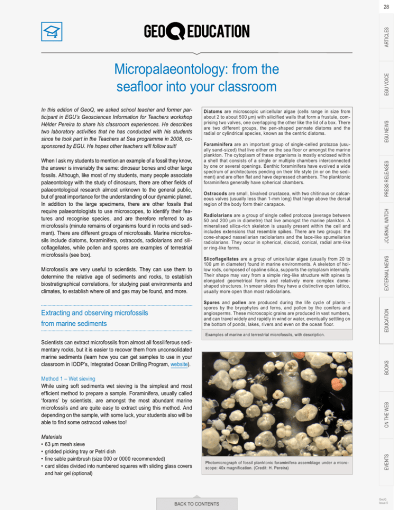
Radiolarians are a group of single celled protozoa (average between 50 and 200 μm in diametre) that live amongst the marine plankton. A mineralised silica-rich skeleton is usually present within the cell and includes extensions that resemble spikes. There are two groups: the cone-shaped nassellarian radiolarians and the lace-like spumellarian radiolarians. They occur in spherical, discoid, conical, radial arm-like or ring-like forms. Microfossils are very useful to scientists. They can use them to determine the relative age of sediments and rocks, to establish biostratigraphical correlations, for studying past environments and climates, to establish where oil and gas may be found, and more. Slicoflagellates are a group of unicellular algae (usually from 20 to 100 μm in diameter) found in marine environments. A skeleton of hollow rods, composed of opaline silica, supports the cytoplasm internally. Their shape may vary from a simple ring-like structure with spines to elongated geometrical forms and relatively more complex domeshaped structures. In smear slides they have a distinctive open lattice, usually more open than most radiolarians. Extracting and observing microfossils from marine sediments Spores and pollen are produced during the life cycle of plants – spores by the bryophytes and ferns, and pollen by the conifers and angiosperms. These microscopic grains are produced in vast numbers, and can travel widely and rapidly in wind or water, eventually settling on the bottom of ponds, lakes, rivers and even on the ocean floor. Examples of marine and terrestrial microfossils, with description. ON THE WEB Method 1 – Wet sieving While using soft sediments wet sieving is the simplest and most efficient method to prepare a sample. Foraminifera, usually called ‘forams’ by scientists, are amongst the most abundant marine microfossils and are quite easy to extract using this method. And depending on the sample, with some luck, your students also will be able to find some ostracod valves too! Materials • 63 μm mesh sieve • gridded picking tray or Petri dish • fine sable paintbrush (size 000 or 0000 recommended) • card slides divided into numbered squares with sliding glass covers and hair gel (optional) EGU NEWS BOOKS Scientists can extract microfossils from almost all fossiliferous sedimentary rocks, but it is easier to recover them from unconsolidated marine sediments (learn how you can get samples to use in your classroom in IODP’s, Integrated Ocean Drilling Program, website). PRESS RELEASES Ostracods are small, bivalved crustacea, with two chitinous or calcareous valves (usually less than 1-mm long) that hinge above the dorsal region of the body form their carapace. JOURNAL WATCH Foraminifera are an important group of single-celled protozoa (usually sand-sized) that live either on the sea floor or amongst the marine plankton. The cytoplasm of these organisms is mostly enclosed within a shell that consists of a single or multiple chambers interconnected by one or several openings. Benthic foraminifera have evolved a wide spectrum of architectures pending on their life style (in or on the sediment) and are often flat and have depressed chambers. The planktonic foraminifera generally have spherical chambers. EXTERNAL NEWS When I ask my students to mention an example of a fossil they know, the answer is invariably the same: dinosaur bones and other large fossils. Although, like most of my students, many people associate palaeontology with the study of dinosaurs, there are other fields of palaeontological research almost unknown to the general public, but of great importance for the understanding of our dynamic planet. In addition to the large specimens, there are other fossils that require palaeontologists to use microscopes, to identify their features and recognise species, and are therefore referred to as microfossils (minute remains of organisms found in rocks and sediment). There are different groups of microfossils. Marine microfossils include diatoms, foraminifera, ostracods, radiolarians and silicoflagellates, while pollen and spores are examples of terrestrial microfossils (see box). Diatoms are microscopic unicellular algae (cells range in size from about 2 to about 500 μm) with silicified walls that form a frustule, comprising two valves, one overlapping the other like the lid of a box. There are two different groups, the pen-shaped pennate diatoms and the radial or cylindrical species, known as the centric diatoms. Photomicrograph of fossil planktonic foraminifera assemblage under a microscope: 40x magnification. (Credit: H. Pereira) BACK TO CONTENTS EVENTS In this edition of GeoQ, we asked school teacher and former participant in EGU’s Geosciences Information for Teachers workshop Hélder Pereira to share his classroom experiences. He describes two laboratory activities that he has conducted with his students since he took part in the Teachers at Sea programme in 2008, cosponsored by EGU. He hopes other teachers will follow suit! EDUCATION Micropalaeontology: from the seafloor into your classroom EGU VOICE ARTICLES 28 GeoQ Issue 5 ARTICLES 29 Method 2 – Preparing smear slides An alternative method to observe not only foraminifera, but also other groups of microfossils like diatoms, radiolarians and silicoflagellates, and that requires an extremely small sample (no bigger than a head of a pin), is the preparation of smear slides. Materials • glass slides • labels • toothpicks • cover slips Suggested procedure Take a microscope glass slide in hand, label it with information about the sediment sample and place a few drops of distilled water in its center. Collect a tiny fraction of the sediment sample with a toothpick, and mix it into the water on the glass slide. Spread the wet sediment with the toothpick until it forms a thin translucent coat across the glass slide, and then carefully place a cover slip on the slide. Smear slides can then be examined under a common or a petrographic light microscope (see figure on top of this page). While the identification of microfossils is not easy, you can ask your students to identify and label each microfossil on the slide using the descriptions in the box on the previous page and the educational resources provided at the end of this article. Hélder Pereira Teacher at Escola Secundária de Loulé, Portugal References Armstrong, H. A. and Brasier, M. D.: Microfossils, Blackwell Publishing, 2005 Fatela, F. and Taborda, R.: Confidence limits of species proportions in microfossil assemblages, Marine Micropaleontology, 45(2), 169–174, 2002 Haq, B. U. and Boersma, A.: Introduction to Marine Micropaleontology, Elsevier Science, 1998 Van der Zwaan, G.J., Jorissen, F.J., and De Stigter, H.C.: The depth dependency of planktic/benthonic foraminiferal ratios: constraints and applications, Marine Geology, 95, 1–16, 1990 Online educational resources on marine sediments and microfossils Teachers at Sea programme aboard the R/V Marion Dufresne during the MD168 AMOCINT cruise IODP sample request for teaching and educational purposes Microfossils: The Ocean’s Storytellers poster, with activities on the back Fossil Fun: A Microfossil Matching game, fact sheet and microfossil key Microfossil Image Recovery And Circulation for Learning and Education Diatom identification guide & ecological resource Foraminifera Gallery – illustrated catalog Radiolaria.org online database Sédiments océaniques et paléoclimats (in French) Projecto LAboratório Oceano (LABO) (in Portuguese) BACK TO CONTENTS PRESS RELEASES JOURNAL WATCH Lastly, students can use the fine sable paintbrush to pick out a selection of specimens from the tray. The selected specimens can then be mounted, using hair gel as glue, for permanent reference in card slides divided into numbered squares with sliding glass covers. Acknowledgments I would like to thank the Institut Paul-Emile Victor (IPEV) and the EGU for sponsoring my participation in the Teachers at Sea programme during the MD168 AMOCINT cruise. A special thank you goes to Carlo Laj, the coordinator of the programme, and to all of those who participated and facilitated our work while we were onboard the RV Marion Dufresne. EXTERNAL NEWS In scientific studies, to ensure statistical validity, usually 300 specimens are counted in randomly chosen grid squares from the tray, but counts of about 100 specimens should be sufficient. EDUCATION where the planktonic fraction %P = 100 x P/(P + B), with P being the amount of planktonic foraminifera and B the amount of benthic foraminifera. By doing any of these lab activities your students will realise that, as incredible it might seem, by studying the fossilised remains of these tiny organisms, scientists can obtain important information about our planet at the time of their deposition on the seafloor. BOOKS Depth = e3,59 + (0,04 x %P) Photomicrograph of fossil radiolarians under a microscope: 100x magnification (Credit: H. Pereira) ON THE WEB A simple quantitative analysis to estimate the planktonic/benthic ratio and infer water depth at the time of deposition can also be undertaken using the following regression: EVENTS The dried sand sized fraction should be poured onto a picking tray so that no sediment grains cover other grains and your students can examine the sample. They can manipulate the sediments on the tray using a fine sable paintbrush and try to distinguish planktonic from benthic foraminifera species based on their shell morphology (see figure on previous page). EGU NEWS EGU VOICE Suggested procedure This method basically consists of the following steps: disaggregating the sample (about 10 cm3), removing fine sediment by soaking and washing it with water through a 63 µm mesh sieve, drying the sand sized fraction in an oven (at 40 to 50 °C) and observe it under a binocular stereo microscope (with a magnification of 10x to 40x). GeoQ Issue 5
