FOCUS | FEATURE
Sequencing with graphene pores
Solid-state nanopores are often used for biomolecular analysis, but have so far been unable to sequence DNA.
Marija Drndić asks whether nanopores made in graphene could fulfil all of the requirements needed for sequencing.
IMAGE COURTESY OF ROBERT JOHNSON
S
olid-state nanopores can be used
to detect, analyse and manipulate
nanoscale objects, and could be used in
applications such as nanoparticle analysis and
water filtration. They are also of particular
interest in the sequencing of DNA. In a
typical experiment, a DNA molecule is
threaded through a nanoscale hole in a
membrane under an applied voltage, and
the ion current flowing through this pore is
monitored; when the DNA nucleotides enter
the pore they block the current by different
amounts, and this signal could then be used
to identify the individual bases and in turn the
DNA sequence.
The field of solid-state nanopores emerged
from work on Coulter counters1, ion channels
and protein pores2–5, and recently proof-ofprinciple DNA sequencing measurements
have been reported using ionic currents and
protein pores6–8. Compared with protein
nanopores, solid-state nanopores offer
potential advantages such as robustness, size
tunability and compatibility with large-scale
chip manufacturing. But solid-state nanopores
cannot yet be used to identify individual
bases. This is due, in part, to the fact that the
membranes used are too thick to probe the
local structure of the DNA; they are typically
silicon nitride membranes with thicknesses of
more than 10 nm, which is equivalent to about
30 DNA bases. As a result, researchers have
turned to thin membranes such as graphene,
which, because of its atomic thickness, could
potentially yield distinct current signals from
one or a few DNA bases.
In 2010, three independent research
groups — led by Jene Golovchenko,
Cees Dekker and myself — reported
threading double-stranded DNA molecules
through nanopores in suspended graphene
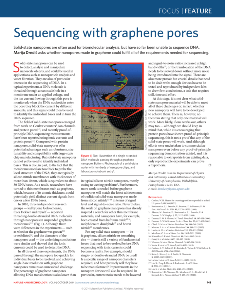
membranes9–11 (Fig. 1). Although there
were differences in the experiments — such
as whether the graphene was grown9,11
or exfoliated12, and the diameters of the
nanopores used — the underlying conclusions
were similar and showed that the ionic
currents could be used to detect the DNA.
In all three of these experiments, the DNA
passed through the nanopore too quickly for
individual bases to be resolved, and achieving
single-base resolution with graphene
nanopores remains an unresolved challenge.
The percentage of graphene nanopores
allowing DNA translocation is also lower than
Figure 1 | Top: Illustration of a single-stranded
DNA molecule passing through a graphene
nanopore. Bottom: Photograph of a solid-state
wafer with hundreds of nanopore chips, and
laboratory notebook entry9.
in typical silicon nitride nanopores, mostly
owing to wetting problems9. Furthermore,
more work is needed before graphene
nanopores will match the latest achievements
of traditional solid-state nanopores made
from silicon nitride12–14 in terms of signal
level and signal-to-noise ratio. Nevertheless,
the work on graphene nanopores has already
inspired a search for other thin membrane
materials, and nanopores have, for example,
been created from hafnium oxide15,16,
molybdenum disulphide17 and boron
nitride18 membranes.
For any solid-state nanopores — be
it graphene, silicon nitride or something
else — there is still a variety of fundamental
issues that need to be resolved before DNA
sequencing with ionic currents could
become a reality. For example, should
single- or double-stranded DNA be used?
Is a specific range of nanopore diameters
required, and how precisely will they have
to be manufactured? Improvements in the
nanopore devices will also be required. In
particular, current noise needs to be lowered
NATURE NANOTECHNOLOGY | VOL 9 | OCTOBER 2014 | www.nature.com/naturenanotechnology
© 2014 Macmillan Publishers Limited. All rights reserved
and signal-to-noise ratios increased at high
bandwidths19, or the translocation of the DNA
needs to be slowed down without more noise
being introduced into the signal. There are
also more prosaic but crucial details that need
to be dealt with: enough devices have to be
tested and reproduced by independent labs
to draw firm conclusions, a task that requires
skill, time and effort.
At this stage, it is not clear what solidstate nanopore material will be able to meet
all of these challenges or, in fact, whether
new nanopores will have to be developed
to achieve them. There is, however, no
theorem stating that only one material will
work. More likely, if one works out, others
may too — although we should keep in
mind that, while it is encouraging that
protein pores have shown proof-of-principle
sequencing, this is not a proof that any of the
solid-state pores will work. And although
efforts were undertaken to commercialize
nanopores even before any proof-of-principle
sequencing demonstration existed, and it is
reasonable to extrapolate from existing data,
only reproducible experiments can prove
a hypothesis.
❐
Marija Drndić is in the Department of Physics
and Astronomy, David Rittenhouse Laboratory,
University of Pennsylvania, Philadelphia,
Pennsylvania 19104, USA.
e-mail: drndic@physics.upenn.edu
References
1. Coulter, W. H. Means for counting particles suspended in a fluid.
US patent 2,656,508 (1953).
2. Kasianowicz, J. J., Brandin, E., Branton, D. & Deamer, D. W.
Proc. Natl Acad. Sci. USA 93, 13770–13773 (1996).
3. Akeson, M., Branton, D., Kasianowicz, J. J., Brandin, E. &
Deamer, D. W. Biophys. J. 77, 3227–3233 (1999).
4. Deamer, D. W. & Akeson, M. Trends Biotechnol. 18, 147–151 (2000).
5. Deamer, D. W. & Branton, D. Acc. Chem. Res. 35, 817–825 (2002).
6. Cherf, G. M. et al. Nature Biotechnol. 30, 344–348 (2012).
7. Manrao, E. A. et al. Nature Biotechnol. 30, 349–353 (2012).
8. Laszlo, A. H. et al. Nature Biotechnol. 32, 829–833 (2014).
9. Merchant, C. A. et al. Nano Lett. 10, 2915–2921 (2010).
10.Schneider, G. F. et al. Nano Lett. 10, 3163–3167 (2010).
11.Garaj, S. et al. Nature 467, 190–193 (2010).
12.Wanunu, M. et al. Nature Nanotech. 5, 807–814 (2010).
13.Venta, K. et al. ACS Nano 7, 4629–4636 (2013).
14.Carlsen, A. T., Zahid, O. K., Ruzicka, J., Taylor, E. W. & Hall, A. R.
ACS Nano 8, 4754–4760 (2014).
15.Shim, J., Rivera, J. A. & Bashir, R. Nanoscale
5, 10887–10893 (2013).
16.Larkin, J. et al. ACS Nano 7, 10121–10128 (2013).
17.Liu, K., Feng, J., Kis, A. & Radenovic, A. ACS Nano
8, 2504–2511 (2014).
18.Liu, S. et al. Adv. Mater. 25, 4549–4554 (2013).
19.Rosenstein, J. K., Wanunu, M., Merchant, C. A., Drndić, M. &
Shepard, K. L. Nature Methods 9, 487–492 (2012).
743

