APPLICATION NOTE No. 140 I June 2013
Polar body biopsy – Advantages of the
Eppendorf micromanipulation system
Markus Montag
University Gynecological Hospital Heidelberg, Department of Gynecological Endocrinology and Fertility Disorders,
Heidelberg, Germany
Abstract
A well-established technique which is used in
preimplantation genetic diagnostics (PGD) is polar body
(PB) biopsy. The polar bodies of the oocyte are extruded
at the conclusion of the meiotic division; normally the first
polar body is noted after ovulation; the second polar body
is observed 2–3 h following entry of the sperm into the
oocyte. Removal of the first and second polar bodies takes
place 6–12 hours after the performance of intracytoplasmic
sperm injection (ICSI). The biopsy of the polar bodies
is followed by detection of certain chromosomes using
fluorescence in-situ hybridization (FISH) or detection of
all chromosomes by comparative genomic hybridization.
Using the Eppendorf micromanipulation system with
manual injectors and the electronic TransferMan® 4m
micromanipulators in combination with the OCTAX Laser
Shot™ System, fast and sensitive handling of the oocyte
and polar bodies can be guaranteed.
Introduction
Over the past few decades the mean age of women conceiving
their first child has steadily increased. However, advanced
maternal age lowers the chance for pregnancy and increases
the risk of miscarriage once a woman is pregnant. One major
problem strongly correlated to maternal age is the occurrence
of numerical chromosomal abnormalities in human oocytes.
In women who are 40 years old and older, up to 70 % of
their oocytes can be chromosomally abnormal [1, 2]. In the
context of assisted reproduction treatment, it is possible to
identify and exclude such oocytes, thereby increasing the
success rate. The underlying technique is the biopsy of the
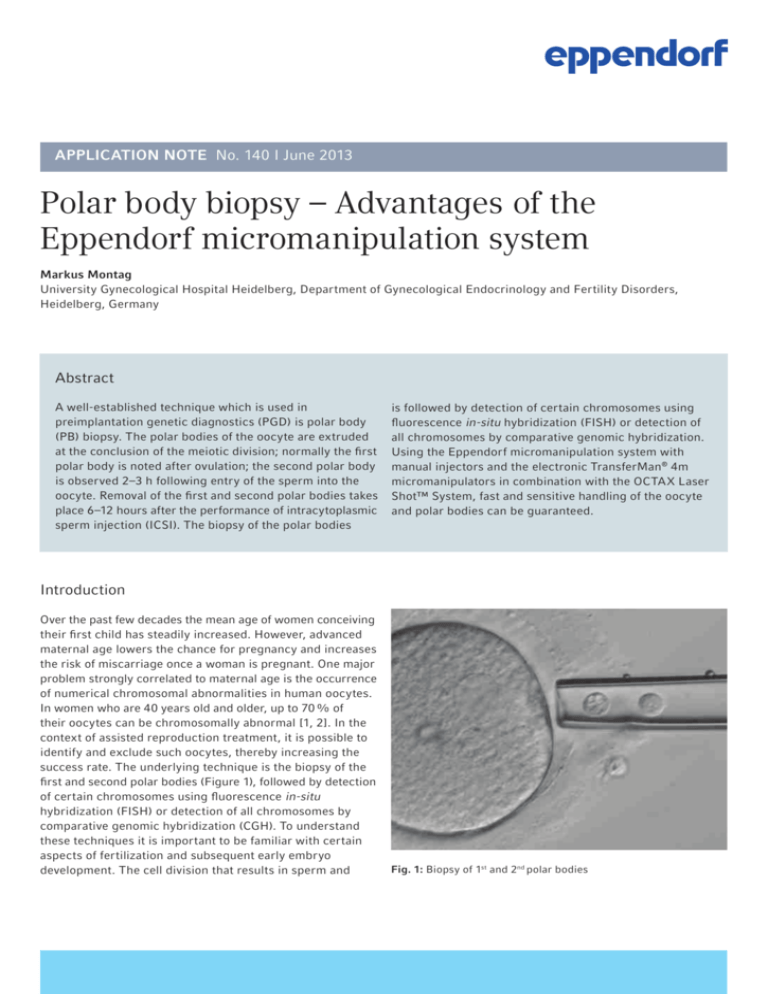
first and second polar bodies (Figure 1), followed by detection
of certain chromosomes using fluorescence in-situ
hybridization (FISH) or detection of all chromosomes by
comparative genomic hybridization (CGH). To understand
these techniques it is important to be familiar with certain
aspects of fertilization and subsequent early embryo
development. The cell division that results in sperm and
Fig. 1: Biopsy of 1st and 2nd polar bodies
APPLICATION NOTE I No. 140 I Page 2
oocytes containing 23 chromosomes each is called meiosis.
In their resting state, eggs exist in a state of arrested meiosis
and still contain all 23 paired chromosomes. During
ovulation, meiosis resumes, and the egg extrudes one set
of its 23 chromosomes in a small structure called the first
polar body. Soon after fertilization occurs, a second polar
body, containing 23 maternal chromatids, is expelled. On
the day after oocyte retrieval, both polar bodies are visible
by light microscopy in the normally fertilized oocyte.
Polar bodies with missing or excess chromosomal material
are indicative of an oocyte that contains either an excess
of chromosomes, which, after fertilization, results in an
embryo with a trisomy, or of an oocyte which is missing
chromosomes, which, again after fertilization, results in an
embryo with a monosomy. Thus, polar body biopsy provides
an indirect diagnosis of the oocyte for aneuploidy testing.
This method can also be applied to couples with a balanced
translocation of the mother and to couples who are aware
of maternal predisposition for a genetic disease that may
manifest itself in the child.
Polar body biopsy was first presented in 1990 [3], and
several aspects of this technique have been technically
refined including the use of lasers and electronic
micromanipulators to facilitate the biopsy procedure [4].
Materials and Methods
Compared to ICSI, polar body biopsy requires additional
micromanipulation steps; thus it greatly benefits from the
proper instrumentation, especially the Eppendorf
TransferMan 4m (Figure 2) with its special features which
render the procedure as economic as possible. The use of
application specific masks of the TransferMan 4m
micromanipulator with predefined as well as freely definable
softkey functions eases the individual workflow process.
The unique DualSpeed joystick enables precise and intuitive
movement along all three axes when handling the oocyte as
well as dynamic movements while covering greater distances
(see Figure 3). This helps reducing exposure time of the
oocytes to stressful conditions outside the incubator, and it
minimizes the risk of losing the material of interest.
speed
[µm/s]
1
2
0
Fig. 3: Movement in direct correlation with the deflection of the
joystick. 1: proportional kinetics, 2: area of dynamic movement.
The ratio of the dynamic movement may be adjusted according to
the proportional range.
Fig. 2: Eppendorf Workstation for IVF
APPLICATION NOTE I No. 140 I Page 3
Devices for polar body biopsy
>Inverted microscope with a heated plate and modulation contrast objectives
>OCTAX Laser Shot™ System (OCTAX, MTG)
>2 TransferMan 4m micromanipulators (Eppendorf)
>CellTram® Air microinjector for holding the embryo
(Eppendorf)
>CellTram® vario microinjector for removal and transfer of the polar body (Eppendorf)
>Polar Body Biopsy Tip MML (Eppendorf)
>Holding capillary, e.g. VacuTips (Eppendorf)
Polar body biopsy
The most important feature of the TransferMan 4m for polar
body biopsy is its ability to store several freely definable
capillary positions. Up to five positions can be stored.
For the polar body biopsy procedure, as it is performed in
our laboratory, one needs to store three user-defined
positions; hence the use of the application mask »Cell Transfer«
with three softkeys dedicated to position storage is
recommended. In addition, using this mask, a vertical limit
can be defined to avoid capillary breakage (see Figure 4).
One softkey is free for individual settings.
array-CGH, position 2B for easily replacing the culture dish
with a glass slide, and position 3B for releasing the polar
bodies into the water droplets on the slide for later FISH.
Additionally, two positions are reserved for the manipulator
used for holding the oocyte: position 1H for holding the
oocyte during biopsy and position 2H for the changing of
droplets in the dish. Once stored, these positions can be
automatically activated by pressing the relevant position
button on the device. Alternatively, the positions can be
relocated in succession by clicking the joystick key twice
(double-click). This set-up enables a fast and economic
change from one capillary working position to another, but
most importantly it controls the capillary that holds the
aspirated polar bodies, thus helping to reduce the rate of
lost polar bodies to less than 0.5 %. An overview of the
different positions required during the biopsy procedure is
shown in Figure 5a–b.
Fig. 5a: Stored positions for polar body biopsy.
Fig. 4: Application mask »Cell transfer« with three softkeys: for
position storage and one for definition of a vertical limit (Z-axis
limit) to avoid capillary breakage. One softkey is free for individual
programming.
The polar body biopsy is performed in a culture dish. A
biopsy capillary is used to transfer the polar bodies directly
into a drop of water on a glass slide for the purpose of FISH
analysis or into another droplet in the dish for later transfer
to PCR tubes if array-CGH is to be performed. The three
user-defined capillary positions are used as follows: position
1B for biopsy/release into adjacent droplets in the case of
Fig. 5b: Stored positions for polar body biopsy.
APPLICATION NOTE I No. 140 I Page 4
For polar body biopsy we recommend setting up a dish with
a single droplet that contains PVP and two/three rows of
droplets for FISH/array-CGH, 3–5 µL each, which contain
buffered culture medium. The buffered culture medium is
required for maintenance of the proper pH during
manipulation outside the incubator. PVP is used for rinsing
the biopsy capillary, which helps to avoid polar bodies
sticking to the inner glass wall. The left row of droplets is
used for the oocytes (one per droplet) and the right row
for sampling of the polar bodies after biopsy. In the case of
array-CGH two rows for polar body sampling are required
as polar bodies 1 and 2 need to be processed separately. In
preparation for biopsy, an oocyte is gently aspirated by the
holding capillary and affixed as closely as possible to the
bottom of the dish. This position is stored as position 1H.
Rotation of the oocyte may be necessary in order to obtain
proper alignment of the first and second polar bodies in one
focal plane (Figure 6a). This focal plane defines position 1B
for the biopsy capillary, and, once adjusted, this position
should also be stored. Following laser-assisted opening of
the zona pellucida, the biopsy capillary is pushed through
the opening of the zona towards the polar body next to the
capillary opening. By rotating the knob of CellTram vario
both polar bodies are slowly aspirated into the capillary
(Figure 6b). To slowly push the capillary through to the polar
bodies, slight suction is usually helpful. Once both polar
bodies are completely aspirated, the capillary is removed
from the zona and the oocyte is released from the holding
capillary.
If several oocytes need to be biopsied, the first and second
polar bodies can be temporarily stored in the neighboring
medium droplets while the biopsy of the next oocyte is
performed as previously described. Once all polar bodies are
biopsied, it is advisable to first place the oocytes back into
the incubator. To do so, both capillaries are brought into a
Fig. 6a: First and second polar bodies are aligned properly in one
focal plane
position outside and far above the culture dish by
activating the »home« key, and if required, the motors are
swiveled out to the side of the microscope. Now the dish
can be removed safely so that the oocytes may be
transferred to another culture dish.
Transfer of polar bodies to a slide for later FISH analysis
The dish still holding the polar bodies is placed back on the
microscope stage, the motors are swiveled back in and the
biopsy capillary is lowered automatically by pressing the
»home« key again. Next the capillary can be moved directly
into position 1B by pressing »Pos1«. The polar bodies corresponding to oocyte 1 are then aspirated into the biopsy
capillary. Next, the capillary is moved to position 2B (by
pressing »Pos 2«), the culture dish is removed, and a glass
slide holding a 0.2 µL droplet of pure water is placed under
the capillary. The biopsy capillary still holding the polar
bodies is lowered into the water droplet so that it just touches the glass surface. This position is stored as position 3B.
The first and second polar bodies are carefully released into
the droplet, and the capillary is first drawn back and then
brought into position 2B by pressing »Pos 2«. The small
volume ensures that the polar body will attach to a small
area on the slide and the fluid will dry fast, thereby reducing the risk of dislocation from the slide. Even so, the drying
process must be observed under a stereo microscope, and
the final location of the polar body after air-drying must
be circled on top of the slide using a diamond marker. This
procedure can be repeated until all polar bodies have been
transferred to the slide. With some experience, 4 to 6 polar
bodies can be placed within an area of approx. 10 mm, each
encircled using a diamond marker [5]. Because all relevant
capillary positions have been stored during the first round,
further manipulation of polar bodies is less time consuming.
Fig. 6b: Both polar bodies are slowly aspirated into the capillary.
APPLICATION NOTE I No. 140 I Page 5
Transfer of polar bodies into a PCR tube for later
array-CGH analysis
For the transfer into PCR tubes polar bodies must be
transferred one by one into PCR tubes pre-filled with PBS.
As PCR tubes are narrow, the transfer cannot be performed
using the biopsy capillary; instead the use of stripper tips
is recommended. A detailed description of the transfer as
well as the array-CGH procedure is provided by Magli et al.,
2012 [6].
Fluorescence in-situ hybridization (FISH)
For FISH analysis, the dried polar bodies are fixed by the
addition of 2 x 10 µL ice-cold methanol: acetic acid (3 : 1),
followed by incubation in methanol at room temperature
for another 5 min. The slides are dried, and the FISH probe
for chromosomal detection is directly applied to the slide,
which is then covered by a cover slip and sealed with rubber
cement. The slide is placed into a Thermomixer ® comfort
with exchangeable thermoblock for slides. Co-denaturation
of the probe and the genomic DNA as well as subsequent
hybridization is performed using the Thermomixer at the
time and temperature settings indicated by the manufacturer of the chromosome probe. Following hybridization,
unbound probe is washed off, and the FISH signals can be
evaluated using a fluorescence light microscope equipped
with appropriate filter sets (Figure 7).
Fig. 7: FISH signals of chromosomes 13, 18, 21, 22
Results and Discussion
Each chromosome should show two signals in the 1st polar
body and one signal in the 2nd polar body. Approx. 70 % of
all chromosomal disorders are detectable in the 1st polar
body; however, a disorder may occur in the formation of the
2nd polar body.
A frequent problem when judging FISH results is the
occurrence of chromatin degeneration which can be
identified by speckled signals. Interestingly, this phenomenon
occurs most often when using LSI probes. Nevertheless,
it is still possible to draw conclusions about the respective
chromosomes, since early segregation of chromatids means
that the regions with speckled signals are also separated.
The main problem with polar body biopsy is the fragmentation
of polar bodies, particularly the first polar body, which can
be seen in Figure 6b. Since each fragment can contain
chromosomes, it is crucial that all fragments be removed
during biopsy. Here, sensitive handling is required which
can be perfectly executed with the intuitive proportional
fine control of the TransferMan 4m micromanipulator in
combination with the fine wheel of the CellTram vario. Any
movement of the hand is directly transferred to the biopsy
capillary, without any delay or even tiniest stutter.
In addition to the removal of all fragments, identifying the
number and re-location of the fragments is critical, because
they can disconnect while drying and move to different
areas of the slide. Therefore it is absolutely necessary to
compile a drawing. Otherwise the risk is quite high that
signals in small fragments are disregarded which will lead to
a false diagnosis.
Depending on the results of the FISH or the array-CGH
analysis, chromosomally normal oocytes can be selected for
further culture and transfer.
APPLICATION NOTE I No. 140 I Page 6
Literature
[1] Hassold T, Chiu D. Maternal age-specific rates of numerical chromosome abnormalities with special reference to
trisomy. Human Genet.; 1985, 70(1):11–7.
[2] Geraedts J, Montag M, Magli MC, Repping S, Handyside A, Staessen C, Harper J, Schmutzler A, Collins J, Goossens V,
van der Ven H, Vesela K, Gianaroli L. Polar body array CGH for prediction of the status of the corresponding oocyte. Part I: clinical results. Human Reprod.; 2011, 26(11):3173–80
[3] Verlinsky Y, Ginsberg N, Lifchez A, Valle J, Moise J, Strom CM. Analysis of the first polar body: preconception genetic diagnosis. Human Reprod.; 1990, 5(7):826–9.
[4] Montag M, van d, V, Delacretaz G, Rink K, van d, V. Laser-assisted microdissection of the zona pellucida facilitates polar body biopsy. Fertil Steril.; 1998, 69(3):539–42.
[5] Montag M, van der Ven K, van der Ven H. Polar body biopsy. In: Gardner, Weissmann, Howles, Shoham, editors.
Textbook of Assisted Reproductive Techniques: Laboratory and Clinical Perspectives. Lancaster: Taylor & Francis
Medical Text Books; 2004, p. 391–404.
[6] Magli MC, Montag M, Köster M, Muzi L, Geraedts J, Collins J, Goossens V, Handyside AH, Harper J, Repping S,
Schmutzler A, Vesela K, Gianaroli L. Polar body array CGH for prediction of the status of the corresponding oocyte. Part II: technical aspects. Human Reprod.; 2011, 26(11):3181–5.
APPLICATION NOTE I No. 140 I Page 7
Ordering Information
Description
1
2
Order no. international
Order no. North America
TransferMan 4m , Proportional micromanipulator for suspension cells
5193 000.012
5193 000.020
CellTram® Air1, Manual pressure device for the reliable holding of suspended cells
(e.g. oocytes)
5176 000.017
920002021
CellTram® vario1, Manual hydraulic microinjector, with gears 1:1 and 1:10
5176 000.033
920002111
VacuTip1,2, 25 glass capillaries for holding large cells (e.g. oocytes), sterilized,
tip angle 35°
5175 108.000
930001015
Polar Body Biopsy Tip MML1,2, 25 glass capillaries for laser supported polar body
biopsy after Markus Montag
5175 210.000
Not available
Polar Body Biopsy Tip FCH1,2, 25 glass capillaries for laser supported polar body
biopsy after Fertility Center Hamburg
5175 230.000
Not available
Themomixer® comfort, without exchangeable thermoblock
5355 000.011
022670107
Exchangeable Slide Thermoblock, for 4 slides, for hybridization experiments
5368 000.010
022670590
®
1
This product is registered in Europe as a medical device (according to Medical Device MDD 93/42/EEC).
Proven non-cytotoxic by mouse embryo development test.
Your local distributor: www.eppendorf.com/contact
Eppendorf AG · 22331 Hamburg · Germany
eppendorf@eppendorf.com · www.eppendorf.com
www.eppendorf.com
OCTAX Laser Shot™ System is a trademark of OCTAX, MTG, Germany.
Eppendorf®, the Eppendorf logo, TransferMan®, CellTram®, and Thermomixer® are registered trademarks of Eppendorf AG, Hamburg, Germany.
All rights reserved, including graphics and images. Copyright © 2013 by Eppendorf AG, Hamburg, Germany.

