OUTLINE FOR SURFACE ANATOMY OF THE PULMONARY SYSTEM
advertisement
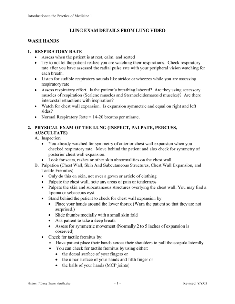
Introduction to the Practice of Medicine 1 LUNG EXAM DETAILS FROM LUNG VIDEO WASH HANDS 1. RESPIRATORY RATE · Assess when the patient is at rest, calm, and seated · Try to not let the patient realize you are watching their respirations. Check respiratory rate after you have assessed the radial pulse rate with your peripheral vision watching for each breath. · Listen for audible respiratory sounds like stridor or wheezes while you are assessing respiratory rate · Assess respiratory effort. Is the patient’s breathing labored? Are they using accessory muscles of respiration (Scalene muscles and Sternocleidomastoid muscles)? Are there intercostal retractions with inspiration? · Watch for chest wall expansion. Is expansion symmetric and equal on right and left sides? · Normal Respiratory Rate = 14-20 breaths per minute. 2. PHYSICAL EXAM OF THE LUNG (INSPECT, PALPATE, PERCUSS, AUSCULTATE) A. Inspection · You already watched for symmetry of anterior chest wall expansion when you checked respiratory rate. Move behind the patient and also check for symmetry of posterior chest wall expansion. · Look for scars, rashes or other skin abnormalities on the chest wall. B. Palpation (Chest Wall, Skin And Subcutaneous Structures, Chest Wall Expansion, and Tactile Fremitus) · Only do this on skin, not over a gown or article of clothing · Palpate the chest wall, note any areas of pain or tenderness · Palpate the skin and subcutaneous structures overlying the chest wall. You may find a lipoma or sebaceous cyst. · Stand behind the patient to check for chest wall expansion by: · Place your hands around the lower thorax (Warn the patient so that they are not surprised.) · Slide thumbs medially with a small skin fold · Ask patient to take a deep breath · Assess for symmetric movement (Normally 2 to 5 inches of expansion is observed) · Check for tactile fremitus by: · Have patient place their hands across their shoulders to pull the scapula laterally · You can check for tactile fremitus by using either: · the dorsal surface of your fingers or · the ulnar surface of your hands and fifth finger or · the balls of your hands (MCP joints) H:\Ipm_1\Lung_Exam_details.doc -1- Revised: 8/8/03 Introduction to the Practice of Medicine 1 · · Use both hands, simultaneously checking upper, mid, and lower fields, and then check laterally posteriorly. Right and left sides should feel the same. Note any area of asymmetry and pay close attention to that area with further exam maneuvers Ask the patient to repeat “Ninety-nine, Ninety-nine . . .” every time you reposition your hands C. Percussion · This is a difficult technique to master. With percussion, you can determine if the tissues deep to the area of percussion are air filled, fluid filled, or solid tissue by the pitch, duration, and intensity of the percussed sound. · Only do this on skin, not over a gown or article of clothing · Room must be quiet for you to appreciate the percussed sound · Technique of percussion: · Hyperextend your index finger at the PIP and DIP joints and flex the index finger about 45 degrees at the MCP joint. · Place the DIP joint of your index finger on the patient’s upper chest in an intercostal space · No other part of your hand should be in contact with the patient’s skin · Place your other hand near this hand · Use your other hand’s index or middle finger to quickly percuss at the DIP joint in contact with the patient’s skin. Use two or three brisk percussion strikes (flexing your hand at the wrist to percuss) and then quickly withdraw the percussing finger from the DIP · Always begin at the apex of the lungs · Always compare right side to left side at each level, then move inferiorly to percuss middle, lower and lateral posterior lung fields · Common errors of percussion: · Examiner places their entire hand on patient’s chest · Examiner’s DIP, PIP, and MCP joints of their middle or index finger is flat against the patient’s chest · Examiner percusses/strikes at their DIP with the other hand from too far away from the chest wall · Examiner does not strike vigorously enough to make an audible sound · Examiner does not assure the room is quiet enough to hear the percussed sound · Examiner percusses one entire lung, and then percusses the other lung · Examiner does not ask the patient to cross their arms across their chest to move their scapula laterally D. Auscultation · Only do this on skin, not over the patient’s gown or article of clothing · Ask the patient to cross their arms across their chest to move their scapulae laterally · Ask the patient to breathe with their mouth open · Ask the patient to breathe normally. If you cannot hear breath sounds, ask the patient to take deeper breaths. H:\Ipm_1\Lung_Exam_details.doc -2- Revised: 8/8/03 Introduction to the Practice of Medicine 1 · · · · · · · Use the diaphragm of your stethoscope Always begin at a lung apex Listen for an entire respiratory cycle at every location. Do NOT move your stethoscope while the patient is still exhaling. After exhalation is complete, quickly move your stethoscope. Compare right side to left side at every level that you auscultate Move inferiorly; auscultate then at mid, lower, and lateral lung fields. Then auscultate anteriorly If you hear a type of breath sound in an unexpected location, proceed to check for 3 transmitted breath sounds (vocal fremitus) in that area using your stethoscope: · Ask the patient to repeat “Ninety-nine” as you auscultate in this area (evaluate for bronchophony) · Ask the patient to whisper “Ninety-nine” as you auscultate in this area (evaluate for whispered pectoriloquy) · Ask the patient to say “EEEEEE” while you listen in this area (evaluate for egophony) Common errors of auscultation: · Auscultating one entire lung, and then moving to the other lung · Auscultating over a patient’s gown or article of clothing · Beginning auscultation inferiorly at the lower lung fields · Moving your stethoscope before each exhalation is complete · Examiner does not make the room quiet enough to hear breath sounds. WASH HANDS H:\Ipm_1\Lung_Exam_details.doc -3- Revised: 8/8/03

