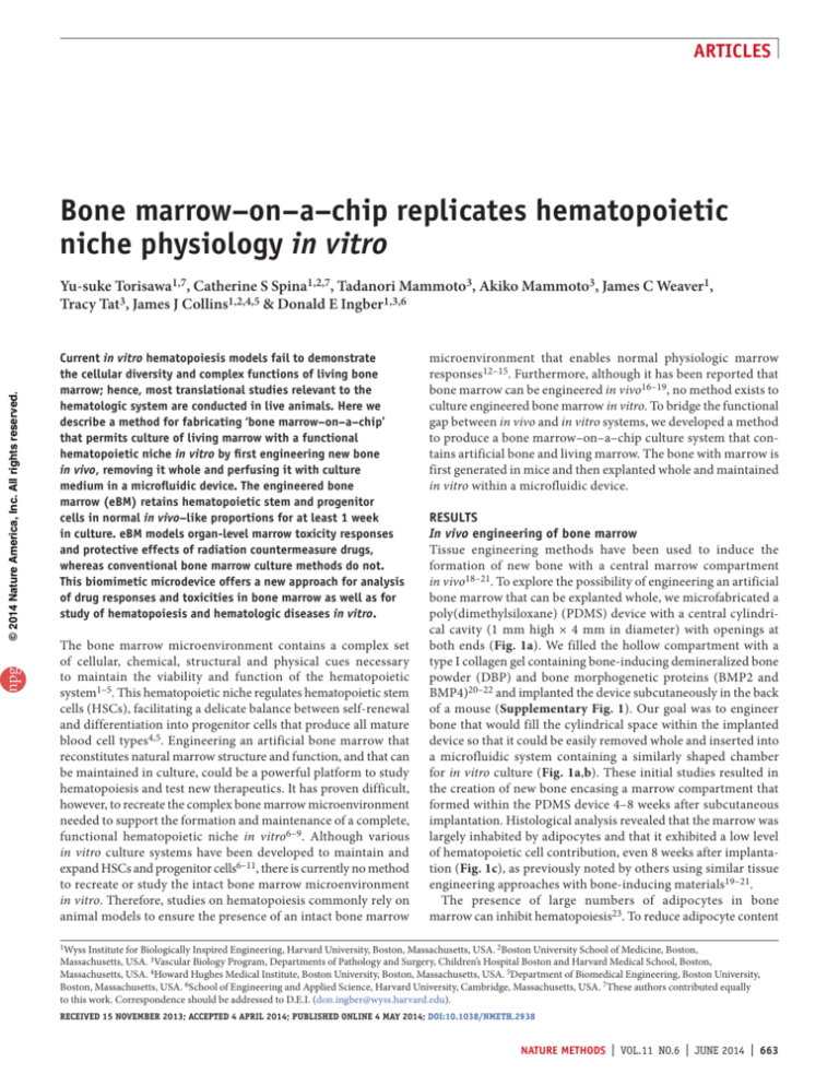
Articles
Bone marrow–on–a–chip replicates hematopoietic
niche physiology in vitro
npg
© 2014 Nature America, Inc. All rights reserved.
Yu-suke Torisawa1,7, Catherine S Spina1,2,7, Tadanori Mammoto3, Akiko Mammoto3, James C Weaver1,
Tracy Tat3, James J Collins1,2,4,5 & Donald E Ingber1,3,6
Current in vitro hematopoiesis models fail to demonstrate
the cellular diversity and complex functions of living bone
marrow; hence, most translational studies relevant to the
hematologic system are conducted in live animals. Here we
describe a method for fabricating ‘bone marrow–on–a–chip’
that permits culture of living marrow with a functional
hematopoietic niche in vitro by first engineering new bone
in vivo, removing it whole and perfusing it with culture
medium in a microfluidic device. The engineered bone
marrow (eBM) retains hematopoietic stem and progenitor
cells in normal in vivo–like proportions for at least 1 week
in culture. eBM models organ-level marrow toxicity responses
and protective effects of radiation countermeasure drugs,
whereas conventional bone marrow culture methods do not.
This biomimetic microdevice offers a new approach for analysis
of drug responses and toxicities in bone marrow as well as for
study of hematopoiesis and hematologic diseases in vitro.
The bone marrow microenvironment contains a complex set
of cellular, chemical, structural and physical cues necessary
to maintain the viability and function of the hematopoietic
system1–5. This hematopoietic niche regulates hematopoietic stem
cells (HSCs), facilitating a delicate balance between self-renewal
and differentiation into progenitor cells that produce all mature
blood cell types4,5. Engineering an artificial bone marrow that
reconstitutes natural marrow structure and function, and that can
be maintained in culture, could be a powerful platform to study
hematopoiesis and test new therapeutics. It has proven difficult,
however, to recreate the complex bone marrow microenvironment
needed to support the formation and maintenance of a complete,
functional hematopoietic niche in vitro6–9. Although various
in vitro culture systems have been developed to maintain and
expand HSCs and progenitor cells6–11, there is currently no method
to recreate or study the intact bone marrow micro­environment
in vitro. Therefore, studies on hematopoiesis commonly rely on
animal models to ensure the presence of an intact bone marrow
microenvironment that enables normal physiologic marrow
responses12–15. Furthermore, although it has been reported that
bone marrow can be engineered in vivo16–19, no method exists to
culture engineered bone marrow in vitro. To bridge the functional
gap between in vivo and in vitro systems, we developed a method
to produce a bone marrow–on–a–chip culture system that contains artificial bone and living marrow. The bone with marrow is
first generated in mice and then explanted whole and maintained
in vitro within a microfluidic device.
RESULTS
In vivo engineering of bone marrow
Tissue engineering methods have been used to induce the
formation of new bone with a central marrow compartment
in vivo18–21. To explore the possibility of engineering an artificial
bone marrow that can be explanted whole, we microfabricated a
poly(dimethylsiloxane) (PDMS) device with a central cylindrical cavity (1 mm high × 4 mm in diameter) with openings at
both ends (Fig. 1a). We filled the hollow compartment with a
type I collagen gel containing bone-inducing demineralized bone
powder (DBP) and bone morphogenetic proteins (BMP2 and
BMP4)20–22 and implanted the device subcutaneously in the back
of a mouse (Supplementary Fig. 1). Our goal was to engineer
bone that would fill the cylindrical space within the implanted
device so that it could be easily removed whole and inserted into
a microfluidic system containing a similarly shaped chamber
for in vitro culture (Fig. 1a,b). These initial studies resulted in
the creation of new bone encasing a marrow compartment that
formed within the PDMS device 4–8 weeks after subcutaneous
implantation. Histological analysis revealed that the marrow was
largely inhabited by adipocytes and that it exhibited a low level
of hematopoietic cell contribution, even 8 weeks after implantation (Fig. 1c), as previously noted by others using similar tissue
engineering approaches with bone-inducing materials19–21.
The presence of large numbers of adipocytes in bone
marrow can inhibit hematopoiesis23. To reduce adipocyte content
1Wyss Institute for Biologically Inspired Engineering, Harvard University, Boston, Massachusetts, USA. 2Boston University School of Medicine, Boston,
Massachusetts, USA. 3Vascular Biology Program, Departments of Pathology and Surgery, Children’s Hospital Boston and Harvard Medical School, Boston,
Massachusetts, USA. 4Howard Hughes Medical Institute, Boston University, Boston, Massachusetts, USA. 5Department of Biomedical Engineering, Boston University,
Boston, Massachusetts, USA. 6School of Engineering and Applied Science, Harvard University, Cambridge, Massachusetts, USA. 7These authors contributed equally
to this work. Correspondence should be addressed to D.E.I. (don.ingber@wyss.harvard.edu).
Received 15 November 2013; accepted 4 April 2014; published online 4 may 2014; doi:10.1038/nmeth.2938
nature methods | VOL.11 NO.6 | JUNE 2014 | 663
npg
© 2014 Nature America, Inc. All rights reserved.
Articles
Figure 1 | In vivo bone marrow engineering.
(a) Workflow to generate a bone marrow–on–a–chip
system in which eBM is formed in a PDMS device
in vivo and is then cultured in a microfluidic
system. (b) Top, PDMS device containing boneinducing materials in its central cylindrical
chamber before implantation. Center, formed
white cylindrical bone with pink marrow visible
within eBM 8 weeks (wk) after implantation.
Bottom, bone marrow chip microdevice used to
culture the eBM in vitro. Scale bars, 2 mm.
(c) Low- (left) and high-magnification views (right)
of histological hematoxylin-and-eosin–stained
sections of the eBM formed in the PDMS device
with two openings (top) or one lower opening
(center) at 8 weeks following implantation
compared with a cross-section of bone marrow
in a normal adult mouse femur (bottom).
Scale bars, 500 and 50 µm for low and high
magnification views, respectively. (d) Threedimensional reconstruction of micro-computed
tomography (micro-CT) data from eBM 8 weeks
after implantation (average bone volume was
2.95 ± 0.25 mm3; n = 3). Scale bar, 1 mm.
a
Subcutaneous
implantation
Bone-inducing
materials
b
8 weeks
In vivo engineering
of bone marrow
0 wk
eBM
Insert eBM
8 wk
c
Two openings 8 wk
d
eBM 8 wk
Femur
in the marrow, we sealed the top of the central cavity in the
implanted device by adding a solid layer of PDMS to restrict
access of cells or soluble factors from the overlying adipocyte-rich
hypodermis to the bone-inducing materials while maintaining
accessibility to the underlying muscle tissue through the lower
opening (Fig. 1a and Supplementary Fig. 1). Subcutaneous
implantation of this improved PDMS device resulted in the
formation of a cylindrical disk of white, bone-like tissue containing a central region of blood-filled marrow over a period of
8 weeks (Fig. 1b and Supplementary Fig. 2). Histological analysis
confirmed the presence of a shell of cortical bone of relatively
uniform thickness surrounding marrow that was dominated by
hematopoietic cells and that contained few adipocytes (Fig. 1c).
Comparison of histological sections of the eBM to sections from
an intact femur confirmed that the morphology of the eBM was
nearly identical to that of natural bone marrow (Fig. 1c).
Micro-computed tomographic (micro-CT) analysis of the eBM
demonstrated that the newly formed cortical shell of bone also
contained an ordered internal trabecular network that closely
resembles the intricate architecture found in normal adult mouse
vertebrae (Supplementary Fig. 3) and that is known to be supportive of HSCs24 (Fig. 1d). Compositional analysis using energydispersive X-ray spectroscopy (EDS) also showed that the calcium
and phosphorous content of the eBM were indistinguishable from
that of natural trabecular bone (Supplementary Fig. 3).
Characterization of engineered bone marrow
Interactions between CXCL12 expressed on the surfaces of various
cell types in the bone marrow (such as osteoblasts25, perivascular
endothelial and perivascular stromal cells26) and its cognate receptor CXCR4 on the surfaces of HSCs and hematopoietic progenitor
cells are critical for the recruitment, retention and maintenance of
HSCs26–28. Immunohistochemical analysis confirmed that both of
these key hematopoietic regulators were expressed in their normal
positions in the eBM: CXCL12 localized to cells lining the inner
surface of the bone and blood vessels, and CXCR4 was expressed
664 | VOL.11 NO.6 | JUNE 2014 | nature methods
eBM 8 wk
by clusters of lymphoid cells in the endosteal and perivascular
niches (Fig. 2a–d). We also confirmed that key hematopoietic
niche cells3 including perivascular nestin+ cells and leptin receptor+ cells, as well as CD31+ vascular endothelial cells, resided in
their normal positions (Supplementary Fig. 4).
To rigorously characterize the hematopoietic content of
the engineered marrow, we harvested cells from the eBM
immediately after surgical removal and analyzed them by flow
cytometry. The cellular components of the marrow contained
within the eBM were compared to hematopoietic populations
isolated from femur bone marrow and peripheral blood from
the same mice (Fig. 2e,g). Devices harvested 4 and 8 weeks after
implantation contained all blood cell types, including HSCs
that are not recognized by a mixture of Lin antibodies that recognize
mature, lineage-restricted blood cells (Lin−Sca1+cKit+CD34+/−,
Lin−Sca1+cKit+CD150+/−CD48−/+) and hematopoietic progenitor cells identified by four different marker sets (Lin−Sca1+,
Lin−cKit+, Lin−CD34+, Lin−CD135+), as well as mature erythrocytes (Ter119+), lymphocytes (T cells, CD45+CD3+; B cells,
CD45+CD19+) and myeloid cells (CD45+Mac1+/−Gr1+/−). The
eBM harvested 4 weeks after implantation did not appear to
be fully developed, as indicated by a lower proportion of HSCs
and hematopoietic progenitor cells compared to that in normal
marrow (Fig. 2f,h). However, cells harvested from the eBM
8 weeks after implantation exhibited a completely normal distribution of HSCs, hematopoietic progenitors and differentiated
blood cells from all lineages that was nearly identical to that displayed by natural bone marrow (Fig. 2f,h and Supplementary
Figs. 5 and 6).
In summary, our modified strategy for eBM produced a cylindrical disk of cortical and trabecular bone (Supplementary Fig. 3)
containing marrow with a hematopoietic cell composition nearly
identical to that of natural bone marrow. The presence of key
cellular and molecular components of the hematopoietic niche
suggests that the cellular content of the eBM closely resembles
the natural bone environment.
mBM
0.14
0.09
0.61
0.43
0.30
eBM/CXCR4
eBM/CXCL12
Lin–Sca1+
eBM
8 wk
Side scatter
0.08
Lin–CD34+
Lin-cKit
0.38
0.59
f
Distribution (%)
d
npg
Femur/CXCR4
0.35
Side scatter
Progen.
HSCs
mPB
(–RBC)
eBM
4 wk
eBM
8 wk
mBM
In vitro culture of engineered bone marrow
To determine whether the eBM could maintain a functional hemato­
poietic system in vitro, we surgically removed the eBM formed
8 weeks after implantation from the mouse, punctured it in multiple
places with a surgical needle to permit fluid access and cultured it
in another clear PDMS microfluidic device containing a similarly
shaped cylindrical central chamber that is separated from overlying and underlying microfluidic channels by porous membranes
(Fig. 1a,b). To maintain the cellular viability of the eBM, we perfused culture medium through the top and bottom channels using
a syringe pump at an optimal rate (1 µl/min) (Supplementary
Fig. 7) once the eBM was inserted into the central chamber and
the surrounding porous membranes and microchannel layers were
attached. The eBM was cultured in vitro for 4 or 7 d within the bone
marrow–chip microsystem (Fig. 1b), which covers a time period
that is commonly used to test for drug efficacies and toxicities
in vitro29,30. The cultured bone and marrow retained their morphology during this time, including the distribution of CXCL12expressing stromal cells (Supplementary Fig. 8). Stroma-supported
culture systems represent the current benchmark for maintaining
survival of HSCs and hematopoietic progenitor cells in vitro7,31.
Distribution (%)
CD45+CD19+
CD45+CD19+
CD45+Mac1+
CD45+Mac1+
+
Ter119
Figure 2 | Localization of
1.4
27
33
g
h 100
12
T cells
cytokines and hematopoietic cell
90
B cells
56
1.4
mBM
composition of the eBM.
80
(a–d) Immunohistochemical
70
Myeloid
1.7
60
analysis of ligand-receptor pair
CD45+
CD45+Gr1+
CD45+CD3+
RBCs
50
CXCL12 and CXCR4 in eBM compared
40
35
2.0
30
to in uncultured mouse femur bone
14
30
marrow (mBM). (a,b) Anti-CXCL12
eBM
58
2.0
20
staining of the eBM (a) and mBM (b). 8 wk
10
(c,d) Anti-CXCR4 staining of eBM (c)
1.5
0
mPB
eBM
eBM
mBM
and mBM (d). Scale bars for a–d,
CD45+
CD45+Gr1+
CD45+CD3+
4 wk
8 wk
25 µm. (e) Flow cytometric analysis
of HSCs and hematopoietic progenitor (progen.) cells within the Lin − cell subpopulation isolated from mBM and eBM isolated 8 weeks (wk)
after subcutaneous implantation. Numbers inside individual gates indicate the proportion of these cells as a percentage of the total cell population
isolated from whole bone marrow. (f) Distribution of HSCs (Lin−Sca1+cKit+, red) and hematopoietic progenitor cells (Lin−Sca1+, cyan; Lin−cKit+, purple;
Lin−CD34+, green; Lin−CD135+, blue) as quantified by flow cytometric analysis of mBM (n = 6), eBM at 4 (n = 5) or 8 weeks (n = 5) after implantation,
or mouse peripheral blood (mPB) (n = 1) that underwent erythrocyte lysis to facilitate detection of rare HSCs. (g,h) Flow cytometry plots (g) and
distribution (h) of matured, lineage-restricted cell types including erythrocytes (Ter119 +, blue), myeloid cells (CD45+Mac1+, orange; CD45+Gr1+, green;
CD45+Mac1+Gr1+, purple), B cells (CD45+CD19+, cyan) and T cells (CD45+CD3+, red) in mBM (n = 6), eBM at 4 weeks (n = 5), eBM at 8 weeks (n = 5)
and intact mPB (n = 1). Error bars, s.e.m.
Ter119+
© 2014 Nature America, Inc. All rights reserved.
Femur/CXCL12
2.0
1.8
1.6
1.4
1.2
1.0
0.8
0.6
0.4
0.2
0
0.39
Side scatter
Side scatter
Lin-cKit
b
Lin–CD135+
e
Lin–CD135+
c
Lin–CD34+
a
Lin–Sca1+
Articles
Thus, we used flow cytometric analysis to compare the hematopoietic cellular composition of the cultured bone marrow–on–a–chip
to that of marrow isolated from mouse femur cultured for the same
amount of time on a stromal ‘feeder’ cell layer (Supplementary
Fig. 9). Because past work has shown that the addition of cytokines
is required to maintain or expand HSCs and their progenitors6,7,
and because serum can suppress the marrow-reconstituting activity of HSCs32, the stroma-supported cultures were maintained in
serum-free medium supplemented with cytokines (mSCF, mIL-11,
mFLt-3 ligand and hLDL) that have been shown by others to more
efficiently maintain and expand both HSCs and hematopoietic
progenitor cell populations in vitro33. Our analysis revealed that
there was no significant difference in cell viability after 4 or 7 d
of culture in the microfluidic eBM device compared to the static
stroma-supported culture (Supplementary Fig. 10). However,
bone marrow cultured on stroma exhibited a significant decrease
(P < 0.0005) in the number of long-term HSCs (Lin−CD150+ CD48−
cells) and a concomitant increase (P < 0.0005) in hematopoietic
progenitor cells (Lin−CD34+, Lin−Sca1+, Lin−cKit+) relative to cells
freshly isolated from natural mouse bone marrow (Fig. 3a,b). Thus,
the long-term HSCs, which are the only cells capable of long-term
nature methods | VOL.11 NO.6 | JUNE 2014 | 665
Articles
a
b
P < 0.003
c
T cells
B cells
P < 0.02
2
1
0
0.15
0.10
0.05
m
BM
m
BM
D
4
m
BM
D
7
eB
M
eB
M
D
4
eB
M
D
7
eB
M
D
7
M
D
4
eB
M
eB
m
BM
m
BM
D
4
m
BM
D
7
0
HSCs
1.5
1.0
0.5
0
eBM
eBM eBM
D4 D7
With
cytokines
eBM eBM
D4 D7
Without
cytokines
666 | VOL.11 NO.6 | JUNE 2014 | nature methods
Distribution (%)
+
self-renewal and multilineage potential, appeared to be differentiating into more specialized progenitor cells in the static stromasupported culture system, as previously reported6–9. In contrast,
the number and distribution of HSCs and hematopoietic progenitor cells in the eBM cultured for up to 7 d on-chip were maintained
in similar proportions to those of freshly harvested bone marrow
(Fig. 3a). The bone marrow–on–a–chip enabled maintenance of
a significantly higher proportion of long-term HSCs while more
effectively maintaining the distribution of mature blood cells compared to the stroma-supported cultures (Fig. 3b,c). Interestingly,
although the proportions of hematopoietic cells were retained
over this culture period, there was no significant difference in the
number or viability of cells cultured on-chip for 7 d compared to
4 d; hence, the HSCs and hematopoietic progenitor cells appeared
to remain relatively quiescent in the marrow-on-a-chip micro­
device. Moreover, although addition of exogenous (and expensive)
cytokines, including mSCF, mIL-11, mFLt-3 ligand and hLDL,
are critical for maintenance of these cell populations in conventional stroma-supported cultures6,7,33, their removal from culture
medium had little effect on the distribution of HSCs and hemato­
poietic progenitors in the cultured eBM (Fig. 3d). Thus, the eBM
contained a functional hematopoietic niche that behaved in an
autonomous fashion to support the continued survival of these
critical blood-forming stem and progenitor cells in vitro. Blood
cell populations could be maintained in normal proportions for at
least 1 week under microfluidic flow in vitro, even in the absence
of exogenous cytokines.
To confirm that the HSCs and hematopoietic progenitor cells
retained in the cultured bone marrow–on–a–chip remained truly
of GFP+CD45+ cells
Figure 3 | In vitro microfluidic culture of eBM within the bone marrow–on–a–chip.
T cells
(a) Abundance of HSCs (Lin−Sca1+cKit+, red) and hematopoietic progenitor
e 100 mBM
f
eBM D4
B cells
100
(progen.) cells (Lin−Sca1+, cyan; Lin−cKit+, purple; Lin−CD34+, green; Lin−CD135+,
90
90
Myeloid
blue) in uncultured mouse femur bone marrow (mBM), femur bone marrow cultured
80
80
70
for 4 and 7 d in a stroma-supported culture system (D4 and D7), fresh uncultured
70
60
60
eBM, and eBM after 4 and 7 d of culture on-chip (n = 7 for all conditions).
50
50
(b) Abundance of long-term HSCs (Lin−CD150+CD48−) present in mBM, mBM after
40
40
30
4 and 7 d in stroma-supported culture, eBM, and eBM after culture for 4 and 7 d
30
20
20
(n = 7). Statistical analysis was conducted using a two-tailed t-test assuming
10
10
independent samples with equal variance. Whiskers show the minimum and
0
0
mBM eBM mBM eBM
6
wk
16
wk
+
maximum values. (c) Abundance of erythrocytes (Ter119 , blue), myeloid cells
D4
D4
Post
transplant
(CD45+Mac1+/−Gr1+/−, green), B cells (CD45+CD19+, purple) and T cells (CD45+CD3+,
(6 wk)
(16 wk)
orange) in the mBM and eBM populations at the time of isolation compared to 4 and 7 d of culture (n = 6).
Post transplant
(d) Abundance of HSCs and hematopoietic progenitor cells from fresh, uncultured eBM compared to 4 and 7 d of culture with
and without supplemental cytokines (SCF, IL-11, Flt-3, LDL) (n = 6). (e) Extent of bone marrow engraftment in lethally irradiated mice transplanted
with 2.5 × 10⁵ GFP+ cells from uncultured mBM or isolated from the eBM following 4 d of in vitro culture on-chip. Engraftment is presented as percentage
of GFP+ cells in the lymphoid population (CD45+) of peripheral blood measured 6 and 16 weeks (wk) after transplantation to confirm retention of
functional hematopoietic progenitor cells and HSCs, respectively (n = 3). (f) Distribution of differentiated blood cells within the engrafted CD45 +
population from mBM and eBM D4 transplants at 6 and 16 weeks after intravenous injection into lethally irradiated mice. Differentiated cell types
include T cells (CD45+CD3+, purple), B cells (CD45+CD19+, green) and myeloid cells (CD45+Mac1+, blue; CD45+Gr1+, red) (n = 3). Error bars, s.e.m.
Engraftment (GFP %)
© 2014 Nature America, Inc. All rights reserved.
npg
Progen.
2.0
Distribution (%)
3
0.20
100
90
80
70
60
50
40
30
20
10
0
m
BM
m
BM
D
4
m
BM
D
7
eB
M
eB
M
D
eB 4
M
D
7
HSCs
4
Long-term HSCs (%)
(Lin–CD150+CD48–)
Distribution (%)
0.25
Progen.
5
Distribution (%)
6
d
Myeloid
RBCs
functional, we evaluated their self-renewal and differentiation
capabilities by testing engraftment and hematopoietic reconstitution potential following transplantation into lethally irradiated,
syngeneic recipient mice. Cells within the marrow compartments
of eBMs that were formed in GFP-expressing animals and cultured
on-chip for 4 d were harvested and transplanted into γ-irradiated
mice; results were compared to those from irradiated mice
transplanted with cells from freshly harvested bone marrow from
mouse femur. Total engraftment was assessed in the peripheral
blood of recipient mice 6 and 16 weeks after transplantation to
confirm the presence of functional short- and long-term HSCs,
respectively. Cells harvested from the eBM after 4 d in culture
on-chip successfully engrafted the mice at a similar rate to that of
freshly isolated, uncultured bone marrow (Fig. 3e) and repopulated all differentiated blood cell lineages (Fig. 3f), showing 70%
and 85% engraftment by 6 and 16 weeks after transplantation,
respectively. These data confirmed that the hematopoietic compartment of the eBM retained fully functional, self-renewing,
multipotent HSCs after it was cultured in the microfluidic bone
marrow chip for 4 d in vitro.
In vitro model for radiation toxicity
The functionality and organ-level responsiveness of the bone
marrow–on–a–chip were tested by exposing the eBM to varying doses of γ-radiation to determine whether this method
could be used as an in vitro model for radiation toxicity, which
currently can only be studied in live animals. Live mice, eBMs
cultured on-chip and marrow cells maintained in stromasupported culture were exposed to 1- and 4-Gy doses of γ-radiation,
Articles
–5
0.1
0
0
1
1.0
–5
0.5
0
1
e
(%)
Gr1
P < 10
–5
P < 0.01
0
1
4
+/–
P < 0.01
–6
+
P < 0.02
P < 10
P < 10
0.2
0
4
80
70
60
50
40
30
20
10
0
–5
P < 0.03
0
–5
P < 10
P < 10
P < 10
–
Lin cKit
P < 10
1.0
–8
P < 0.02
–7
P < 10
P < 0.002
γ-radiation (Gy)
–
1
γ-radiation (Gy)
4
–5
+
Lin CD34
–5
–6
0
4
f
–8
P < 10
1
γ-radiation (Gy)
Myeloid
P < 10
–6
+/–
P < 10
0.3
γ-radiation (Gy)
Lymphoid
16
14
12
10
8
6
4
2
0
0.4
0.1
0
4
CD45 Mac1
+
CD45 CD3
+/–
CD19
+/–
(%)
d
© 2014 Nature America, Inc. All rights reserved.
0.5
P < 10
γ-radiation (Gy)
npg
–6
+
P < 10
P < 10
1.5
–
–6
P < 10
0.2
Progenitors
0.6
–
+
–
2.0
–9
Distribution (%)
0.3
c
Progenitors
P < 10
Lin cKit (%)
In vivo
eBM
Dish
+
–4
P < 10
+
Lin Sca1 cKit (%)
b
HSCs
0.4
Lin CD34 (%)
a
+
HSCs
P < 0.04
P < 0.008
0.8
0.6
0.4
0.2
0
1 Gy
1 Gy
With
G-CSF
4 Gy
4 Gy
With
G-CSF
γ-radiation
Figure 4 | Radiation toxicity of the bone marrow–on–a–chip. (a–e) Effects of γ-radiation on the abundance of HSCs (a), hematopoietic progenitors (b,c),
lymphoid cells (d) and myeloid cells (e) within bone marrow freshly isolated from femurs of living mice (in vivo), eBM cultured on-chip for 4 d (eBM)
or 4-d-old stroma-supported cultures (Dish) (n = 5). (f) Effect of G-CSF on the abundance of HSCs (Lin−Sca1+cKit+ cells, black) and hematopoietic
progenitor cells (Lin−cKit+, light gray; Lin−CD34+, dark gray) in eBM cultured on-chip for 4 d (n = 5). Statistical analysis was conducted using a
two-tailed t-test assuming independent samples with equal variance. Error bars, s.e.m.; P values above individual bars represent comparisons
between in vivo and experimental samples; brackets represent comparisons between experimental samples.
which have been shown to produce marrow toxicity in mice34,
and were maintained in culture. In measurements made 4 d
after radiation exposure, we detected a statistically significant,
radiation dose–dependent decrease in the proportion of HSCs,
hematopoietic progenitors, lymphoid cells and myeloid cells
(Fig. 4a–e), which closely mimics what is observed in the bone
marrow of live irradiated mice. Interestingly, the proportion of
HSCs (Lin−Sca1+cKit+) or progenitors (Lin−CD34+) observed
in eBM after exposure to 1- and 4-Gy doses of γ-radiation were
nearly identical to the proportions measured in whole marrow
from live mice that underwent similar irradiation. In contrast,
the proportion of HSCs and progenitors were significantly lower
(P < 0.05) in the stroma-supported culture after 4-Gy irradiation
compared to after 1-Gy irradiation. Various types of marrow cells
(HSCs, progenitors, lymphoid cells and myeloid cells) cultured on
stroma also exhibited suppressed responses, and all were significantly more resistant (P < 0.05) to the effects of radiation toxicity
(Fig. 4a–e and Supplementary Fig. 11).
To further evaluate the functional relevance and power of our
system, we tested the effects of administering granulocyte colony–
stimulating factor (G-CSF), which has been shown to accelerate recovery and prevent potentially lethal bone marrow failure
following radiation exposure in vivo35. When G-CSF was added
to the eBM cultured on-chip 1 d after exposure to γ-radiation,
samples analyzed 3 d later demonstrated a significant increase in
the total number of HSCs (Lin−Sca1+cKit+) and hematopoietic
progenitor cells (Lin−cKit+, Lin−CD34+) compared to untreated
bone marrow chips that were similarly irradiated (Fig. 4f). These
findings suggest that G-CSF induced proliferation of HSCs
and hematopoietic progenitor cells in the bone marrow chip
in vitro, as previously reported in vivo35. These data clearly
demonstrate that the bone marrow–on–a–chip faithfully mimicked the natural physiological response of living bone marrow to
clinically relevant doses of γ-radiation and to a validated radiation
countermeasure drug (G-CSF), whereas conventional stromasupported cultures do not.
DISCUSSION
Our bone marrow–on–a–chip fabrication strategy provides a
proof of concept for the creation of an organ-on-chip device
that reconstitutes and sustains an intact, functional, living bone
marrow when cultured in vitro. This strategy differs substantially from conventional tissue engineering approaches in which
materials or living cells are implanted in vivo without geometric
constraint and without any intent of removing the newly formed
organ and maintaining its viability ex vivo. Although we regenerated the complex structural, physical and cellular microenvironment of whole bone marrow by employing in vivo tissue
engineering techniques, we then leveraged microfluidic strategies to deliver nutrients, chemicals and other soluble signals in
a fashion that supports the continued viability and function
of this engineered organ in vitro. This also differs from most
organ-on-chip methods that use microengineering and micro­
fluidics approaches to model tissue architecture, cell-cell relationships, chemical gradients and the mechanical microenvironment
and then populate the devices with cultured cell lines or isolated
stem cells36.
Our in vivo engineering approach enabled us to reconstitute
hematopoietic niche physiology and restore complex tissuelevel functions of natural bone marrow. The eBM autonomously
nature methods | VOL.11 NO.6 | JUNE 2014 | 667
npg
© 2014 Nature America, Inc. All rights reserved.
Articles
produces the factors necessary to support the maintenance and
function of the hematopoietic system in vitro, which offers a
major practical advantage over existing culture systems in that
expensive growth supplements can be removed from the culture
medium or greatly reduced. Another advantage is that the bone
marrow–on–a–chip supports HSCs and progenitor cells in normal in vivo–like proportions relative to the other hematopoietic
cell populations and maintains their spatial positions within a
fully formed three-dimensional bone marrow niche in vitro.
These features of the bone marrow–on–a–chip are likely key to
its ability to preserve complex functionalities of the whole organ
that cannot be replicated by conventional stroma-supported
cultures. Notably, the use of microfluidics also enables analysis of
responses under flow, which is important for both the regulation
of marrow physiology5,6,10,37,38 and the study of pharmacokinetic
and pharmacodynamic behaviors of drugs that are critical for
evaluation of their clinical behavior.
The eBM cultured on-chip mimics complex tissue-level
responses to radiation toxicity normally observed only in vivo and
to a therapeutic countermeasure agent (G-CSF) that is known to
accelerate recovery from radiation-induced toxicity in patients39.
Thus, this biomimetic microsystem could serve as a valuable
in vitro replacement for whole animals in the testing and development of drugs and other medical countermeasures that might
protect against radiation poisoning in the future. The completeness of our organ mimic permits us to recapitulate the physiologic
responses of the whole hematopoietic niche to clinically relevant
cues (such as cytokines, drugs and radiation), whereas conventional cell cultures do not. This finding underscores the novelty of
maintaining functional marrow containing multiple components
of the hematopoietic niche in vitro rather than merely culturing
particular hematopoietic cell types.
The bone marrow–on–a–chip provides an interesting alternative to animal models because it offers the ability to manipulate
individual hematopoietic cell populations (such as genetically or
using drugs), or to insert other cell types (such as tumor cells)
in vitro, before analyzing the response of the intact marrow to
relevant clinical challenges, including radiation or pharmaceuticals. It also might be possible to generate human bone marrow
models: for example, an eBM could be engineered in immunocompromised mice (such as NOD.Cg-PrkdcscidIl2rgtm1Wjl/Sz;
NSG) that have their endogenous marrow cells replaced with
human hematopoietic cells.
The ability to produce trabecular bone with architectural
and compositional properties similar to those of natural bone
offers a way to produce bones of predefined size and shape,
and it could represent a new method for the study of bone
biology, remodeling and pathophysiology in vitro. Therefore,
the bone marrow–on–a–chip is a powerful method to accelerate
discovery and development in a wide range of biomedical
fields ranging from hematology, oncology and drug discovery to
tissue engineering.
Methods
Methods and any associated references are available in the online
version of the paper.
Note: Any Supplementary Information and Source Data files are available in the
online version of the paper.
668 | VOL.11 NO.6 | JUNE 2014 | nature methods
Acknowledgments
We thank G.Q. Daley for guidance and helpful discussions and P.L. Wenzel,
N. Arora, E. Jiang, A. Jiang, B. Mosadegh, D. Huh, A. Bahinski and
G.A. Hamilton for their technical assistance and advice. This work was
supported by the Wyss Institute for Biologically Inspired Engineering at
Harvard University, the Defense Advanced Research Projects Agency under
Cooperative Agreement Number W911NF-12-2-0036, and the US Food and
Drug Administration (FDA) HHSF223201310079C.
AUTHOR CONTRIBUTIONS
Y.-s.T., C.S.S. and D.E.I. conceived of the experiments; Y.-s.T. and
C.S.S. performed the experiments, designed research and analyzed data
with assistance from T.M., A.M., J.C.W., T.T., J.J.C. and D.E.I. Y.-s.T., C.S.S. and
D.E.I. wrote the manuscript.
COMPETING FINANCIAL INTERESTS
The authors declare no competing financial interests.
Reprints and permissions information is available online at http://www.nature.
com/reprints/index.html.
1. Sacchetti, B. et al. Self-renewing osteoprogenitors in bone marrow
sinusoids can organize a hematopoietic microenvironment. Cell 131,
324–336 (2007).
2. Chan, C.K.F. et al. Endochondral ossification is required for haematopoietic
stem-cell niche formation. Nature 457, 490–494 (2009).
3. Méndez-Ferrer, S. Mesenchymal and haematopoietic stem cells form a
unique bone marrow niche. Nature 466, 829–834 (2010).
4. Orkin, S.H. & Zon, L.I. Hematopoiesis: an evolving paradigm for stem cell
biology. Cell 132, 631–644 (2008).
5. Wang, L.D. & Wagers, A.J. Dynamic niches in the origination and
differentiation of haematopoietic stem cells. Nat. Rev. Mol. Cell Biol. 12,
643–655 (2011).
6. Di Maggio, N. et al. Toward modeling the bone marrow niche using
scaffold-based 3D culture systems. Biomaterials 32, 321–329 (2011).
7. Takagi, M. Cell processing engineering for ex-vivo expansion of
hematopoietic cells. J. Biosci. Bioeng. 99, 189–196 (2005).
8. Nichols, J.E. et al. In vitro analog of human bone marrow from
3D scaffolds with biomimetic inverted colloidal crystal geometry.
Biomaterials 30, 1071–1079 (2009).
9. Cook, M.M. et al. Micromarrows-three-dimensional coculture of
hematopoietic stem cells and mesenchymal stromal cells. Tissue Eng.
Part C Methods 18, 319–328 (2012).
10. Csaszar, E. et al. Rapid expansion of human hematopoietic stem cells
by automated control of inhibitory feedback signaling. Cell Stem Cell 10,
218–229 (2012).
11. Boitano, A.E. et al. Aryl hydrocarbon receptor antagonists promote the
expansion of human hematopoietic stem cells. Science 329, 1345–1348
(2010).
12. Cao, X. et al. Irradiation induces bone injury by damaging bone
marrow microenvironment for stem cells. Proc. Natl. Acad. Sci. USA 108,
1609–1614 (2011).
13. Askmyr, M., Quach, J. & Purton, L.E. Effects of the bone marrow
microenvironment on hematopoietic malignancy. Bone 48, 115–120
(2011).
14. Greenberger, J.S. & Epperly, M. Bone marrow-derived stem cells and
radiation response. Semin. Radiat. Oncol. 19, 133–139 (2009).
15. Meads, M.B., Hazlehurst, L.A. & Dalton, W.S. The bone marrow
microenvironment as a tumor sanctuary and contributor to drug
resistance. Clin. Cancer Res. 14, 2519–2526 (2008).
16. Scotti, C. et al. Engineering of a functional bone organ through
endochondral ossification. Proc. Natl. Acad. Sci. USA 110, 3997–4002
(2013).
17. Lee, J. et al. Implantable microenvironments to attract hematopoietic
stem/cancer cells. Proc. Natl. Acad. Sci. USA 109, 19638–19643 (2012).
18. Reddi, A.H. & Huggins, C. Biochemical sequences in the transformation of
normal fibroblasts in adolescent rats. Proc. Natl. Acad. Sci. USA 69,
1601–1605 (1972).
19. Krupnick, A.S., Shaaban, S., Radu, A. & Flake, A.W. Bone marrow tissue
engineering. Tissue Eng. 8, 145–155 (2002).
20. Chen, B. et al. Homogeneous osteogenesis and bone regeneration by
demineralized bone matrix loading with collagen-targeting bone
morphogenetic protein-2. Biomaterials 28, 1027–1035 (2007).
21. Schwartz, Z. et al. Differential effects of bone graft substitutes on
regeneration of bone marrow. Clin. Oral Implants Res. 19, 1233–1245
(2008).
22. Ekelund, A., Brosjö, O. & Nilsson, O.S. Experimental induction of
heterotopic bone. Clin. Orthop. Relat. Res. 263, 102–112 (1991).
23. Naveiras, O. et al. Bone-marrow adipocytes as negative regulators of the
haematopoietic microenvironment. Nature 460, 259–263 (2009).
24. Xie, Y. et al. Detection of functional haematopoetic stem cell niche using
real-time imaging. Nature 457, 97–101 (2009).
25. Calvi, L.M. et al. Osteoblastic cells regulate the haematopoietic stem cell
niche. Nature 425, 841–846 (2003).
26. Ding, L. & Morrison, S.J. Haematopoietic stem cells and early lymphoid
progenitors occupy distinct bone marrow niches. Nature 495, 231–235
(2013).
27. Zou, Y.-R. et al. Function of the chemokine receptor CXCR4 in
haematopoiesis and in cerebellar development. Nature 393, 595–599
(1998).
28. Peled, A. et al. The chemokine SDF-1 stimulates integrin-mediated arrest
of CS34+ cells on vascular endothelium under shear flow. J. Clin. Invest.
104, 1199–1211 (1999).
29. Olaharski, A.J. et al. In vitro to in vivo concordance of a high throughput
assay of bone marrow toxicity across a diverse set of drug candidates.
Toxicol. Lett. 188, 98–103 (2009).
30. Hoeksema, K.A. et al. Systematic in-vitro evaluation of the NCI/NIH
Developmental Therapeutics Program Approved Oncology Drug Set for the
identification of a candidate drug repertoire for MLL-rearranged leukemia.
OncoTargets and Therapy 4, 149–168 (2011).
31. Dexter, T.M., Wright, E.G., Krizsa, F. & Lajtha, L.G. Regulation of
haemopoietic stem cells proliferation in long term bone marrow cultures.
Biomedicine 27, 344–349 (1977).
32. Bryder, D. & Jacobsen, E.W. Interleukin-3 supports expansion of long-term
multilineage repopulating activity after multiple stem cell divisions
in vitro. Blood 96, 1748–1755 (2000).
33. Miller, C.L. & Eaves, C.J. Expansion in vitro of adult murine hematopoietic
stem cells with transplantable lympho-myeloid reconstituting ability.
Proc. Natl. Acad. Sci. USA 94, 13648–13653 (1997).
34. Williams, J.P. et al. Animal models for medical countermeasures to
radiation exposure. Radiat. Res. 173, 557–578 (2010).
35. Cary, L.H., Ngudiankama, B.F., Salber, R.E., Ledney, G.D. & Whitnall, M.H.
Efficacy of radiation countermeasures depends on radiation quality.
Radiat. Res. 177, 663–675 (2012).
36. Huh, D. et al. Microengineered physiological biomimicry: organs-in-chips.
Lab Chip 12, 2156–2164 (2012).
37. Schwartz, R.M., Palsson, B.O. & Emerson, S.G. Rapid medium perfusion
rate significantly increase the productivity and longevity of human
bone marrow cultures. Proc. Natl. Acad. Sci. USA 88, 6760–6764
(1991).
38. Wendt, D., Stroebel, S., Jakob, M., John, G.T. & Martin, I. Uniform
tissues engineered by seeding and culturing cells in 3D scaffolds
under perfusion at defined oxygen tensions. Biorheology 43, 481–488
(2006).
39. Hérodin, F. & Drouet, M. Cytokine-based treatment of accidentally
irradiated victims and new approaches. Exp. Hematol. 33, 1071–1080
(2005).
npg
© 2014 Nature America, Inc. All rights reserved.
Articles
nature methods | VOL.11 NO.6 | JUNE 2014 | 669
npg
© 2014 Nature America, Inc. All rights reserved.
ONLINE METHODS
Animals. CD-1 mice were purchased from Charles River
Laboratories and C57BL/6 mice and C57BL/6-Tg(UBCGFP)30Scha/J mice were from Jackson Laboratories. All animal
studies were reviewed and approved by the Animal Care and Use
Committee of Children’s Hospital Boston. For both implantation of devices and transplant experiments, no randomization
or blinding was completed. All transplanted mice and those
implanted with devices were equivalent (age, sex, strain).
Bone-inducing materials. Demineralized bone powder (DBP) was
prepared from femurs harvested from CD-1 mice18. The femurs
were washed in sterile water, extracted with absolute ethanol
and dehydrated with ether. The bones were crushed with a mortar and pestle and demineralized in 0.5 N HCl (50 mL/g) for 3 h
at room temperature. After demineralization, DBP was washed
with sterile water, extracted with absolute ethanol, dehydrated
with ether and passed through a sieve with 250-µm pores. 3 mg
DBP mixed with 30 µL solution of type I collagen gel (3 mg/
mL, Cellmatrix Type I-A, Nitta Gelatin), 100 ng BMP2 (Alpha
Diagnostic Intl.) and 100 ng BMP4 (Alpha Diagnostic Intl.) was
placed in the central cylindrical cavity (1 mm high × 4 mm in
diameter) of a device (1 mm high × 8 mm in diameter) (Fig. 1a,b),
which was fabricated from poly(dimethylsiloxane) (PDMS)
formed from prepolymer (Sylgard 184, Dow Corning) at a ratio
of 10:1 base to curing agent and using biopsy punches (Miltex). To
seal the top of the central cylindrical cavity, we bonded together a
solid layer of PDMS (0.5 mm thick) and the device using a plasma
etcher (SPI Plasma-Perp II, Structure Probe) in air for 30 s.
Implantation of bone-inducing materials. The PDMS devices
filled with the bone-inducing materials were implanted subcutaneously on the backs of 8- to 12-week-old CD-1 mice or C57BL/6Tg(UBC-GFP)30Scha/J mice and harvested 4 or 8 weeks after
implantation (Supplementary Fig. 1). Because implantation
of devices was completed in all animals procured or bred, a
randomization procedure was not performed. The PDMS device
with two openings permitted access between the bone-inducing
materials and both the underlying muscle and overlying skin
(Fig. 1a and Supplementary Fig. 1). The PDMS device with
a single lower opening permitted access to only the muscle.
Histology and immunohistochemistry. Engineered bone
marrow (eBM) harvested from mice 8 weeks after implantation,
and femurs from the same mice, were collected and fixed in 4%
paraformaldehyde at 4 °C for 24 h. Tissues were transferred into
70% ethanol and stored at room temperature until processed.
Tissues were embedded in paraffin and sectioned for subsequent
hematoxylin and eosin (H&E) staining, immunohistochemistry
using anti-CXCL12 (SDF-1 beta; eBioscience, #14-7991-83)
or anti-CXCR4 (UMB2; Novus Biologicals, #NBP1-95362)
polyclonal antibodies, followed by anti-rabbit IgG conjugated
to HRP (Vector Laboratories). Slides were counterstained with
hematoxylin. Tissues were also embedded in Tissue-Tek O.C.T.
(Sakura Finetek) and sectioned for subsequent immunofluorescence analysis using anti-CD31 (Abcom, #ab28364), anti-nestin
(Abcam, #ab6142) or anti-leptin receptor (Abbiotec, #250739),
followed by anti-rabbit IgG conjugated to Alexa Fluor 488 (Life
nature methods
Technologies, #A11034) or anti-mouse IgG conjugated to Alexa
Fluor 488 (Life Technologies, #A21202).
Flow cytometry. To evaluate the distribution of the various
hematopoietic cell populations, flow cytometric analysis was
performed using a five-laser Fortessa flow cytometer (Becton
Dickinson). Cell types were evaluated on the basis of expression of
surface antigens that are characteristic for HSCs, long-term HSCs,
hematopoietic progenitor cells and multiple differentiated blood
cell lineages. The differential distribution was evaluated according to percentage of whole bone marrow. Because the HSCs and
hematopoietic progenitor cell populations represent a small percentage of the total, representative flow cytometry plots are shown
after excluding mature lineage-restricted (Lin) cells. The numbers
shown on the FACS plots indicate the percent contribution relative
to the whole–bone marrow population. For harvesting of bone
marrow cells, the eBM was removed from the PDMS devices, cut
into small pieces and digested using 1 mg/mL collagenase (Roche)
for 30 min. Bone marrow cells harvested from eBM and normal
mouse femur were stained in cold PBS containing 3% FBS and
0.05% sodium azide for 30 min with antibodies directed against
(i) eFluor 450 hematopoietic lineage cocktail (1:5, eBioscience,
#88-7772-72), APC Sca1 (1:333, eBioscience, #17-5981-82, clone
D7), APC-eFluor780 cKit (1:160, eBioscience, #47-1172-82,
clone ACK2), FITC CD34 (1:50, eBioscience, #11-0341-82,
clone RAM34) or APC CD34 (1:10, eBioscience, #50-0341-82,
clone RAM34), and PE CD135 (1:20, eBioscience, 12-1351-82,
clone A2F10) to identify HSCs (Lin−Sca1+cKit+) and progenitor
cells (Lin−Sca1+, Lin−cKit+, Lin−CD34+, Lin−CD135+); (ii) lineage cocktail (1:5, eBioscience, #88-7772-72), PE CD150 (1:40,
eBioscience, #12-1501-82, clone 9D1) and APC CD48 (1:160,
eBioscience, #17-0481-82, clone HM48-1) to identify long-term
HSCs (Lin−CD150+CD48−); or (iii) APC-eFluor780 Ter119 (1:40,
eBioscience, #47-5921-82, clone TER-119), Pacific Blue CD45
(1:200, eBioscience, #48-0451-82, clone 30-F11), APC-Cy7 CD3
(1:20, BD Pharmingen, #560590, clone 17A2), APC CD19 (1:80,
BD Pharmingen, #550991, clone 1D2), APC-Cy7 Mac1 (1:160,
eBioscience, #47-0112-82, clone M1/70) and APC Gr1 (1:160,
eBioscience, #17-5931-82, clone RB6-8C5) to identify erythrocytes (Ter119), leukocytes (CD45), B cells (CD19), T cells (CD3)
and myeloid cells (Mac1 and Gr1). Cellular viability was evaluated
using propidium iodide (25 ng/mL, Millipore).
In vitro microfluidic culture on-chip. The cylindrical eBM was
removed from the PDMS device, pierced multiple (4–6) times with
a surgical needle (32 gauge) and cultured in a similarly shaped
central chamber within a microfluidic chip device (Fig. 1a,b)
that was separated from overlying and underlying microfluidic channels (200 µm high) by porous PDMS membranes
(20-µm thick with 100-µm pores). The microfluidic channels were
molded against master molds made by standard photolithography using the negative photoresist SU-8 (MicroChem). PDMS
membranes were made by spin-coating a PDMS layer on a silanized glass slide (50 mm × 75 mm) at 1,500 r.p.m. for 60 s and
then curing in an 80 °C oven for at least 2 h. An array of 100-µm
pores with a 100-µm pitch was made on a PDMS membrane using
a laser cutter (Versal Laser VL-300, Universal Laser Systems).
Microfluidic channel layers and porous PDMS membranes were
doi:10.1038/nmeth.2938
npg
© 2014 Nature America, Inc. All rights reserved.
bonded together using a plasma etcher in air for 30 s. To allow
introduction of solution into the channels, we punched access
holes through the top channel layer with a 2-mm biopsy punch,
and the inlets and the outlets were connected with tubes (i.d. =
1/32 inch). The microfluidic device was oxidized using a plasma
etcher in air for 10 min to make the PDMS surface hydrophilic.
The eBM was inserted into the central chamber (which was
bonded to the bottom channel layer) before attachment of the
top channel layer. The microfluidic device was placed between
two acrylic plates (30 mm in diameter) made by a laser cutter and
immobilized using screws (Fig. 1b). To maintain cellular viability
of the eBM, we perfused culture medium (SFEM basal medium,
StemCell Technologies) containing cytokines32,33 (50 ng/mL
mouse SCF, 100 ng/mL mouse IL-11, 100 ng/mL mouse FLt-3,
and 20 µg/mL human LDL, StemCell Technologies) through
the top and bottom channels (1 µL/min, 0.005 dyn/cm2) using
a syringe pump (BS-8000, Braintree Scientific).
Stroma-supported bone marrow cell culture. Bone marrow
stromal cells were harvested from 8- to 12-week-old C57BL/6
mice, resuspended in DMEM medium (Gibco) containing 20%
FBS (Gibco), GlutaMAX (Gibco) and 100 units/mL penicillinstreptomycin (Gibco), and were cultured in the same medium on
tissue culture plates (Falcon), with the medium changed every
other day to create stromal feeder layers. After the adherent
monolayer became established (about 3 weeks), the cells were
irradiated with 12 Gy. Bone marrow cells harvested from femurs
of C57BL/6-Tg(UBC-GFP)30Scha/J mice were cultured on this
bone marrow stromal cell layer using the same culture medium
used in the microfluidic culture.
Bone marrow transplantation. Bone marrow transplantation
was performed on 8-week-old C57BL/6 mice exposed to two
doses of radiation measuring 6 Gy separated by 2–3 h. Because
these mice were purchased from a vendor and randomized upon
arrival, a randomization procedure was not conducted. The bone
marrow cells were harvested from femurs of C57BL/6-Tg(UBCGFP)30Scha/J mice or from eBM produced in similar GFP-labeled
mice after 4 d of microfluidic culture. 2.5 × 10⁵ bone marrow cells
were delivered by intravenous (i.v.) tail-vein injection within 12 h
of lethal irradiation. Engraftment was measured 6 weeks and
16 weeks after transplant using retro-orbital bleeds and flow
cytometric analyses.
Micro-computed tomographic (micro-CT) analysis. eBM
harvested from mice 4 and 8 weeks after device implantation
doi:10.1038/nmeth.2938
were fixed for 48 h in 4% paraformaldehyde and stored in 70%
ethanol at 4 °C. Vertebrae harvested from the same mice imme­
diately after device removal were handled similarly. Both the eBM
and vertebrae were imaged (in 70% ethanol) with an XRA-002
X-Tek MicroCT system. X-ray transmission images were acquired
at 55 kV and 200 µA, and the 3D reconstructions were performed
using CT-Pro (Nikon Metrology); surface renderings were generated using VGStudio Max.
Compositional backscattered scanning electron (BSE) micrographs and elemental mapping. eBM and vertebrae harvested
and fixed as described for micro-CT were serially dehydrated into
100% ethanol and then embedded in Spurr’s resin and sectioned
at the desired imaging plane using a slow-speed diamond saw.
The resulting sections were polished with silicon carbide papers
down to P1200, sputter coated with gold and examined using a
Tescan Vega-3 scanning electron microscope equipped with a
Bruker X-Flash 530 energy-dispersive spectrometer (EDS). All
EDS spectra and elemental maps were acquired at 20-keV accelerator voltage. For calculating elemental composition of both the
sectioned implant and vertebra samples, ten point spectra from
the surface of each sample were acquired, and the percent phosphorous and calcium content was determined by averaging the
obtained values ± s.e.m.
g-radiation. Freshly harvested eBM made in C57BL/6-Tg(UBCGFP)30Scha/J (Jackson Laboratories) mice, 1 × 10⁷ mouse femur
bone marrow cells maintained in stroma-supported culture and
8-week-old C57BL/6-Tg(UBC-GFP)30Scha/J were exposed
to one dose of γ-irradiation (Cs-137) at 1 Gy or 4 Gy. 96 h
after irradiation, marrow from the eBM cultured on-chip, bone
marrow in stroma-supported culture and bone marrow from the
femurs of live mice were collected for flow cytometric analysis.
500 U/mL granulocyte colony–stimulating factor (G-CSF, SigmaAldrich) was added in the culture medium containing cytokines
24 h after exposure to γ-irradiation. After 72 h in culture on-chip
with G-CSF, marrow from the eBM was collected for flow
cytometric analysis.
Statistics. Sample size for in vitro and in vivo experiments
was determined on the basis of a minimum of n = 3 biological
replicates. Statistical differences were analyzed by Student’s t-test.
All statistical evaluation was conducted using a two-tailed t-test,
assuming independent samples of normal distribution with equal
variance. P < 0.05 was deemed statistically significant; all error
bars indicate s.e.m.
nature methods


