Chapter 18
advertisement
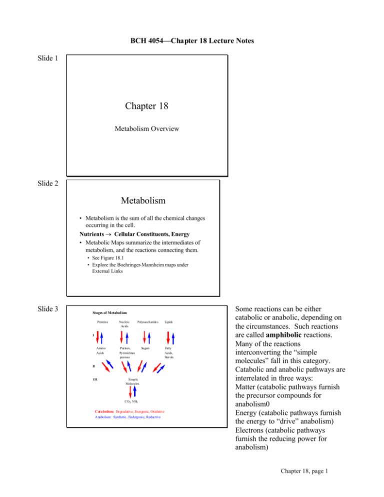
BCH 4054—Chapter 18 Lecture Notes Slide 1 Chapter 18 Metabolism Overview Slide 2 Metabolism • Metabolism is the sum of all the chemical changes occurring in the cell. Nutrients → Cellular Constituents, Energy • Metabolic Maps summarize the intermediates of metabolism, and the reactions connecting them. • See Figure 18.1 • Explore the Boehringer-Mannheim maps under External Links Slide 3 Stages of Metabolism Proteins Nucleic Ac ids Amino Acids Purines, Pyrimidines pentose Polysaccharide s Lipids I Sugars II III Simple Molecules CO2, NH3 Cata bolism: Degradative, Exergonic, Oxidative Anabolism: Synthetic, Endergoni c, Reductive Fatty Acids, Sterols Some reactions can be either catabolic or anabolic, depending on the circumstances. Such reactions are called amphibolic reactions. Many of the reactions interconverting the “simple molecules” fall in this category. Catabolic and anabolic pathways are interrelated in three ways: Matter (catabolic pathways furnish the precursor compounds for anabolism0 Energy (catabolic pathways furnish the energy to “drive” anabolism) Electrons (catabolic pathways furnish the reducing power for anabolism) Chapter 18, page 1 Slide 4 Topology of Metabolic Pathways • There are four ways that pathways can be organized “topologically” 1. 2. 3. 4. Linear Branched Cyclic Equilibrium pool Linear pathways convert one compound through a series of intermediates to another compound. An example would be glycolysis, where glucose is converted to pyruvate. Slide 5 Pathway Topology, con’t. 1. Linear Pathway: A B C D E F 2. Branched Pathway: C A B X or A B C Q R S M N O Q X D E Y Z R S Y Slide 6 Pathway Topology, con’t. 3. Cyclic Pathway B X C Z A Branched pathways may either be divergent (an intermediate can enter several linear pathways to different end products) or convergent (several precursors can give rise to a common intermediate). Biosynthesis of purines and of some amino acids are examples of divergent pathways. There is usually some regulation at the branch point. The conversion of various carbohydrates into the glycolytic pathway would be an example of convergent pathways. In a cyclic pathway, intermediates are regenerated, and so some intermediates act in a catalytic fashion. In this illustration, the cyclic pathway carries out the net conversion of X to Z. The Tricarboxylic Acid Cycle is an example of a cyclic pathway. D E Chapter 18, page 2 Slide 7 Pathway Topology, con’t. 4. Equilibrium Pool Z B X A C P M D W N Y A pool of compounds in equilibrium with each other provides the intermediates for converting compounds to a variety of products, depending on what is fed “into” the pool and what is “withdrawn” from the pool. The phosphogluconate pathway is an example of such a pool of intermediates. The pathway can convert glucose to CO2 , hexoses to pentoses, pentoses to hexoses, pentoses to trioses, etc. depending on what the cell requires in a particular situation. NADPH as a source of reducing power for anabolic reactions is also a main product of the phosphogluconate pathway. Slide 8 Enzyme Organization in Pathways • Some pathway enzymes are organized in a multienzyme complex. • The complex may consist of dissociable subunits, multiple domains on a single polypeptide chain, or a membrane associated complex. • See Figure 18.5 • Enzymes that are isolated as individual soluble entities may in some cases be associated in the cell. • This arrangement allows metabolic channeling, where the product of one step does not have to diffuse through the cell to find the next enzyme. Slide 9 Cellular Compartmentalization • Some pathways are compartmentalized in the cell. The necessity for intermediates to cross membrane boundaries between cellular compartments adds another layer of complexity to the regulation of and interactions between metabolic pathways. • TCA cycle occurs in mitochondria, glycolysis is in the cytoplasm • Fatty acid oxidation is in mitochondria, fatty acid synthesis is in the cytoplasm • Formation of urea uses enzymes in both compartments Chapter 18, page 3 Slide 10 Energetics of Pathways • If reaction through a pathway occurs spontaneously, the overall process must have a negative ∆G. • In fact, each step in the pathway must have a negative ∆G. • Remember: • ∆G= ∆Go’ + RTlnQ, or ∆G = RT ln(Q/K) • For each step, Q must be < K. Slide 11 Energetics of Pathways, con’t. • If Q is only a little smaller than K (i.e. larger than about 0.05 K), this step of the pathway is near equilibrium. There is nothing special about the number 0.05, it only represents an approximate distinction between the two extremes. The ∆G when Q = 0.05 K is –7.4 kJ/mol. • The rate through such a step depends on substrate and product concentrations, but doesn’t change much with a change in the activity of the enzyme. Slide 12 Energetics of Pathways, con’t. • If Q is less than about 0.05 K, this step of the pathway is removed from equilibrium. • The rate through such a step will vary by changing the activity of the enzyme. • Such a step is a rate limiting step. • Most pathway regulation occurs at such steps. • (Regulation at equilibrium steps does occur, usually by availability of a coenzyme cosubtrate.) Chapter 18, page 4 Slide 13 Anabolic and Catabolic Pathways Differ • Because ∆G must always be negative, the path from A → X must differ from the path from X → A. • Either completely different (Figure 18.7a) • Or different in at least one step (Figure 18.7b) • In the latter case, it is usually the steps removed from equilibrium which are different. Slide 14 Role of ATP in Metabolism • ATP (and closely related compounds with high negative free energies of hydrolysis) is considered the energy currency of the cell. • Catabolic reactions generate ATP. • Photosynthesis stores some light energy ATP. • ATP coupling helps make some anabolic reactions spontaneous. • The relative concentrations of ATP, ADP, and AMP regulate many enzymes where these nucleotides serve as allosteric effectors. Slide 15 Role of Nicotinamide Nucleotides in Metabolism • See Figure 18.19 for the structure of NAD+ and NADP + • Reduction adds a hydrogen atom and two electrons to the nicotinamide ring to form NADH and NADPH. • Hydrogen addition and removal is stereospecific. NAD+ was first called cozymase, the dialyzable cofactor needed for yeast extracts to carry out fermentation. When its structure was determined, it was first named diphosphopyridine nucleotide (DPN +). The dinucleotide nomenclature was adopted for consistency with naming of other compounds such as flavin adenine dinucleotide (FAD). Some enzymes add and remove the pro-R hydrogen, some add and remove the pro-S hydrogen. Chapter 18, page 5 Slide 16 Nicotinamide Nucleotides • NAD+ and NADP + are coenzyme cosubstrates. • NAD+ is the electron acceptor in most catabolic oxidation reactions. • NADH reoxidation by the electron transport chain is a major source of ATP production. • NADPH is the electron donor for most anabolic reduction reactions. Slide 17 Vitamins and Coenzymes • Many vitamins are components of important coenzymes. • Chapter 18 shows the structure, key reactions, and vitamin components of most coenzymes. • Review these coenzymes and be able to: • • • • We will return to discussion of individual coenzymes as we encounter them in metabolism. To be prepared for those discussions, it will be helpful if you become familiar with their structures now. Recognize the structure. Describe the chemical change the coenzyme undergoes. Classify as cosubstrate or prosthetic group. Name the vitamin component. Slide 18 Summary of Coenzymes Coenzyme Vitamin Class Figure Thiamine Pyrophosphate Thiamine (B1) Prosthetic Group 18.17 NAD + and NADP + Niacin Cosubstrate 18.19 FAD and FMN Riboflavin (B2) Prosthetic Group 18.21 and 18.22 Pyridoxal Phosphate Pyridoxine (B6) Prosthetic Group 18.25 and 18.27 Deficiency of thiamine (vitamin B1 ) is found in beriberi. Deficiency of niacin (nicotinic acid and nicotinamide) is found in pellegra (humans) and blacktongue (dogs). Chapter 18, page 6 Slide 19 Summary of Coenzymes, con’t. Coenzyme Vitamin Class Figure Coenzyme A Pantothenic Acid (B 3) Cosubstrate 18.23 Phosphopantetheine Pantothenic Acid (B 3) Prosthetic Group 18.23 Prosthetic Group 18.28 Cosubstrate 18.30 5’-Deoxyadenosyl- Cyanocobalamin Cobalamin (B 12) Ascorbic Acid Vitamin C Slide 20 Summary of Coenzymes, con’t. Coenzyme Vitamin Class Figure Biotin Biotin Prosthetic Group 18.32 Lipoic Acid Not a Vitamin Prosthetic Group 18.33 Tetrahydrofolate Folic Acid (B-complex) Prosthetic Group 18.35 Slide 21 Summary of Coenzymes, con’t. Coenzyme Vitamin Class Figure Retinal Retinol (A) Prosthetic Group 18.36 1,25-DihydroxyVitamin D 3 Ergo- and Cholecalciferol (D) Hormonelike action 18.37 Alpha Tocopherol (E) Antioxidant 18.38 Phylloquinone (K) Carboxylation Cofactor 18.39 and 18.40 Both coenzyme A and phosphopantetheine are carriers of acyl groups which are attached in thiolester linkage to the terminal SH. The thiol esters ha ve high negative free energies of hydrolysis, and they also help to labilize the hydrogens on the alpha carbon. 5’Deoxyadenosylcobalamin has a carbon-cobalt bond, and it is the making and breaking of this bond which is involved in its mechanism of action. Ascorbic acid deficiency is found in the disease scurvy. Ascorbic acid is a necessary cofactor in hydroxylation and proper maturation of collagen. Biotin cured dermatitis and paralysis in rats fed large amounts of egg white (called egg white syndrome ). A protein in egg white called avidin binds biotin very tightly and was responsible for the deficiency. Vitamins A, D, K and E are fatsoluble vitamins. Deficiency of Vitamin A can lead to night blindness. Deficiency of vitamin D leads to rickets in children, or a weakness in bones known as osteomalacia in adults. Vitamin E is a family of substances like alpha-tocopherol that are potent antioxidants. Vitamin K is a cofactor in the carboxylation of glutamyl residues in several blood clotting proteins. Vitamins D, E, and K don’t fit the “cosubstrate/prosthetic group” classification scheme. Chapter 18, page 7
