Genetix is an independent business
unit within Leica Microsystems
®
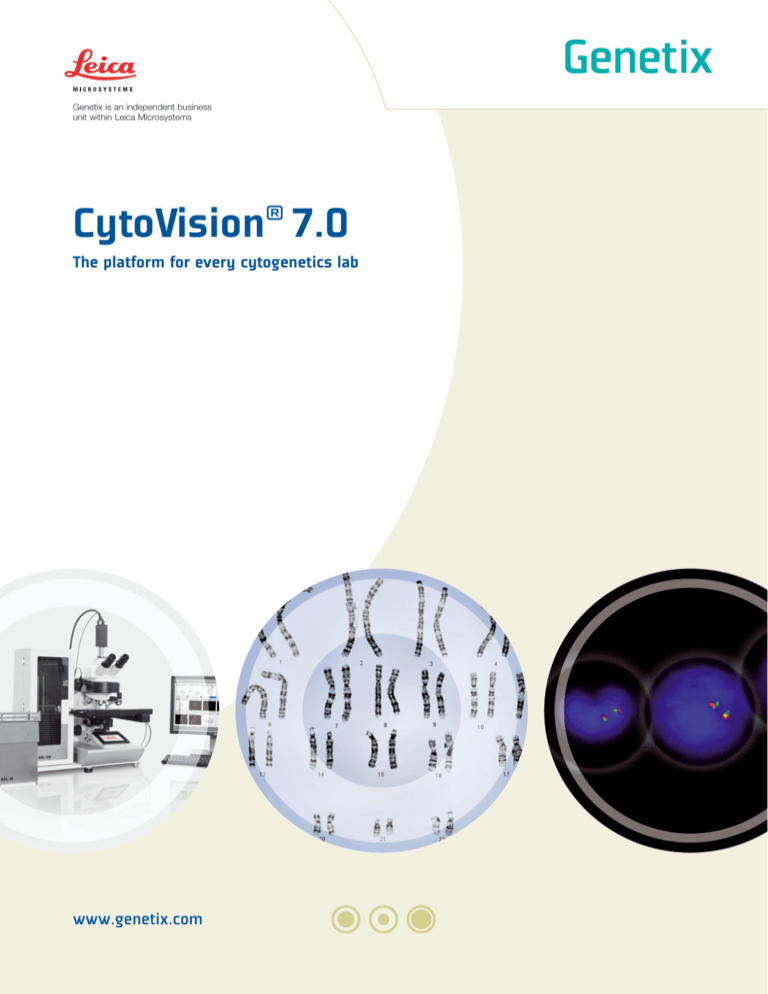
CytoVision 7.0
The platform for every cytogenetics lab
www.genetix.com
A solution for every cytogenetics lab
A proven, future-proof platform
Used in over 2000 laboratories worldwide, the
CytoVision® platform provides the convenience and
comfort of on-screen analysis together with flexibility
in software and hardware configurations.
CytoVision supports the simplest, single application
workstation through to fully networked, multi-application,
nationwide programs.
New
The new CytoVision 7.0 software, optimized
for the latest Windows® 7 operating system,
ensures future-proof compatibility. This latest version
features enhanced FISH imaging and includes support
for the Leica DM6000B microscope to ensure high
quality optics.
Current users of CytoVision should contact their local
representative to discuss the potential for upgrading.
Reduce reporting times, improve consistency, free skilled staff for quality analysis
Many facilities face growing demands to increase
productivity and efficiency, reduce backlogs and speed
up reporting.
Unattended scanning and capture using the unique
combination of CytoVision software and GSL slide
loaders enable cytogeneticists to fulfil these demands.
Unattended scanning, location and capture
Slide loader and microscope controlled by CytoVision
software: samples scanned at low power (10X), cells
located, oil dispensed automatically, cells captured
(63X /100X)
“Spending ages scanning slides is a thing of the past!”
Technologist at Cytogenetics Laboratory, Yorkhill Hospital, Glasgow, UK
02
A choice of application
modules within CytoVision
A broad range of application
modules ensures full flexibility
to meet changing laboratory
requirements.
Karyotyping
FISH
Scanning platform
Metaphase finding
Spot counting
CGH
le
Multip tion
a
applic ules
mod
M-FISH
Flexible Karyotyping
Focuses experts on
critical analysis steps
Reduces reporting
and turnaround times
Operator actions recorded
for every case
Operator results archived
and reports generated
“With work force reduction of 480 hours, the TAT was reduced
by 0.3 days without compromising abnormality rate.”
Christine M. Higgins, MMI Human Genetics Lab, Omaha, NE, USA.
03
Bright field analysis on-screen and in comfort
CytoVision analysis modules support a variety of applications including metaphase finding and karyotyping for
human and non-human cell lines. In the example shown here, a simple interface leads the analyst through each
stage of karyotyping and analysis.
Select the best captured cells from the Organize screen
Every cell is shown in a gallery.
Count and karyotype on the Analyze screen
Analysis of cells automatically tracked.
Digital record of count and results kept for security.
Progress tracked during analysis as each class is
marked off.
04
Select and analyze cells
at desktop review stations
Ensure quality control using Clear Cells screen – easily check classes
Each class is marked with clearly visible homologues.
Use karyotypes to clear classes and scroll through
remaining cells to check any unclear classes.
A record of karyotypes or metaphases is kept for each
cell to ensure sufficient homologues are checked.
Review case summary on the Report screen
Digital records contain cell coordinates, images
and results.
Images and reports can be annotated and printed
or exported to a LIMS.
“I much prefer analyzing chromosomes on my large screen… easier
on the eyes, more comfortable on the body and, more importantly,
I am happier analyzing chromosomes when they are paired up side
by side than at opposite ends of a metaphase.”
Technologist at Cytogenetics Laboratory, Yorkhill Hospital, Glasgow
05
On-screen analysis brings FISH
out of the dark room
Fluorescence analysis modules in CytoVision support a variety of applications including
FISH, spot counting/interphase FISH, CGH and M-FISH
Automated slide loaders enable unattended scanning, location and capture of fluorescence images,
minimizing fading problems, and sending images to review stations for on-screen analysis. Simple interface
screens guide the operator through the analysis workflow, as illustrated by this cellular FISH example.
Capture high quality images
Low power scanning (10X) locates interphase cells.
Frames are then captured at high power with DAPI
masking of probe signals to eliminate extraneous
fluorescence outside of cells. Captured frames are
displayed for manual or automatic scoring.
Capture high quality images
+
Dynamic Z-stack multi-plane
imaging captures images of counter
stains and probes through each cell.
Virtual microscopy enables dynamic
focusing, up and down through the
image.
Image stack
06
Composite image
Select and analyze cells
at desktop review stations
Choose scoring process
Automatic:
Display scored cells in a grid for easy viewing and
sorting of cells assigned to each class.
Manual:
Create assay, name classes and assign to function
keys. Class totals are updated as cells are scored.
Produce customizable reports for export
Add annotations
Include scores, patient data and images
07
Improvements in productivity and efficiencyextracts of reports from around the world
The West of Scotland Regional Cytogenetics Service, U.K. provides a region-wide service for analysis of
around 2800 post-natal blood samples per year. Each slide is scanned for up to a maximum of 300 metaphases
and the 50 best cells are captured for analysis.
Reduced backlog of cases
• Increase in quality of urgent and routine samples
• Decrease in reporting times
• Increase in % of cases reported within
guideline times
• Increase in success rate, decrease in poor
quality rate
Data courtesy of L. Monkman, J. Colgan, L. Crawford, M. Campbell,
G. Lowther Cytogenetics Laboratory, Duncan Guthrie Institute of Medical
Genetics, Yorkhill Hospital, Glasgow
The South West Thames Regional Genetics Service, part of the St. George’s Healthcare NHS Trust in the UK,
had a routine blood sample backlog for nearly 4 years, due to staff shortages.
Backlog cleared 8 months ahead of schedule
Within 30 days:
• CytoVision and GSL-120 integrated
into in-house LIMS
• Classifiers designed for specific cultures
• Staff fully trained
Within 4 months:
• Fully automated workflow from scan to report for
blood and pre-natal samples
• Backlog reduced from 500 to 70 cases
• Reporting times dropped from 116 days to 28 days
• Decrease in inscription errors
• Improved staff morale
8
Data courtesy of Victoria J. Anthony-Dubernet, Clinical Cytogeneticist,
South West Thames Regional Genetics Service, St. Georges Hospital,
London
Select and analyze cells
at desktop review stations
The University of Nebraska Medical Center in Omaha, Nebraska compared work hours, TAT and abnormality
rate between 60 day periods before and after implementation of a GSL-120 slide loader for unattended scanning,
location and capture.
• Work hours cut by 480 hours
• Turnaround time (TAT) reduced by 0.3 days
• Abnormality rate maintained
Data courtesy of Bhavana J. Dave, PhD, FACMG, Munroe Meyer Institute,
NE, USA.
Sonora Quest Laboratories in Tempe, Arizona evaluated efficiency gains by collecting time requirements for
scanning, capture and karyotyping of 220 neoplastic cases and comparing with time required to run 152 cases
using CytoVision and GSL-120.
• Reduced time for case completion
by 31.06 mins (20.45%)
• Decreased failure rate to 1.5%
Process before GSL-120
Scan and analyze 20 cells: 126 min.
Capture min. 5 pictures: 12.8 min.
• Removed human inattention
from equation
• Located metaphases on slides
151.9 min. per case
(n=152)
Karyotype full case (~2-5 karyotypes per case): 13.1 min.
Process after GSL-120 implementation
Scan, analyze 20 cells capture and cut:
a technologist may assume to
be empty
120.8 min. per case
(n=220)
Data courtesy of Leah Weems, BS, CLSp (CG) Angela Fylak,
BA., Sonora Quest Laboratories, Tempe, Arizona
Thank
You
Genetix would like to thank everyone featured here and on our web site for their kind
permission to present excerpts from their work.
9
A solution for every cytogenetics lab
Configurations to match applications, throughput and networking requirements
Building from CytoVision software for review, workstation solutions can be configured to match the applications,
throughput, imaging and automation requirements of any cytogenetics lab.
Ready to meet increasing caseloads and facilitate workload balancing
The CytoVision platform enables expansion from a single review station to fully automated scanning and capture
stations with review stations supported by server networks and LIMS connections.
This table gives a general guideline to creating optimal CytoVision configurations.
Your local representative will be happy to assist in finalizing your exact requirements.
One step slide loading, scanning
and capture
Scanning and capture
with manual oiling
Software
CytoVision 7.0 using Windows 7 operating system
CytoVision 7.0 using Windows 7 operating system
Microscope
Leica DM6000B
Leica DM6000B
Digital cameras
1600 x 1200 pixels
1600 x 1200 pixels
Slide loader or motorized stage
GSL-120 (up to 120 slides per run) or GSL-10
(10 slides per run)
8-bay motorized stage
Bar code reader and oiler
integrated
n.a.
Application options
Karyotyping
FISH
Karyotyping
FISH
Monitor
24" LCD
24" LCD
System architecture
(1 to 9 stations)
Internal domain server
SQL CaseBase
Data archive: 2TB using internal 4TB RAID5 array
Internal domain server
SQL CaseBase
Data archive: 0.5TB using internal 1TB RAID1 array
System architecture
(10 or more stations)
External domain server
SQL CaseBase
Data Server: 2TB using 4TB RAID5 array
External domain server
SQL CaseBase
Data Server: 2TB using 4TB RAID5 array
UPS
Standard
Option
10
Standard capture station
with on-screen analysis
Review station for on-screen analysis
Software license for review station
CytoVision 7.0 using Windows 7 operating system
CytoVision 7.0 using Windows 7 operating system
User-provided computer
(subject to minimum specification)
Leica DM2500
Leica DM6000B
Other manufacturer microscopes subject
to specification
n.a.
n.a.
1300 x 1024 pixels
n.a.
n.a.
n.a.
n.a.
n.a.
n.a.
n.a.
n.a.
Karyotyping
FISH
MFISH
CGH
Flexible Karyotyping
Karyotyping
FISH
MFISH
CGH
Flexible Karyotyping
Karyotyping
FISH
MFISH
CGH
Flexible Karyotyping
24" or 22" LCD flat panel
24" or 22" LCD flat panel
n.a.
Internal domain server
SQL CaseBase
Data archive: 0.5TB using internal 1TB RAID1 array
n.a.
n.a.
External domain server
SQL CaseBase
Data Server: 2TB using 4TB RAID5 array
n.a.
n.a.
Option
n.a.
n.a.
11
Genetix provides unrivalled solutions based on imaging excellence
and intelligent image analysis
Headquartered in New Milton, UK, Genetix is an
independent business unit within Leica Microsystems,
providing scientists with unrivalled solutions that utilize
imaging and intelligent image analysis. The company’s
CytoVision platform is used for chromosomal
investigations in cytogenetics laboratories in over 80
countries worldwide. In basic research, pharmaceutical
and biotherapeutic development, Genetix systems
continue to establish industry standards in areas such
as picking microbial colonies for genomic studies or
screening and selection of mammalian cell lines. Other
systems use imaging platforms to monitor cell growth,
evaluate cellular responses and quantify protein
production. Through its expertise in robotics, cell
and molecular biology, image analysis and
interpretation, supported by a strong IP portfolio,
Genetix is committed to the continual development
of innovative solutions.
Intended Use (US)
Please refer to www.genetix.com for most recent ‘intended use’ statements
in your region.
In the US, CytoVision® Karyotyper and CEP XY are For In Vitro Diagnostic
Use. All diagnostic decisions are made by the qualified clinician.
For more information, visit
www. genetix.com
All other applications are for Research Use Only. Not for Use in Diagnostic
Procedures.
In the EU, Canada and Thailand, only CytoVision Karyotyper is For In Vitro
Diagnostic Use. All diagnostic decisions are made by the qualified
clinician.
CytoVision® is a registered trademark of Genetix Corp.
Genetix reserves the right to amend specifications without notice.
All Rights Reserved. © Genetix Corp 2010
All other applications are For Research Use Only.
All third party trademarks are the property of their respective owners.
Genetix Corp.
Genetix Europe Ltd.
120 Baytech Dr
San Jose, CA 95134, USA
Tel +1 408-719-6400
Unit 7 Queens Park, Queensway
Team Valley Gateshead, NE11 0QD
Tel +44 (0) 191 497 7020
www.genetix.com
01LBL1016.A2
Intended Use (EU, Canada and Thailand)

