April 2012
Arrays Complete the Picture in Cytogenetics Lab
Cincinnati Children’s Cytogenetics Laboratory provides researchers and clinicians with more
information through karyotyping, FISH, and Illumina® microarrays.
Introduction
Cytogenetics, the study of chromosomal structure and function, was
once focused solely on identifying congenital disorders such as Down
syndrome. Its scope has expanded over the last 50 years, fueled by
advances in a variety of tools that have enabled clinical cytogeneticists
to delve deeper into the genetic basis of many diseases, including
cancer, where genetic mutations are acquired. Several of these tools
can be found in the cytogenetics laboratory at Cincinnati Children’s
Hospital and Medical Center, where they process patient samples received
from across the Ohio River Valley region and as far away as Canada.
Although it is one of the top U.S. pediatric hospitals, Cincinnati
Children’s cytogenetics laboratory also accepts adult patient samples
from the region, particularly for cancer studies. It uses a trio of
cytogenetics tools to diagnose congenital syndromes and perform
cytogenetic testing for disease research. Karyotyping, the pairing
and ordering of chromosomes, provides a low-resolution genomewide view of a patient’s chromosomes, enabling the laboratory’s
clinical cytogeneticists to detect genetic anomalies involving several
megabases or more of DNA. A powerful molecular technique,
fluorescent in situ hybridization (FISH), enables them to locate the
position of specific DNA sequences to identify gene deletions,
duplications, and translocations associated with disease. The highest
resolution tool at their disposal is the Infinium® HumanOmni1-Quad
BeadChip. Using this assay, a genome-wide profile of chromosomal
aberration and structural variation of over one million markers derived
from the Human HapMap and 1000 Genomes Project will be obtained.
Cincinnati Children’s Cytogenetics Laboratory is an Illumina Certified
Service Provider (CSPro®). We recently had an opportunity to speak
with Teresa Smolarek, Ph.D., Director, and Sarah Zimmerman, Ph.D.,
Assistant Director of Cincinnati Children’s Cytogenetics Laboratory
to learn more about how they are using microarrays to enhance
cytogenetics studies.
Q: What different types of testing do you perform in
your laboratory?
Sarah Zimmerman (SZ): There are approximately 35 people in the lab
performing chromosome analysis using a combination of karyotyping,
FISH, and microarray analysis, enabling us to perform a range of
clinical and research testing. Most of our samples are from patients
with genetic abnormalities that are present at birth. We also perform
testing on prenatal samples, looking for subtle abnormalities, as well
as oncology samples, where we’re looking for gene alterations in
tumor and cancer cells.
Teresa Smolarek (TS): We have been very proactive in educating
our physicians and clinicians in the region about the types of
cytogenetic tests we offer. As we replaced bacterial artificial
chromosome (BAC) arrays with microarrays, we met with our
clinicians to educate them about the platform and the information it
could provide. We started with the HumanCNV370-Duo BeadChip,
moved to the Human610-Quad, and finally the HumanOmni1-Quad.
With each change in platform, we communicated the added value
the BeadChips offered, such as genome-wide coverage of diseaseassociated regions. It’s given us a reason to keep in contact with
clinicians and answer any questions they may have.
SZ: We’re a cytogenetics laboratory, but also a reference laboratory
for the region, analyzing pediatric and adult patient samples for
physicians, universities, and other hospitals. We routinely communicate
with pediatric and adult oncology groups, as well as pathologists in
our community, letting them know about the cytogenetics testing
we can perform on blood samples, as well as bone marrow and
lymph nodes. At the recent Cancer Consortia meeting, many people
were talking about replacing FISH with microarrays to obtain higherresolution data. That’s definitely something we can support.
Q: Why did you switch from BAC arrays to SNP microarrays?
TS: BAC arrays provided a view of what was occurring at the
molecular level, but were limited in their genome coverage, resulting
in an inability to detect deletions and amplifications in many parts of
the genome. We initially looked at oligonucleotide arrays, but realized
that we needed the higher resolution of SNP arrays and their ability
to detect copy-neutral loss of heterozygosity (LOH) that’s important
for oncology and cases of consanguinity.
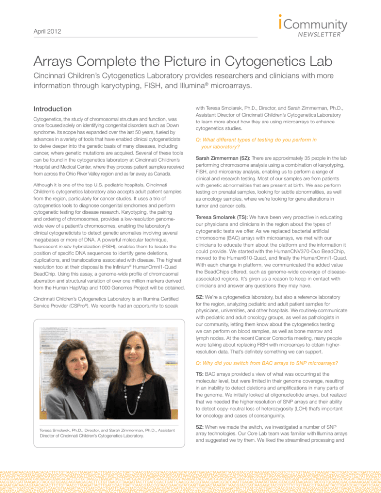
Teresa Smolarek, Ph.D., Director, and Sarah Zimmerman, Ph.D., Assistant
Director of Cincinnati Children’s Cytogenetics Laboratory.
SZ: When we made the switch, we investigated a number of SNP
array technologies. Our Core Lab team was familiar with Illumina arrays
and suggested we try them. We liked the streamlined processing and
April 2012
data output of the Infinium assays, and were pleased with the ability
of the array to detect LOH events and profile somatic mosaicism
that occurs when tumor cell populations have a different genetic
makeup than adjacent non-tumor cells. We upgraded from an Illumina
BeadArray™ Reader to the HiScan® system, which reduced our
processing time in half and significantly increased our testing capacity.
what we saw when FISH began to be used in tandem with chromosome studies for cytogenetics testing. For example, a microdeletion
of chromosome 22 first detected using FISH is now used to diagnose
Velo-Cardo-Facial Syndrome (VCFS). If you look hard enough at VCFS
karyotypes you may see a deletion, but it is much easier and faster to
identify using FISH.
TS: We felt comfortable adopting Illumina technology, especially since
we already had someone within our DNA Core Lab who was familiar
with the data output and would be sharing the Illumina scanner with
us. GenomeStudio®, the data analysis software package for the
HiScan, enables us to modify how we analyze the data, providing the
flexibility to show all the copy number changes that exist between
parent and child, or to limit the data output to copy number changes
of a certain size.
Arrays have had a similar impact on the cytogenetics laboratory. When
you look at a standard karyotype you can see certain polymorphisms,
but it wasn’t until we began using microarrays that we realized how
polymorphic our genome really is. Using the array we’re finding that we
can pinpoint copy number changes that might be at the root of a child’s
developmental delay, mental retardation, or dysmorphic features.
“We realized that we needed the
higher resolution of SNP arrays and
their ability to detect copy-neutral
LOH that’s important for oncology
and cases of consanguinity.”
Q: What is the value of all three technologies—karyotyping,
FISH, and microarrays—in performing cytogenetics studies?
SZ: Karyotyping, FISH, and array analysis together provide us with a
comprehensive picture of the human genome. There are things that you
can see with one type of test that you can’t identify with the others. For
example, the SNP array allows us to identify deletions and duplications
that are below the detection limit of resolution of karyotyping. Unlike
FISH, where you have to know which genetic abnormality you’re looking for, the array allows us to look across the whole genome without
a specific target in mind. Since we first started using SNP arrays, we
have detected amplifications, small deletions and duplications, and
mosaicism that might have eluded us had we just used karyotyping
and FISH analyses. These results have been instrumental in helping us
identify genetic abnormalities.
TS: Any disconnect between our karyotyping, FISH, and array data
for a particular sample alerts us to further examine our results. We had
one patient sample where the chromosomes appeared normal, yet the
FISH results and array data came back suggesting a genetic anomaly.
We took another look at the chromosomes and we were able to identify the cryptic abnormality.
Q: How does the addition of microarray technology enable you
to identify genetic mutations?
TS: We’ve seen an increase in testing volume that I think is a result of
a shift in the genetics community and some medical subspecialties,
which are now pushing for the use of arrays in the diagnostic setting to
identify microdeletions and microduplications. I think the shift parallels
It’s had the same impact on our oncology testing, where leukemic
or lymphoma sample karyotypes can be challenging to interpret.
Often it’s clear that the metaphases are abnormal, but they’re so
tight that we just can’t analyze them for copy number changes.
The SNP array provides what some refer to as a ‘virtual karyotype’
to detect copy number changes at a higher resolution than
conventional karyotyping.
Q: How do the quality and clarity of the HumanOmni1-Quad
BeadChip results impact physician decisions or clinical
research efforts?
SZ: Using the HumanOmni1-Quad BeadChip, we can more easily
identify or confirm the presence of genetic abnormalities. There have
been instances where we discovered a genetic abnormality in a patient
where none had been suspected. If it’s the first time we’ve seen the
abnormality, it provides researchers with a starting point. In addition,
the HumanOmni1-Quad has the resolution necessary to identify the
genetic clues as to why some patients would respond to treatment
while others may not.
Q: Have there been instances where SNP arrays were able to
detect a gene defect?
TS: Yes, one of our genetic counselors was conducting a study with
samples from a patient with consanguinity, where the parents were
known to be closely related. We initially used the results from the
SNP arrays to scan for regions of homozygosity. Later, the physician
identified a specific enzyme deficiency in the patient and the microarray was able to identify the causative gene mutation, including which
members of the immediate family also possessed the mutation. We’ve
had a few of these cases in which we were successful in suggesting a
causative gene.
SZ: The HumanCNV370-Duo BeadChip helped identify a disorder
in a six–year old patient whose gestation, delivery, birth weight, and
development were all normal, but who had a two-year history of progressive tachypnea or shortness of breath1. Both parents were healthy
with no history of lung disease. Primary pulmonary alveolar proteinosis
(PAP) was suspected, a rare syndrome characterized by accumulation
of surfactant in the lungs, resulting from the disruption of GM-CSF
signaling. However, the disease typically progresses quickly in the
first 10 years of life and that was not the case in this patient. We used
the array to identify a deletion on the X chromosome in the CSF2RA
gene in the patient, her sister, and their mother that severely reduced
GM-CSF binding and receptor signaling. It turned out that the sister
had poor growth and reduced lung function that had been undiag-
April 2012
nosed. Interestingly, the father and both children had a point mutation
in CSF2RA that increased receptor signaling, providing a molecular
explanation for the slow progression of the disease in both children.
“The SNP arrays have helped us
realize that the human genome is
much more complex and possesses
a lot more copy number changes
than we ever imagined.”
TS: Our cytogenetics laboratory and the Core Lab contributed to
a heterotaxy study in 96 patients to determine the genetic basis of
a severe form of congenital heart disease, where left-right asymmetry during embryonic development leads to a malformation of the
heart2. Using SNP arrays and whole-exome sequencing, a recessive
missense mutation was identified in SHROOM3, a central regulator
of morphogenetic cell shape changes necessary for organogenesis.
SHROOM3 could be a novel target for therapeutic intervention.
Q: Are there ongoing studies where the Illumina arrays are being
used to detect clinically relevant gene mutations?
TS: We are involved in a neurofibromatosis (NF) study in monozygotic
(MZ) or identical twins, where the twins present with different clinical
symptoms. We’re testing the identical twin patients and their parents to
determine whether there are copy number changes that might account
for the different clinical symptoms between the twins.
Q: Do you see cytogenetics testing playing an increasing role in
cancer treatments?
TS: As a participant in the Children’s Oncology Group, we process
genetic samples for their studies. Our clinicians also are approved for
treating patients following the group’s protocols. Clearly, there are copy
number changes we are just beginning to appreciate, but we don’t yet
know how much they are going to change how patients are treated. In
pediatric acute lymphoblastic leukemia (ALL), the risk stratification is
definitely based on genetics, which demonstrates its potential for use
in other forms of cancer.
Q: What do you seen on the horizon in the use of microarrays
and sequencing for cytogenetics testing?
SZ: SNP arrays have helped us realize that the human genome is
much more complex and possesses a lot more copy number changes
than we ever imagined. Our challenge now is to identify which
changes are benign and which are clinically relevant.
TS: While we understand some of the single gene mutations that
are associated with genetic abnormalities or diseases such as VCFS,
we don’t yet know whether the deletion of a 300 kb region on a
chromosome is clinically relevant or not. The hurdle is identifying
enough patients with the same clinical symptoms, something that
various consortia are trying to do. We have the tools to obtain a good
overall picture of what the genomes look like and have the resolution to
identify altered gene regions. At some point, whole-genome sequencing
may become a practical tool in the cytogenetics laboratory, but I don’t
know if that’s five years from now or ten years down the line.
References
1. Suzuki T, Salagami T, Rubin BK, Nogee LM, Wood RE, et al. (2008)
Familial pulmonary proteinosis caused by mutations in CSF2RA.
J Exp Med 205: 2703-2710.
2. Tariq M, Belmont JW, Lalani S, Smolarek T, and Ware SM. (2011)
SHROOM3 is a novel candidate for heterotaxy identified by whole
exome sequencing. Genome Biol 12: R91.
Illumina • 1.800.809.4566 toll-free (U.S.) • +1.858.202.4566 tel • techsupport@illumina.com • www.illumina.com
For research use only
© 2012 Illumina, Inc. All rights reserved.
Illumina, illuminaDx, BaseSpace, BeadArray, BeadXpress, cBot, CSPro, DASL, DesignStudio, Eco, GAIIx, Genetic Energy,
Genome Analyzer, GenomeStudio, GoldenGate, HiScan, HiSeq, Infinium, iSelect, MiSeq, Nextera, Sentrix, SeqMonitor, Solexa,
TruSeq, VeraCode, the pumpkin orange color, and the Genetic Energy streaming bases design are trademarks or registered
trademarks of Illumina, Inc. All other brands and names contained herein are the property of their respective owners.
Pub. No. 070-2012-003 Current as of 11 April 2012

