Nuclear envelope formation by chromatin
advertisement
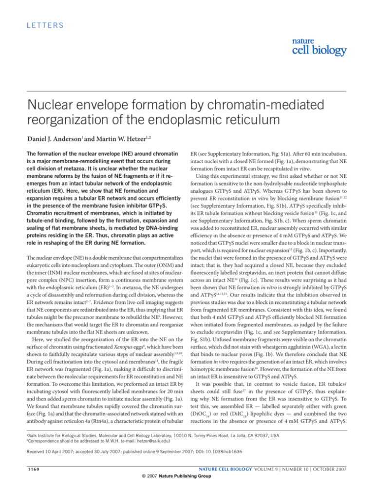
letters Nuclear envelope formation by chromatin-mediated reorganization of the endoplasmic reticulum Daniel J. Anderson1 and Martin W. Hetzer1,2 The formation of the nuclear envelope (NE) around chromatin is a major membrane-remodelling event that occurs during cell division of metazoa. It is unclear whether the nuclear membrane reforms by the fusion of NE fragments or if it reemerges from an intact tubular network of the endoplasmic reticulum (ER). Here, we show that NE formation and expansion requires a tubular ER network and occurs efficiently in the presence of the membrane fusion inhibitor GTPγS. Chromatin recruitment of membranes, which is initiated by tubule-end binding, followed by the formation, expansion and sealing of flat membrane sheets, is mediated by DNA-binding proteins residing in the ER. Thus, chromatin plays an active role in reshaping of the ER during NE formation. The nuclear envelope (NE) is a double membrane that compartmentalizes eukaryotic cells into nucleoplasm and cytoplasm. The outer (ONM) and the inner (INM) nuclear membranes, which are fused at sites of nuclearpore complex (NPC) insertion, form a continuous membrane system with the endoplasmic reticulum (ER)1–3. In metazoa, the NE undergoes a cycle of disassembly and reformation during cell division, whereas the ER network remains intact2–7. Evidence from live-cell imaging suggests that NE components are redistributed into the ER, thus implying that ER tubules might be the precursor membrane to rebuild the NE6. However, the mechanisms that would target the ER to chromatin and reorganize membrane tubules into the flat NE sheets are unknown. Here, we studied the reorganization of the ER into the NE on the surface of chromatin using fractionated Xenopus eggs8, which have been shown to faithfully recapitulate various steps of nuclear assembly3,9,10. During cell fractionation into the cytosol and membranes11, the fragile ER network was fragmented (Fig. 1a), making it difficult to discriminate between the molecular requirements for ER reconstitution and NE formation. To overcome this limitation, we preformed an intact ER by incubating cytosol with fluorescently labelled membranes for 20 min and then added sperm chromatin to initiate nuclear assembly (Fig. 1a). We found that membrane tubules rapidly covered the chromatin surface (Fig. 1a) and that the chromatin-associated network stained with an antibody against reticulon 4a (Rtn4a), a characteristic protein of tubular 1 2 ER (see Supplementary Information, Fig. S1a). After 60 min incubation, intact nuclei with a closed NE formed (Fig. 1a), demonstrating that NE formation from intact ER can be recapitulated in vitro. Using this experimental strategy, we first asked whether or not NE formation is sensitive to the non-hydrolysable nucleotide triphosphate analogues GTPγS and ATPγS. Whereas GTPγS has been shown to prevent ER reconstitution in vitro by blocking membrane fusion11,12 (see Supplementary Information, Fig. S1b), ATPγS specifically inhibits ER tubule formation without blocking vesicle fusion11 (Fig. 1c, and see Supplementary Information, Fig. S1b, c). When sperm chromatin was added to reconstituted ER, nuclear assembly occurred with similar efficiency in the absence or presence of 4 mM GTPγS and ATPγS. We noticed that GTPγS nuclei were smaller due to a block in nuclear transport, which is required for nuclear expansion13 (Fig. 1b, c). Importantly, the nuclei that were formed in the presence of GTPγS and ATPγS were intact; that is, they had acquired a closed NE, because they excluded fluorescently labelled streptavidin, an inert protein that cannot diffuse across an intact NE14 (Fig. 1c). These results were surprising as it had been shown that NE formation in vitro is strongly inhibited by GTPγS and ATPγS11,12,15. Our results indicate that the inhibition observed in previous studies was due to a block in reconstituting a tubular network from fragmented ER membranes. Consistent with this idea, we found that both 4 mM GTPγS and ATPγS efficiently blocked NE formation when initiated from fragmented membranes, as judged by the failure to exclude streptavidin (Fig. 1c, and see Supplementary Information, Fig. S1b). Unfused membrane fragments were visible on the chromatin surface, which did not stain with wheatgerm agglutinin (WGA), a lectin that binds to nuclear pores (Fig. 1b). We therefore conclude that NE formation in vitro requires the generation of an intact ER, which involves homotypic membrane fusion16. However, the formation of the NE from an intact ER is insensitive to GTPγS and ATPγS. It was possible that, in contrast to vesicle fusion, ER tubules/ sheets could still fuse17 in the presence of GTPγS, thus explaining why NE formation from the ER was insensitive to GTPγS. To test this, we assembled ER — labelled separately either with green (DiOC16) or red (DiIC18) lipophilic dyes — and combined the two reactions in the absence or presence of 4 mM GTPγS and ATPγS. Salk Institute for Biological Studies, Molecular and Cell Biology Laboratory, 10010 N. Torrey Pines Road, La Jolla, CA 92037, USA Correspondence should be addressed to M.W.H. (e-mail: hetzer@salk.edu) Received 10 April 2007; accepted 30 July 2007; published online 9 September 2007; DOI: 10.1038/ncb1636 1160 nature cell biology volume 9 | number 10 | OCTOBER 2007 © 2007 Nature Publishing Group letters c 50 ATPγS Post ER 0 4 min GT tE R AT Pγ Sp re ER AT Pγ Sp os tE R ATPγS Pre ER 10 R GTPγS Post ER 20 po s GTPγS Pre ER 30 Co Control Membrane/DNA b 40 GT Pγ S NE-60 Pγ Sp re E NE-1 ntr ol Reconstituted ER Percentage of closed NE Fragmented ER 20 min NPC d Interphase Cross-section Surface Overlay Metaphase Membrane proteins 100 80 60 40 20 0 anti-Rtn2 Pre ER post ER anti-Rtn4 gp210 POM121 Sec61β e Percentage of closed NE a Figure 1 The ER network is required for nuclear-envelope formation. (a) Fragmented endoplasmic reticulum (ER) membranes were stained with DiOC6 (green) and imaged at 4 ºC using confocal microscopy. The ER network was reconstituted by incubating membranes with cytosol for 20 min at 25 ºC. Nuclear envelope (NE) formation was induced by adding nucleoplasmin-decondensed sperm chromatin24; DNA was stained with Hoechst 3222 (blue) and imaged live after 1 min (NE-1) and 60 min (NE-60) incubations. (b) Nuclei were assembled as in a in the absence or presence of 4 mM GTPγS or ATPγS either before (Pre ER) or after (Post ER) ER reconstitution for 90 min. Before GTPγS was added, sperm chromatin was first decondensed in membrane-free cytosol. NPCs were stained with Alexafluor©568 conjugated WGA (red). (c) NEs were formed as described in b around sperm chromatin-containing biotinylated histones14. After 4 min or 20 min, Alexafluor©568 conjugated streptavidin was added, which was excluded only when a closed NE had formed; n = 60. (d) A total of 10 µM anti-Rtn2 or anti-Rtn4 was added to nuclear assembly reactions either before or after ER formation, and the percentage of closed nuclei was determined as described in c; n = 100. (e) HeLa cells were co-transfected with histone H2B-tdTomato and GFP-fused Sec61β (aa 1–65), and imaged live during interphase and mitosis. Additionally, HeLa cells stably expressing POM121–EGFP3 and gp210–EGFP3 were transfected with tdTomato H2B and imaged during interphase and mitosis. Scale bars, 10 µm. The fusion of the two ER populations was specifically blocked by GTPγS but not ATPγS (see Supplementary Information, Fig. S1c). Thus, under these experimental conditions, GTPγS acts as a general inhibitor of ER membrane fusion. Taken together, these results suggest that NE formation can occur either by GTPγS-insensitive fusion events or by fusionindependent reorganization of the ER. One prediction from the latter hypothesis is that the topology of the tubular ER network is required for NE formation. Recently, it was shown that the formation of ER tubules is mediated by Rtn4a18. Consistent with this report, we found that antibodies against Rtn4a, but not Rtn2, blocked ER network formation in vitro (see Supplementary Information, Fig. S1d). We incubated cytosol and membranes in the presence of anti-Rtn4a or anti-Rtn2, and added sperm chromatin. Closed NE formation was specifically inhibited by anti-Rtn4a. When anti-Rtn4 was added to preformed ER, NE formation occurred efficiently (Fig. 1d). Importantly, ER membranes could still fuse in the presence of anti-Rtn4, resulting in the formation of large vesicles17,19,20 (data not shown). These results indicate that the specific membrane nature cell biology volume 9 | number 10 | OCTOBER 2007 © 2007 Nature Publishing Group 1161 letters a DNA Membranes d Membranes Membranes/DNA Time e t=0 0.3 s 7.1 s 9.3 s 4.9 s Membranes b Ilustration x z Patch outline y x 16 s 66 s t=0 10 s 30 s f Membrane patch area (µm2) 6s Lateral tethering Tip binding c 30 28 26 24 22 20 18 16 14 12 10 0 1000 2000 3000 4000 Time (s) Figure 2 Chromatin-mediated reshaping of the ER. (a) Biotinylated 10 kb double-stranded DNA was immobilized on streptavidin-coated cover glass (schematic, left panel) and visualized by Hoechst 3222 (blue, middle panel). After the addition of cytosol and membranes, a nuclear envelope (NE) formed specifically on the two-dimensional (2D) chromatin spot (right panel). Scale bar, 10 µm. (b) Early chromatin/membrane interactions were imaged with time-lapse microscopy on 2D chromatin at a rate of ~4 frames/s. An initial interaction occurred via membrane tubule tips (arrowhead). Tubule ends on the surface of 2D chromatin were imaged in 3D by confocal microscopy (lower right panel). Scale bars, 2 µm. (c) De-condensed sperm chromatin was spun onto coverslips and stained with Syto©62 (blue). Preformed endoplasmic reticulum (ER) network was added and membrane/chromatin interactions were instantaneously imaged. Cross-sections of the chromatin are shown and arrowheads indicate initial membrane contacts to chromatin. Scale bar, 5 µm. (d) NIH3T3 cells were plated onto chambered slides and stained with DiOC6 and Hoechst 3222 in growth medium. Live mitotic cells were imaged with confocal microscopy and 3D reconstructions were generated. Arrowheads indicate tubule-tip binding. NE formation was performed as in a. Confocal images were taken every 10 s. Membrane sheets were schematically outlined (lower panels). Scale bar, 5 µm. (e) NE formation was performed as in a. Confocal images were taken every 10 s. Membrane sheets were outlined (lower panels). Scale bar, 5 µm. (f) Expansion of membrane sheets was monitored by measuring the surface area of a single patch every 10 s. topology of ER tubules is required for NE formation. Additionally, the rate of NE formation in vitro was increased when reactions were started from intact ER versus ER fragments (see Supplementary Information, Fig. S1e). One explanation for this effect is that chromatin recruitment of NE components, such as the nucleoporin POM121, is more rapid from an intact ER network than from ER fragments (see Supplementary Information, Fig. S1f). Our in vitro studies predict that nuclear membrane proteins of the NE are dispersed throughout the ER during mitosis in metazoa, similar to the observed mitotic ER localization of the INM protein lamin B receptor6. To test this, we transfected HeLa cells with GFP-tagged versions of the ER protein Sec61 (aa 1–65) and the two transmembrane nucleoporins POM121 and gp210. As expected, the two nucleoporins localized specifically to the NE during interphase, whereas Sec61 localized to the ER and ONM. During cell division, all three proteins stained the mitotic ER network (Fig. 1e), supporting the idea that NE formation occurs from the tubular mitotic ER network both in vitro and in vivo. To study the mechanism by which the ER is reorganized into the NE, we wanted to analyse this process using time-lapse microscopy. Highresolution live imaging attempts on sperm chromatin and live cells failed due to the highly condensed nature of sperm chromatin, the high density of mitotic ER and technical challenges such as focal drift during image acquisition caused by rapid chromatin decondensation (see Supplementary Information, Movie 1). To overcome these limitations, we developed a NE formation assay — based on the finding that functional nuclei can be assembled around heterologous double-stranded DNA (dsDNA)21. We spotted 10 kb linear DNA, biotinylated on one end, onto streptavidin-coated glass (Fig. 2a). After addition of cytosol, the immobilized DNA assembled into a two-dimensional (2D) chromatin surface with a diameter of 800±200 µm (see Supplementary Information, 1162 nature cell biology volume 9 | number 10 | OCTOBER 2007 © 2007 Nature Publishing Group letters a t=0 b Control c 2 min 15 min Naked DNA Chromatin Cellular cross-section Chromatin surface z x y x Figure 3 NE formation is mediated by protein–DNA interactions. (a) The endoplasmic reticulum (ER) network was formed with heat-inactivated cytosol and added to immobilized DNA and imaged in real-time every 10 s. (b) Immobilized DNA was first incubated with buffer (left and middle panels) or cytosol (right panel), ER network was formed with cytosol (left panel) or heat-inactivated cytosol (centre and right panel) and added to the DNA surface. (c) Schematic illustration of nuclear envelope (NE) formation. The ER network is recruited to chromatin via tubule ends, which anchor tubules on chromatin. Tubule sliding along these immobilized tubules results in membrane network coating of chromatin. A cross-section of chromatin, where ER is drawn in green and DNA in blue, illustrates the tubular network formation on the surface of chromatin (left and centre columns). Surface image of chromatin illustrates membrane tubule flattening into the NE (right column). Scale bars, 5 µm. Fig. S2a). When fluorescently labelled membranes were added, extensive membrane sheets formed specifically on the chromatin-covered surfaces (Fig. 2a). These membrane sheets were composed of two lipid bilayers into which NPCs had assembled, as confirmed by transmission electron microscopy (see Supplementary Information, Fig. S2b). Additionally, NPCs were visualized with fluorescently labelled importin-β, a nuclear transport receptor that stably associates with NPCs13. Pores also contained Tpr, a NE-specific nucleoporin22, suggesting that the formed membranes are not annulate lamellae, NPC-containing ER cisternae23 (see Supplementary Information, Fig. S2b). The ability to form NEs on immobilized 2D chromatin allowed us to monitor membrane dynamics on chromatin at high resolution using time-lapse confocal microscopy (see Supplementary Information, Fig. S2c, Movie 2). When imaging was started immediately after addition of cytosol containing an intact ER network, we observed bright membrane dots of uniform size stably associating with chromatin (Fig. 2b). Three-dimensional (3D) reconstruction revealed that the observed dots did not correspond to vesicles but rather to tubule ends bound to chromatin (Fig. 2b). These ‘anchored’ tubules either bound laterally to chromatin or allowed the recruitment of additional ER tubules to the surface of chromatin, which was a result of tubules rapidly sliding along each other (Fig. 2b, and see Supplementary Information, Movie 3). The addition of nocodazole did not inhibit tubule binding, suggesting that microtubules are not required for NE formation in vitro (data not shown). Tethered tubules, which were, at first, highly dynamic between a few stable interaction points, bound laterally along chromatin and were immobilized on the chromatin surface following further incubation (see Supplementary Information, Fig. S3b, Movie 2). As ER tubule-end binding on chromatin has not been reported before, we wanted to analyse whether or not similar interactions can occur on sperm chromatin. When cytosol with preformed ER was added to decondensed sperm chromatin24, we observed the instantaneous tubuleend binding from the surrounding ER (Fig. 2c). Consistent with the observations on 2D chromatin, these tubules bound laterally and rapidly immobilized the ER network on the chromatin surface (Fig. 2b, see Supplementary Information, Movie 4). Importantly, a similar membrane conformation was observed in vivo. When ER tubules surrounding mitotic chromatin of NHI3T3 cells were stained with DiOC6 or HeLa cells were transfected with Sec61–GFP and imaged by confocal microscopy, single membrane dots were visible on the surface of chromatin in anaphase — which, after 3D reconstitutions of multiple z-sections, were identified as tubule ends (Fig. 2d and data not shown). Furthermore, time-lapse microscopy of Sec61–GFP-expressing HeLa cells revealed that NE formation was initiated by tubule-end binding on telophase chromosomes (Fig. S2d, Movie 1). Taken together, these results show that NE formation is initiated by ER tubule-end binding and subsequent tethering of the ER network on chromatin in vitro and in vivo. Once the ER was immobilized on 2D chromatin, flat sheets formed by lateral growth and expanded steadily over time (Fig. 2e, f). As individual membrane patches were connected with each other and the surrounding ER via tubules, the flattening process reorganized patches into large membrane sheets, which eventually sealed into a continuous NE (see Supplementary Information, Fig. S2e). Importantly, during sheet formation, we did not observe the fusion of tubules, which are immobilized at this stage, or additional recruitment of tubules. Therefore, we conclude that NE formation occurs by chromatin-mediated membrane flattening, which could explain why NE formation occurred efficiently in the presence of GTPγS or ATPγS (Fig. 1b). Taken together, these results suggest that the reorganization of the ER network is sufficient to form the NE. However, homotypic fusion events involved in ER maintenance are likely to be required when tubular connections between sheets break11,16,19. This raises the question of how flattening of ER tubules into NE sheets is mediated. In a recent report, many NE proteins that are dispersed in the ER during mitosis have been shown to possess DNA-binding activity25. These findings prompted us to test whether or not NE formation can occur on protein-free immobilized DNA. nature cell biology volume 9 | number 10 | OCTOBER 2007 © 2007 Nature Publishing Group 1163 letters a Control 900 rpm b Control NE-120 900 rpm NE-180 ER in t ac t6 0 m in NE-60 Cross-sectional area (µm2) d c GTPγS ER gm fra ER intact 60 min 4500 4000 3500 3000 2500 2000 en 1500 d te 60 1000 m in 500 0 NE-60 NE-120 NE-120 ER NE-180 ER fragmented recovered Figure 4 NE expansion requires an intact ER network. (a) Endoplasmic reticulum was preformed for 20 min, then 2 mM GTPγS was added and the membrane network was incubated unperturbed (control) or fragmented at 900 rpm for 60 min. (b) Membrane fusion was tested by combining washed membrane fragments with 5 mM ATP and 5 mM GTP and incubated unperturbed (control), shaken at 900 rpm, or incubated unperturbed with the addition of GTPγS for 60 min. (c) Nuclei were assembled for 60 min (NE-60) and then incubated unperturbed or shaken at 900 rpm for 60 min; both reactions were then incubated an additional 60 min unperturbed. Nuclei were fixed and imaged at each step. (d) Nuclear cross-sectional areas from c were measured to determine the extent of nuclear growth; n < 50. Scale bars, 10 µm. When an intact ER network was formed in the absence of cytosol18 and added to a protein-free DNA, NE formation occurred (Fig. 3a, and see Supplementary Information, Movie 5). We noticed that NE formation was more efficient on protein-free DNA in the absence of cytosol compared with reactions with cytosol or NE formation on pre-formed chromatin (Fig. 3b, and see Supplementary Information, Movie 1), suggesting that nucleosomes do not contribute to membrane recruitment in vitro. Additional evidence for the mechanistic role of DNA binding in ER reorganization on chromatin was obtained by studying tubule alignment and sheet formation. When DNA was spotted at lower concentrations (see Supplementary Information, Fig. S3b), ER tubules failed to immobilize even during prolonged incubations compared with control reactions with higher DNA concentrations (see Supplementary Information, Movie 6). Furthermore, when excess 1 kbp dsDNA was added after the ER was bound to chromatin, flat sheet formation was inhibited (data not shown). ER interactions with chromatin were mediated by proteins, as proteinase K treatment of membranes abolished DNA binding (see Supplementary Information, Fig. S3a). Taken together, these results suggest the following model for NE formation. DNA-binding proteins residing in tubule ends initially bind to chromatin. As the ER is a threedimensional, highly dynamic network with tubules constantly sliding along each other19, additional DNA-binding events are sufficient to stabilize tubules and to rapidly cover the chromatin surface with a membrane network. Additional tethering of DNA-binding proteins results in tubules flattening and thereby the merging of sheets, resulting in a closed NE (Fig. 3c). As the chromatin-bound sheets are connected via tubules with each other and the surrounding ER, lipids required for the NE are not limiting and do not necessitate a separate enzymatic fusion process. However, membrane fusion is required when ER is fragmented (for example, during fractionation) or when tubules break and membrane sheets become disconnected from the ER. Therefore, NE formation requires chromatin-mediated membrane flattening and active maintenance of the ER by homotypic membrane fusion. During the final stages of NE formation, small holes, which are formed by the boundaries of merging sheets, are sealed (Fig. 3c, and see Supplementary Information, Fig. S2e). An attractive possibility is that these holes are occupied by forming NPCs. In support of this idea, we found that NPC-free NEs, which formed around DNA beads in the absence of cytosol, failed to exclude 70 kD, but not 150 kD dextrans (see Supplementary Information, Fig. S3c, d). In contrast, NEs assembled in the presence of cytosol contained NPCs and excluded 70 kD dextrans (data not shown), suggesting that pore assembly might contribute to the final stages of NE sealing. Once nuclei are formed, they grow in size and the NE expands13. It has been suggested that vesicle fusion is involved in this process3,26. To test whether or not the ER network is required for NE expansion or whether vesicle fusion can account for the size increase, we mechanically fragmented the ER network by constant shaking (Fig. 4a). Vesicle fusion was not inhibited under these conditions as large vesicles were generated, which did not form when GTPγS was added (Fig. 4b). Nuclei were assembled for 60 min and then incubated for an additional 60 min in the absence or presence of mechanical stress (Fig. 4c). Although the nuclei expanded dramatically in the control, physical disruption of the ER blocked NE growth. When mechanical stress was stopped, the ER network recovered (Fig. 4c, d) and the NE expanded, showing that the shearing forces did not inactivate the extracts. The nuclei incubated in constant mechanical stress imported fluorescently labelled BSANLS, excluded 70 kD dextran and inserted NPCs (see Supplementary Information, Fig. S4a, b), further strengthening the idea that the observed block in expansion was due to disruption of the NE–ER connections and not due to compromised nuclear function or transport13. 1164 nature cell biology volume 9 | number 10 | OCTOBER 2007 © 2007 Nature Publishing Group letters We have previously shown that NE expansion is blocked by impairing the function of AAA-ATPase p97, which is required for ER network maintenance in vitro27. Consistent with these data, we found that NE formation from pre-formed ER occurs efficiently in the presence of an inhibitory antibody against Ufd1, an adaptor protein of p97 (data not shown). However, ER network formation was blocked after brief mechanical stress in the presence of anti-Ufd1 (see Supplementary Information, Fig. S4c). Importantly, the addition of anti-Ufd1 did not block membrane vesicle fusion (see Supplementary Information, Fig. S4d). When ER reconstitution after mechanical stress was inhibited during NE expansion, nuclei failed to grow (see Supplementary Information, Fig. S4e). Taken together, these results suggest that NE formation and expansion requires an intact ER, which provides the lipids that are necessary to accommodate the increase in NE surface area. In summary, we propose that the intrinsic capacity of the ER to form sheets18 is utilized and coordinated by chromatin (that is, accessible DNA in chromatin) to mediate the rapid conversion of the ER network into NE sheets. DNA-binding proteins, which are dispersed in the ER, target the membranes to chromatin and mediate NE sheet formation. As DNA binding is a feature of many NE proteins and depletion of individual polypeptides failed to block NE formation, this is probably a redundant process25 (data not shown). Interestingly, the recruitment of the nucleoporins POM121 and Ndc1 correlates with the strength of their basic DNA-binding domains25 (data not shown). It remains to be determined whether or not tubule-end binding, alignment and sheet formation is mediated by multiple ER proteins with different DNA-binding affinities or whether structural elements in the ER lumen participate in NE membrane flattening. Our data also suggest that homotypic membrane fusion is not the principal mechanism of NE formation from an intact ER network, but is required for ER reconstitution and maintenance. We cannot exclude the possibility that other membrane structures, such as mitotically generated COPI vesicles20,28, fuse with the ER and contribute to NE formation. However, we propose that the formation, expansion and merging of NE sheets is driven by chromatin-mediated shaping of the ER network using the intrinsic capacity of the tubular ER to form sheets17. Therefore, enzymatic machinery might not be required to reorganize the ER into the NE. In contrast, fusion of separated membrane units (for example, fragmented ER or vesicles) requires fusion machinery, explaining the GTPγS sensitivity of ER reconstitution and other fusion processes. Interestingly, in the absence of RanGTP, a small GTPase required for NE formation12, NE formation from intact ER occurred efficiently, whereas ER fragments immobilized on chromatin did not fuse12 (data not shown). Similarly, nuclear assembly was blocked when chromatin decondensation occurred in the presence of GTPγS, followed by the addition of intact ER (data not shown). This suggests that Ran, which is also involved in nuclear-pore assembly23, and potentially other GTPases, might be involved in chromatin organization prior to NE formation. METHODS Recombinant proteins and antibodies. Importin-β was purified as described previously23. Antibodies against Rtn2 and Rtn4 were used at 10 µM for inhibition and at 0.5 µM for immunofluorescence18. In vitro nuclear and ER assembly. Xenopus egg extracts and sperm chromatin were prepared as described previously12. Membranes were stained with either DiIC18, DiOC16 or DiOC6, as previously described27. Nuclei were formed by either the classical method of combining fragmented ER membranes, cytosol and sperm chromatin on ice; or by first reconstituting the ER network by mixing membranes with cytosol and incubating at room temperature for 10–20 min, then carefully adding sperm chromatin to the intact ER network. Nuclear growth was monitored by measuring cross-sectional areas using Adobe® Photoshop® 7.0 and Microsoft® Excel® 2004. Nuclear exclusion experiments were carried out by first de-condensing sperm chromatin with the addition of heat-inactivated cytosol, along with biotinylated histones. Sperm chromatin containing incorporated biotinylated histones was then combined with membranes and cytosol; Alexa Fluor® 568 conjugated streptavadin (Molecular Probes, Carlsbad, CA) was added at various points during nuclear assembly, and rapidly fixed and quenched with 4% paraformaldehyde along with biotinylated WGA. ER was assembled with Xenopus egg extracts as described previously11. Intact ER network was pipetted with cut-off tips to reduce fragmentation. The ER network and nuclear-assembly reactions were deliberately sheared by shaking in an orbital shaker at 900 rpm or pipetting five times. Import of BSA-NLS and exclusion of dextran were carried out as described previously12. Real-time imaging of nuclear assembly. Sperm chromatin was first de-condensed with heat-inactivated cytosol. A total of 35 mm culture dishes with a cover-slip insert (MatTek, Ashland, MD) were pre-coated with poly-l-lysine; de-condensed chromatin was then spotted on dishes and pelleted at 1000 g for 5 min. Pelleted chromatin was then stained with Syto® 62 (Molecular Probes) and time-lapse microscopy was performed at 25 ºC, with frames being acquired every 2 s. A preformed ER network was then added to capture the initial interactions of nuclear assembly. Two-dimensional nuclear assembly was carried out using biotinylated dsDNA, which was prepared as described previously29. DNA was spotted on streptavadin-coated cover slips (Xenoprobe, Hawthora, NY) and washed three times with TE. Nuclear-assembly reactions were performed in slide chambers of 10 µl in volume on which the DNA-spotted cover slips were sealed. Time-lapse images were acquired and processed as described previously13. In vivo cellular imaging. NIH3T3 cells were grown asynchronously in DMED with 10% fetal calf serum at 37 ºC in 10% CO2 in Lab-Tek®II 8-chamber coverglass slides (Nalge Nunc, Rochester, NY). DiOC16 was added to the growth medium at 0.5 µg ml–1, along with Hoechst 3222 at 1 µg ml–1. Cells were incubated for 10 min, then mitotic cells were imaged live with SP2 confocal microscope (Leica, Heidelberg, Germany). Z-stacks were then analysed in 3D using Imaris (Bitplane, Zurich, Switzerland), in which representative 3D projections were generated. Note: Supplementary Information is available on the Nature Cell Biology website. Acknowledgements Antibodies against Rtn4a and Rtn2 were kindly provided by G. Voeltz and Tpr antibodies by V. Cordes. The histone H2B-tdTomato construct was a kind gift from Gray Person. We thank members of our lab for helpful discussions and T. Hunter for critically reading the manuscript. This work was supported in part by the National Institutes of Health (GM0739994-01). M. W. H is a Pew Scholar. Author contributions Both authors contributed to experimental design and data analysis. D.J.A. performed all experiments. M.W.H. wrote the paper. Published online at http://www.nature.com/naturecellbiology/ Reprints and permissions information is available online at http://npg.nature.com/ reprintsandpermissions/ 1. D’Angelo, M. A. & Hetzer, M. W. The role of the nuclear envelope in cellular organization. Cell Mol. Life Sci. 63, 316–332 (2006). 2. Burke, B. & Ellenberg, J. Remodelling the walls of the nucleus. Nature Rev. Mol. Cell Biol. 3, 487–497 (2002). 3. Hetzer, M., Walther, T. C. & Mattaj, I. W. Pushing the envelope: Structure, function, and dynamics of the nuclear periphery. Annu. Rev. Cell Dev. Biol. 21, 347–380 (2005). 4. Terasaki, M. Dynamics of the endoplasmic reticulum and golgi apparatus during early sea urchin development. Mol. Biol. Cell 11, 897–914 (2000). 5. Zaal, K. J. et al. Golgi membranes are absorbed into and reemerge from the ER during mitosis. Cell 99, 589–601 (1999). 6. Ellenberg, J. et al. Nuclear membrane dynamics and reassembly in living cells: targeting of an inner nuclear membrane protein in interphase and mitosis. J. Cell Biol. 138, 1193–1206 (1997). nature cell biology volume 9 | number 10 | OCTOBER 2007 © 2007 Nature Publishing Group 1165 letters 7. Porter, K. R. & Machado, R. D. Studies on the endoplasmic reticulum. IV. Its form and distribution during mitosis in cells of onion root tip. J. Biophys. Biochem. Cytol. 7, 167–180 (1960). 8. Lohka, M. J. & Masui, Y. The germinal vesicle material required for sperm pronuclear formation is located in the soluble fraction of egg cytoplasm. Exp. Cell Res. 148, 481–491 (1983). 9. Gant, T. M. & Wilson, K. L. Nuclear assembly. Annu. Rev. Cell Dev. Biol. 13, 669–695 (1997). 10.Vasu, S. K. & Forbes, D. J. Nuclear pores and nuclear assembly. Curr. Opin. Cell Biol. 13, 363–375 (2001). 11.Dreier, L. & Rapoport, T. A. In vitro formation of the endoplasmic reticulum occurs independently of microtubules by a controlled fusion reaction. J. Cell Biol. 148, 883–898 (2000). 12.Hetzer, M., Bilbao-Cortes, D., Walther, T. C., Gruss, O. J. & Mattaj, I. W. GTP hydrolysis by Ran is required for nuclear envelope assembly. Mol. Cell 5, 1013–1024 (2000). 13.D’Angelo, M. A., Anderson, D. J., Richard, E. & Hetzer, M. W. Nuclear pores form de novo from both sides of the nuclear envelope. Science 312, 440–443 (2006). 14.Franz, C. et al. MEL-28/ELYS is required for the recruitment of nucleoporins to chromatin and postmitotic nuclear pore complex assembly. EMBO Rep. 8, 165–172 (2007). 15.Boman, A. L., Delannoy, M. R. & Wilson, K. L. GTP hydrolysis is required for vesicle fusion during nuclear envelope assembly in vitro. J. Cell Biol. 116, 281–294 (1992). 16.Powell, K. S. & Latterich, M. The making and breaking of the endoplasmic reticulum. Traffic 1, 689–694 (2000). 17.Shibata, Y., Voeltz, G. K. & Rapoport, T. A. Rough sheets and smooth tubules. Cell 126, 435–439 (2006). 1166 18.Voeltz, G. K., Prinz, W. A., Shibata, Y., Rist, J. M. & Rapoport, T. A. A class of membrane proteins shaping the tubular endoplasmic reticulum. Cell 124, 573–586 (2006). 19.Voeltz, G. K., Rolls, M. M. & Rapoport, T. A. Structural organization of the endoplasmic reticulum. EMBO Rep. 3, 944–950 (2002). 20.Cotter, L., Allen, T. D., Kiseleva, E. & Goldberg, M. W. Nuclear membrane disassembly and rupture. J. Mol. Biol. 369, 683–695 (2007). 21.Forbes, D. J., Kirschner, M. W. & Newport, J. W. Spontaneous formation of nucleus-like structures around bacteriophage DNA microinjected into Xenopus eggs. Cell 34, 13–23 (1983). 22.Onischenko, E. A., Gubanova, N. V., Kieselbach, T., Kiseleva, E. V. & Hallberg, E. Annulate lamellae play only a minor role in the storage of excess nucleoporins in Drosophila embryos. Traffic 5, 152–164 (2004). 23.Walther, T. C. et al. RanGTP mediates nuclear pore complex assembly. Nature 424, 689–694 (2003). 24.Philpott, A. & Leno, G. H. Nucleoplasmin remodels sperm chromatin in Xenopus egg extracts. Cell 69, 759–767 (1992). 25.Ulbert, S., Platani, M., Boue, S. & Mattaj, I. W. Direct membrane protein-DNA interactions required early in nuclear envelope assembly. J. Cell Biol. 173, 469–476 (2006). 26.Burke, B. The nuclear envelope: filling in gaps. Nature Cell Biol. 3, E273–E274 (2001). 27.Hetzer, M. et al. Distinct AAA-ATPase p97 complexes function in discrete steps of nuclear assembly. Nature Cell Biol. 3, 1086–1091 (2001). 28.Liu, J., Prunuske, A. J., Fager, A. M. & Ullman, K. S. The COPI complex functions in nuclear envelope breakdown and is recruited by the nucleoporin Nup153. Dev. Cell 5, 487–498 (2003). 29.Heald, R. et al. Self-organization of microtubules into bipolar spindles around artificial chromosomes in Xenopus egg extracts. Nature 382, 420–425 (1996). nature cell biology volume 9 | number 10 | OCTOBER 2007 © 2007 Nature Publishing Group
