The Eye
advertisement
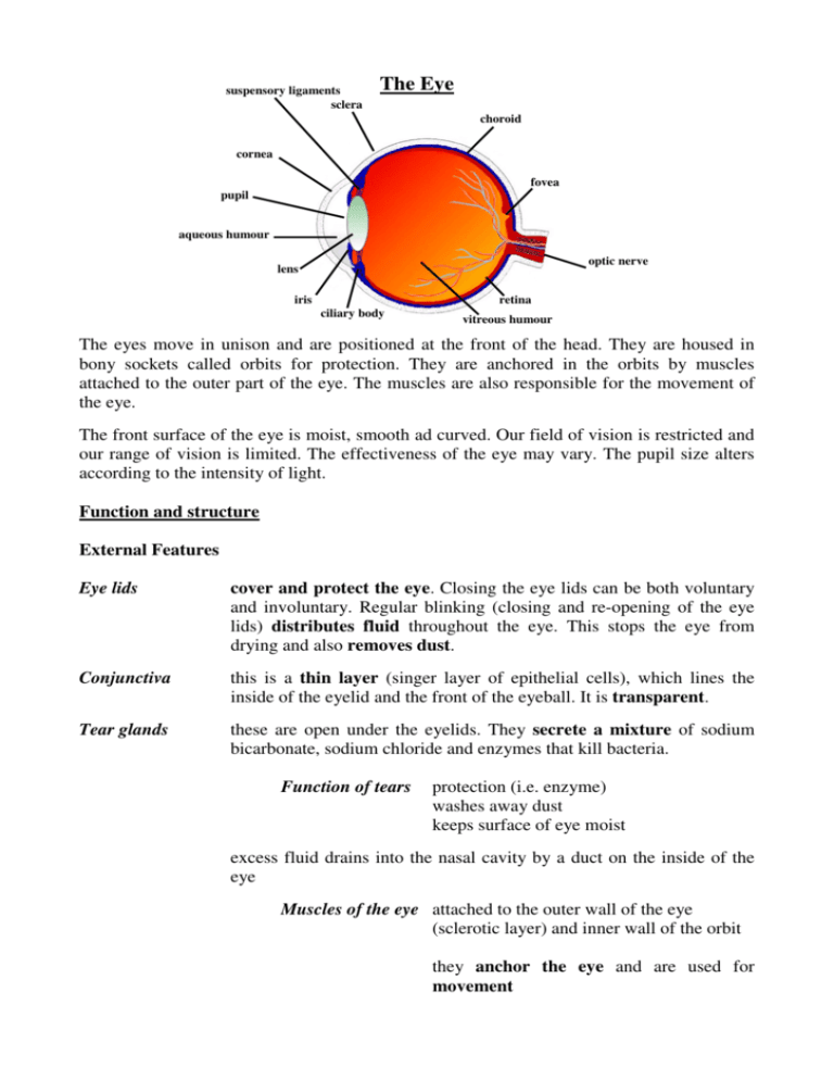
suspensory ligaments sclera The Eye choroid cornea fovea pupil aqueous humour optic nerve lens iris retina ciliary body vitreous humour The eyes move in unison and are positioned at the front of the head. They are housed in bony sockets called orbits for protection. They are anchored in the orbits by muscles attached to the outer part of the eye. The muscles are also responsible for the movement of the eye. The front surface of the eye is moist, smooth ad curved. Our field of vision is restricted and our range of vision is limited. The effectiveness of the eye may vary. The pupil size alters according to the intensity of light. Function and structure External Features Eye lids cover and protect the eye. Closing the eye lids can be both voluntary and involuntary. Regular blinking (closing and re-opening of the eye lids) distributes fluid throughout the eye. This stops the eye from drying and also removes dust. Conjunctiva this is a thin layer (singer layer of epithelial cells), which lines the inside of the eyelid and the front of the eyeball. It is transparent. Tear glands these are open under the eyelids. They secrete a mixture of sodium bicarbonate, sodium chloride and enzymes that kill bacteria. Function of tears protection (i.e. enzyme) washes away dust keeps surface of eye moist excess fluid drains into the nasal cavity by a duct on the inside of the eye Muscles of the eye attached to the outer wall of the eye (sclerotic layer) and inner wall of the orbit they anchor the eye and are used for movement Internal features There are three layers to the outer wall of the eye: Sclerotic coat it is a tough, non-elastic, fibrous layer, which covers the whole of the eye except where the optic nerve joins the back of the eye. It is modified at the front of the eye to form the cornea. Cornea it is a transparent disc (so that light rays can pass through). It has a curved surface and begins the refraction of light. Choroid this is living tissue. It has blood vessels supplying glucose and oxygen to the eye. It is deeply pigmented (in man it is black so that it can absorb all the light. In animals, it is not so pigmented). This absorbs and prevents the reflection of light. Retina this is the inner layer at the back of the eye where the image is formed. It contains two types of light sensitive cells: rods and cones. BUT there are no light sensitive cells where the optic nerve joins the back of the eye. This is known as the blind spot. Rods are sensitive to light of low intensity (black and white image). They are scattered fairly evenly throughout the retina except on the fovea, where only cones are situated. Cones are sensitive to high light intensity (colour image). They are highly concentrated in they in the fovea – where only cones are situated. Other features Blind spot No sensitive cells (i.e. cones/rods) are at the blind spot. This is because all of the axon fires from the sensory receptors join to form the optic nerve, which passes the eye and a hole in the skull to the brain. If an image falls on the blind spot, no impression is recorded because there are no light sensitive cells here. We are not aware of this blank in our vision because we have two eyes, which scan the same field. Aqueous humour a fluid-like medium at the front of the eye between the lens and cornea Vitreous humour a jelly-like medium that makes up the main body of the eye between the lens and the retina. Functions of both humours: refract light to provide image for retina provide food for eye provide pressure outwards to give the eye a shape Crystalline lens the cornea begins the refraction of light and the lens continues this process. The lens is held in position by suspensory ligaments, which are attached to ciliary muscle at the front edge of the choroid. The shape of the lens is altered by the contraction and the relaxation of the muscles of the ciliary body. Ciliary body this has muscle and is positioned at the thickened front end of the choroid. Suspensory ligaments these are non-elastic Focusing on an object “Accommodation” is the ability of the eye to receive light from far or near objects and to focus a sharp image on the retina. Far object (the eye is in its resting state) - the ciliary muscles are relaxed the suspensory ligaments are pulled taut (tight) lens is long and thin – i.e. less refraction Near object - the ciliary muscles are contracted the suspensory ligaments are slack (loose) lens is fatter and shorter – i.e. greater refraction The iris – light and dark The iris is muscle. It is the pigmented/coloured part of the eye, which can be seen (e.g. brown, blue, green etc.). In the centre of the iris is the pupil (the gap). The size of the gap (pupil) is altered by the contraction and relaxation of two muscles (radial and circular) in the iris. The size of the pupil alters in response to the amount of light. In the dark the pupil is larger, in brightness the pupil is smaller. This is to retain a constant amount of light in the eye (homeostasis) so that the eye is not damaged by bright light. This is an example of muscles working in Dim light: radial muscles contract antagonistic harmony. circular muscles relax pupil gets larger The iris remains the same size. Bright light: radial muscles relax circular muscles contract The colour of the iris is pupil gets smaller determined by genes. Fovea (centralis) There is a small depression (dip) in the centre of the retina called the fovea. In this part of the retina, there are no rods – only cones. The fovea has the highest concentration of light sensitive cells in the retina and therefore, gives the best interpretation of the image. The living parts of the eye include: lens cornea They all obtain their nutrients from the humours. choroid conjunctiva Image formation The image formed on the retina is: 1. smaller 2. upside down 3. a real image – it is formed by light rays The brain receives and interprets the image (via nerve impulses from the optic nerve) and converts it into the upright position. Eye Experiments 1. The blind spot experiment: take a long word on a piece of paper. Hold it out in front of you. Close one eye. Read the letters one by one. As you read along the word, when you reach one of the letters, the first letter will disappear but when you continue onto the next letter, the first letter will reappear again. 2. Shut one eye and rotate the other one. Both eyes are moving. 3. Close one eye. Take two objects the same shape and colour, but different in size. Try to get them level. It is difficult because both eyes are needed to judge distance. 4. Angle of the eyes: Tell a partner to focus on a spot far, and then a near spot. Measure the distance between the pupils in both cases. For far focussing, the distance is greater than for near focussing. 5. Effect of light on pupil size: Using a torch/light, see the difference in the size of your partner’s pupil in bright light. 6. Weak/strong eye: look to a tree in the distance. Place a pen directly on top of the tree using one eye. Open the other eye. Do the same with the other eye. Whichever eye moves the pen the least is the strongest.

