The Structure of the Eye The Structure of the Eye
advertisement
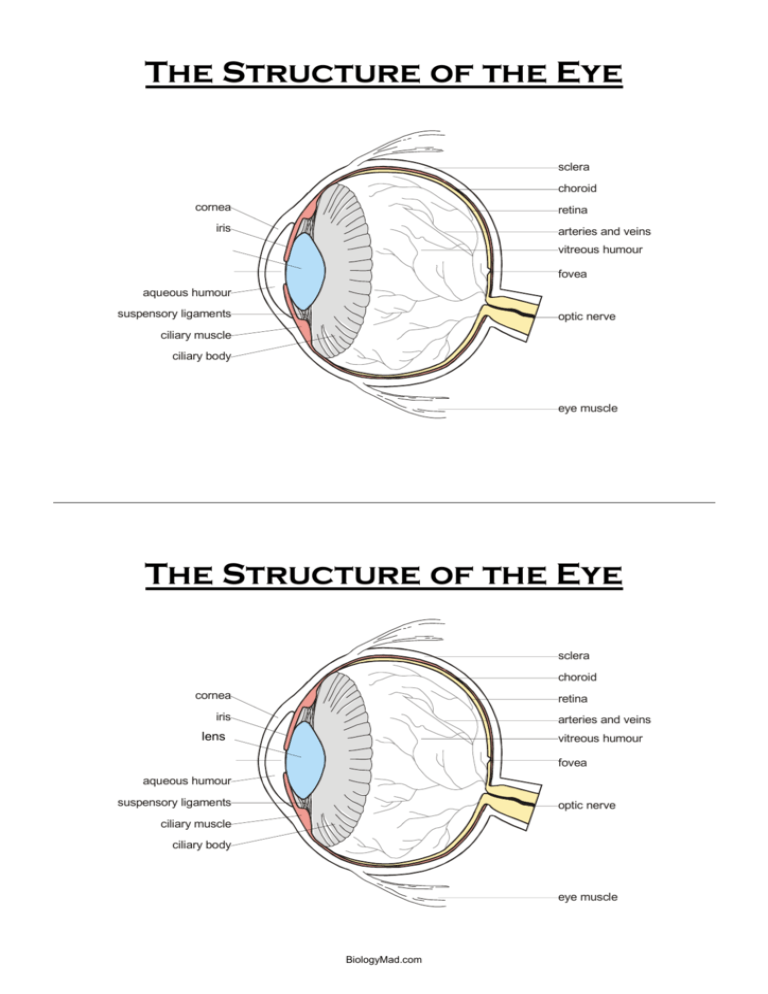
The Structure of the Eye sclera choroid cornea retina iris arteries and veins lens pupil vitreous humour pupil lens fovea aqueous humour suspensory ligaments optic nerve ciliary muscle ciliary body eye muscle The Structure of the Eye sclera choroid cornea retina iris arteries and veins lens pupil vitreous humour pupil lens fovea aqueous humour suspensory ligaments optic nerve ciliary muscle ciliary body eye muscle BiologyMad.com Cut out the white blocks and match them up to each key word to form the correct definition Conjunctiva Has a network of blood vessels to supply nutrients to the cells and remove waste products. It is pigmented that makes the retina appear black, thus preventing reflection of light within the eyeball. Cornea Helps to maintain the shape of the anterior chamber of the eyeball Sclera Is a pigmented muscular structure consisting of an inner ring of circular muscle and an outer layer of radial muscle. Its function is to help control the amount of light entering the eye so that: - too much light does not enter the eye which would damage the retina - enough light enters to allow a person to see Choroid Is where the bundle of sensory fibres form the optic nerve; it contains no light-sensitive receptors Ciliary body Has suspensory ligaments that hold the lens in place. It secretes the aqueous humour, and contains ciliary muscles that enable the lens to change shape, during accommodation (focusing on near and distant objects) Iris A part of the retina that is directly opposite the pupil and contains only cone cells. It is responsible for good visual acuity (good resolution) Pupil Is a thin protective covering of epithelial cells. It protects the cornea against damage by friction (tears from the tear glands help this process by lubricating the surface of the conjunctiva) Lens Is the transparent, curved front of the eye which helps to converge the light rays which enter the eye Retina Is a transparent, jelly-like mass located behind the lens. It acts as a ‘suspension’ for the lens so that the delicate lens is not damaged. It helps to maintain the shape of the posterior chamber of the eyeball Fovea (yellow spot) Is a layer of sensory neurones, the key structures being photoreceptors (rod and cone cells) which respond to light. Contains relay neurones and sensory neurones that pass impulses along the optic nerve to the part of the brain that controls vision Blind spot Is a transparent, flexible, curved structure. Its function is to focus incoming light rays onto the retina using its refractive properties Vitreous humour Is a hole in the middle of the iris where light is allowed to continue its passage. In bright light it is constricted and in dim light it is dilated Aqueous humour Is an opaque, fibrous, protective outer structure. It is soft connective tissue, and the spherical shape of the eye is maintained by the pressure of the liquid inside. It provides attachment surfaces for eye muscles BiologyMad.com Conjunctiva Is a thin protective covering of epithelial cells. It protects the cornea against damage by friction (tears from the tear glands help this process by lubricating the surface of the conjunctiva) Cornea Is the transparent, curved front of the eye which helps to converge the light rays which enter the eye Sclera Is an opaque, fibrous, protective outer structure. It is soft connective tissue, and the spherical shape of the eye is maintained by the pressure of the liquid inside. It provides attachment surfaces for eye muscles Choroid Has a network of blood vessels to supply nutrients to the cells and remove waste products. It is pigmented that makes the retina appear black, thus preventing reflection of light within the eyeball. Ciliary body Has suspensory ligaments that hold the lens in place. It secretes the aqueous humour, and contains ciliary muscles that enable the lens to change shape, during accommodation (focusing on near and distant objects) Iris Is a pigmented muscular structure consisting of an inner ring of circular muscle and an outer layer of radial muscle. Its function is to help control the amount of light entering the eye so that: - too much light does not enter the eye which would damage the retina - enough light enters to allow a person to see Pupil Is a hole in the middle of the iris where light is allowed to continue its passage. In bright light it is constricted and in dim light it is dilated Lens Is a transparent, flexible, curved structure. Its function is to focus incoming light rays onto the retina using its refractive properties Retina Is a layer of sensory neurones, the key structures being photoreceptors (rod and cone cells) which respond to light. Contains relay neurones and sensory neurones that pass impulses along the optic nerve to the part of the brain that controls vision Fovea (yellow spot) A part of the retina that is directly opposite the pupil and contains only cone cells. It is responsible for good visual acuity (good resolution) Blind spot Is where the bundle of sensory fibres form the optic nerve; it contains no light-sensitive receptors Vitreous humour Is a transparent, jelly-like mass located behind the lens. It acts as a ‘suspension’ for the lens so that the delicate lens is not damaged. It helps to maintain the shape of the posterior chamber of the eyeball Aqueous humour Helps to maintain the shape of the anterior chamber of the eyeball BiologyMad.com

