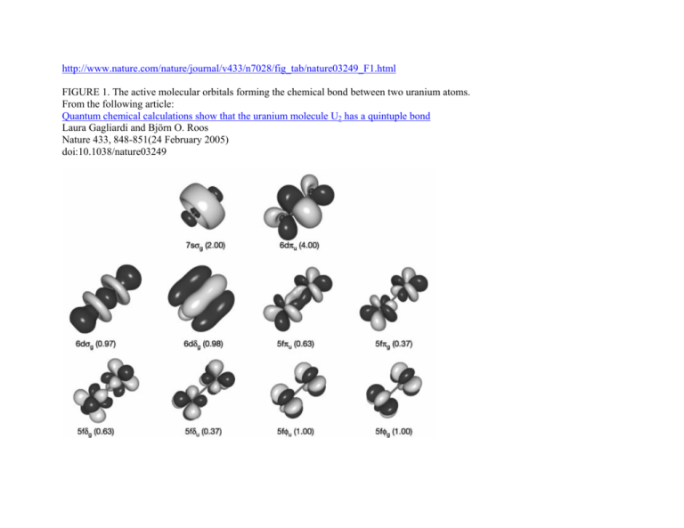
http://www.nature.com/nature/journal/v433/n7028/fig_tab/nature03249_F1.html
FIGURE 1. The active molecular orbitals forming the chemical bond between two uranium atoms.
From the following article:
Quantum chemical calculations show that the uranium molecule U2 has a quintuple bond
Laura Gagliardi and Björn O. Roos
Nature 433, 848-851(24 February 2005)
doi:10.1038/nature03249
http://www.chemistry.mcmaster.ca/esam/Chapter_8/section_3.html
An Introduction to the Electronic Structure of Atoms and Molecules
Dr. Richard F.W. Bader
Molecular Orbitals for Homonuclear Diatomics
While the specific forms of the molecular orbitals (their dependence on ρ and z in a cylindrical coordinate system) are different for
each molecule, their dependence on the angle φ as denoted by the quantum number λ and their g or u behaviour with respect to
inversion are completely determined by the symmetry of the system. These properties are common to all of the molecular orbitals for
homonuclear diatomic molecules. In addition, the relative ordering of the orbital energies is the same for nearly all of the homonuclear
diatomic molecules. Thus we may construct a molecular orbital energy level diagram, similar to the one used to build up the electronic
configurations of the atoms in the periodic table. The molecular orbital energy level diagram (Fig. 8-4) is as fundamental to the
understanding of the electronic structure of diatomic molecules as the corresponding atomic orbital diagram is to the understanding of
atoms.
Fig. 8-4. Molecular orbital energy level diagram for homonuclear diatomic molecules
showing the correlation of the molecular orbitals with the atomic orbitals of the separated atoms. The schematic representation of the molecular orbitals is to
illustrate their general forms and nodal properties (the nodes are indicated by dashed lines). Only one component of the degenerate 1πu and 1πg orbitals is shown.
The second component is identical in form in each case but rotated 90° out of the plane. The ordering of the orbital energy levels shown in the figure holds
generally for all homonuclear diatomic molecules with the exception of the levels for the 1πu and 3σg orbitals, whose relative order is reversed for the molecules
after C2.
Molecular orbitals exhibit the same general properties as atomic orbitals, including a nodal structure. The nodal properties of the
orbitals are indicated in Fig. 8-4. Notice that the nodal properties correctly reflect the g and u character of the orbitals. Inversion of a g
orbital interchanges regions of like sign and the orbital is left unchanged. Inversion of a u orbital interchanges the positive regions
with the negative regions and the orbital is changed in sign.
An orbital of a particular symmetry may appear more than once. When this occurs a number is added as a prefix to the symbol. Thus
there are 1σg, 2σg, 3σg, etc. molecular orbitals just as there are 1s, 2s, 3s, etc. atomic orbitals. The numerical prefix is similar to the
principal quantum number n in the atomic case. As n increases through a given symmetry set, for example, 1σg, 2σg, 3σg, the orbital
energy increases, the orbital increases in size and consequently concentrates charge density further from the nuclei, and finally the
number of nodes increases as n increases. All these properties are common to atomic orbitals as well.
We may obtain a qualitative understanding of the molecular orbital energy level diagram by considering the behaviour of the orbitals
under certain limiting conditions. The molecular orbital must describe the motion of the electron for all values of the internuclear
separation; from R = ∞ for the separated atoms, through R = Re, the equilibrium state of the molecule, to R = 0, the united atom
obtained when the two nuclei in the molecule coalesce (in a hypothetical reaction) to give a single nucleus. Hence a molecular orbital
must undergo a continuous change in form. At the limit of large R it must reduce to some combination of atomic orbitals giving the
proper orbital description of the separated atoms and for R = 0 it must reduce to a single atomic orbital on the united nucleus.
Consider, for example, the limiting behaviour of the 1σg orbital in the case of the hydrogen molecule. The most stable state of H2 is
obtained when both electrons are placed in this orbital with paired spins giving the electronic configuration 1σg2. For large values of
the internuclear separation, the hydrogen molecule dissociates into two hydrogen atoms. Thus the limiting form of the 1σg molecular
orbital for an infinite separation between the nuclei should be a sum of 1s orbitals, one centred on each of the nuclei. If we label the
two nuclei as A and B we can express the limiting form of the 1σg orbital as
where lsA is a 1s orbital centred on nucleus A, and lsB is a ls orbital centred on nucleus B. This form for the 1σg orbital predicts the
correct density distribution for the system at large values of R. Squaring the function (lsA + lsB) we obtain for the density
The first two terms denote that one electron is on atom A and one on atom B, both with 1s atomic density distributions. The cross
term 2 × lsA × lsB obtained in the product is zero since the distance between the two nuclei is so great that the overlap of the orbitals
vanishes. Notice as well that the function (lsA + lsB) has the same symmetry properties as does the 1σg molecular orbital; it is
symmetric with respect to both a rotation about the line joining the nuclei and to an inversion of the coordinates at the mid-point
between the nuclei. The 1σg orbital for the molecule is said to correlate with the sum of 1s orbitals, one on each nucleus, for the
separated atom case.
Consider next the limiting case of the separated atoms for the helium molecule. Of the four electrons present in He2 two are placed
in the 1σg orbital and the remaining two must, by the Pauli exclusion principle, be placed in the next vacant orbital of lowest energy,
the1σu orbital. The electronic configuration of He2 is thus 1σg21σu2. The 1σg orbital will correlate with the sum of the 1s orbitals for
the separated helium atoms. Of the two electrons in the1σg molecular orbital one will correlate with the 1s orbital on atom A and the
other with the 1s orbital on atom B. Since each helium atom possesses two 1s electrons, the 1σu orbital must also correlate its
electrons with 1s atomic functions on A and B. In addition, the correlated function in this case must be of u symmetry. A function with
these properties is
The limiting density distribution obtained by squaring this function places one electron in a 1s atomic distribution on A, the other in a
1s atomic distribution on B. The sum of the limiting charge densities for the 1σg and 1σu molecular orbitals places two electrons in 1s
atomic charge distributions on each atom, the proper description of two isolated helium atoms.
Every diatomic homonuclear molecular orbital may be correlated with either the sum (for σg and πu orbitals) or the difference (for σu
and πg orbitals) of like orbitals on both separated atoms. By carrying out this correlation procedure for every orbital we may construct
a molecular orbital correlation diagram (Fig. 8-4) which relates each of the orbital energy levels in the molecule with the correlated
energy levels in the separated atoms. It is important to note that the symmetry of each orbital is preserved in the construction of this
diagram. Consider, for example the molecular orbitals which correlate with the 2p atomic orbitals. The direction of approach of the
two atoms defines a new axis of quantization for the atomic orbitals. The 2p orbital which lies along this axis is of σ symmetry while
the remaining two 2p orbitals form a degenerate set of π symmetry with respect to this axis. The sum and difference of the 2pσ
orbitals on each centre correlate with the 3σg and 3σu orbitals respectively, while the sum and the difference of the 2pπ orbitals
correlate with the πu and πg orbitals (Fig. 8-5).
Fig. 8-5. The correlated separated atom forms of the 3σg, 3σu, 1πu and 1πg molecular orbitals. The nodal planes are indicated by dashed lines. Only one
component of each π orbital is shown.
For large values of the internuclear distance, each molecular orbital is thus represented by a sum or a difference of atomic orbitals
centred on the two interacting atoms. As the atoms approach one another the orbitals on each atom are distorted by polarization and
overlap effects. In general, the limiting correlated forms of the molecular orbitals are not suitable descriptions of the molecular
orbitals for finite internuclear separations.
The correlation of the molecular orbitals with the appropriate atomic orbitals of the united atom is left as a problem for the reader
(Problem 2).
We are now in a position to build up and determine the electronic configurations of the homonuclear diatomic molecules by adding
electrons two at a time to the molecular orbitals with the spins of the electrons paired, always filling the orbitals of lowest energy first.
We shall, at the same time, discuss the effectiveness of each orbital in binding the nuclei and make qualitative predictions regarding
the stability of each molecular configuration.
www.cartage.org.lb/.../Molecular/Molecular.htm
Hydrogen. The two electrons in the hydrogen molecule may both be accommodated in the 1σg orbital if their spins are paired and the
molecular orbital configuration for H2 is 1σg2. Since the 1σg orbital is the only occupied orbital in the ground state of H2, the density
distribution shown previously in Fig. 6-2 for H2 is also the density distribution for the 1σg orbital when occupied by two electrons. The
remarks made previously regarding the binding of the nuclei in H2 by the molecular charge distribution apply directly to the properties
of the 1σg charge density. Because it concentrates charge in the binding region and exerts an attractive force on the nuclei the 1σg
orbital is classified as a bonding orbital.
Excited electronic configurations for molecules may be described and predicted with the same ease within the framework of
molecular orbital theory as are the excited configurations of atoms in the corresponding atomic orbital theory. For example, an
electron in H2 may be excited to any of the vacant orbitals of higher energy indicated in the energy level diagram. The excited
molecule may return to its ground configuration with the emission of a photon. The energy of the photon will be given approximately
by the difference in the energies of the excited orbital and the 1σg ground state orbital. Thus molecules as well as atoms will exhibit a
line spectrum. The electronic line spectrum obtained from a molecule is, however, complicated by the appearance of many
accompanying side bands. These have their origin in changes in the vibrational energy of the molecule which accompany the change
in electronic energy.
Helium. The electronic configuration of He2 is 1σg2 1σu2. A σu orbital, unlike a σg orbital, possesses a node in the plane midway
between the nuclei and perpendicular to the bond axis. The 1σu orbital and all σu orbitals in general, because of this nodal property,
cannot concentrate charge density in the binding region. It is instead concentrated in the antibinding region behind each nucleus (Fig.
8-6).
Fig. 8-6. Contour maps of the doubly-occupied 1σg and 1σu molecular orbital charge densities and of the total molecular charge distribution of He2 at R = 2.0 au.
A profile of the total charge distribution along the internuclear axis is also shown. Click here for contour values.
The σu orbitals are therefore classified as antibonding. It is evident from the form of density distribution for the 1σu orbital that the
charge density in this orbital pulls the nuclei apart rather than drawing them together. Generally, the occupation of an equal number of
σg and σu orbitals results in an unstable molecule. The attractive force exerted on the nuclei by the charge density in the σg orbitals is
not sufficient to balance both the nuclear force of repulsion and the antibinding force exerted by the density in the σu orbitals. Thus
molecular orbital theory ascribes the instability of He2 to the equal occupation of bonding and antibonding orbitals. Notice that the
Pauli exclusion principle is still the basic cause of the instability. If it were not for the Pauli principle, all four electrons could occupy a
σg-type orbital and concentrate their charge density in the region of low potential energy between the nuclei. It is the Pauli principle,
and not a question of energetics, which forces the occupation of the 1σu antibonding orbital.
The total molecular charge distribution is obtained by summing the individual molecular orbital densities for single or double
occupation numbers as determined by the electronic configuration of the molecule. Thus the total charge distribution for He2 (Fig. 8-6)
is given by the sum of the 1σg and 1σu orbital densities for double occupation of both orbitals. The adverse effect which the nodal
property of the 1σu orbital has on the stability of He2 is very evident in the total charge distribution. Very little charge density is
accumulated in the central portion of the binding region. The value of the charge density at the mid-point of the bond in He2 is only
0.164 au compared to a value of 0.268 au for H2.
We should reconsider in the light of molecular orbital theory the stability of He2+ and the instability of the hydrogen molecule with
parallel spins, cases discussed previously in terms of valence bond theory. He2+ will have the configuration 1σg2 1σu1. Since the 1σu
orbital is only singly occupied in He2+, less charge density is accumulated in the antibinding regions than is accumulated in these same
regions in the neutral molecule. Thus the binding forces of the doubly-occupied 1σg density predominate and He2 is stable. The
electron configuration of H2 is 1σg1()1σu1() when the electronic spins are parallel. The electrons must occupy separate orbitals because
of the Pauli exclusion principle. With equal occupation of bonding and antibonding orbitals, the H2 ()species is predicted to be
unstable.
Lithium. The Li2 molecule with the configuration 1σg21σu22σg2 marks the beginning of what can be called the second quantum shell in
analogy with the atomic case. Since the 1σu antibonding orbital approximately cancels the binding obtained from the 1σg bonding
orbital, the bonding in Li2 can be described as arising from the single pair of electrons in the 2σg orbital. Valence bond theory, or a
Lewis model for Li2, also describes the bonding in Li2 as resulting from a single electron pair bond. This is a general result. The
number of bonds predicted in a simple Lewis structure is often found to equal the difference between the number of occupied bonding
and antibonding orbitals of molecular orbital theory.
The forms of the orbital density distributions for Li2 (Fig. 8-7) bear out the prediction that a single electron pair bond is responsible
for the binding in this molecule.
Fig. 8-7. Contour maps of the doubly-occupied 1σg, 1σu and 2σg molecular orbital charge densities for Li2 at R = 5.051 au, the equilibrium internuclear
separation. Click here for contour values. The total molecular charge distribution for Li2 is shown in Fig. 7-3.
The 1σg and 1σu density distributions are both strongly localized in the regions of the nuclei with spherical contours characteristic of
1s atomic distributions. The addition of just the doubly-occupied 1σg and 1σu orbital densities in Li2 will yield a distribution which
resembles very closely and may be identified with the doubly-occupied 1s or inner shell atomic densities on each lithium nucleus.
Only the charge density of the pair of valence electrons in the 2σg orbital is delocalized over the whole of the molecule and
accumulated to any extent in the binding region.
Thus there are cases where the molecular orbitals even at the equilibrium bond length resemble closely their limiting atomic forms.
This occurs for inner shell molecular orbitals which correlate with the inner shell atomic orbitals on the separated atoms. Inner shell 1s
electrons are bound very tightly to the nucleus as they experience almost the full nuclear charge and the effective radii of the 1s
density distributions are less than the molecular bond lengths. Because of their tight binding and restricted extension in space, the
inner electrons do not participate to any large extent in the binding of a molecule. Thus with the exception of H2 and He2 and their
molecular ions, the 1σg and 1σu molecular orbitals degenerate into non-overlapping atomic-like orbitals centred on the two nuclei and
are classed as nonbonding orbitals.
Beryllium. The configuration of Be2 is 1σg21σu22σg22σu2 and the molecule is predicted to form a weakly bound van der Waals
moecule.
Oxygen. Since the method of determining electronic configurations is clear from the above examples, we shall consider just one more
molecule in detail, the oxygen molecule. Filling the orbitals in order of increasing energy the sixteen electrons of O2 are described by
the configuration 1σg21σu22σg22σu23σg21πu41πg2. The orbital densities are illustrated in Fig. 8-8.
Fig. 8-8. Contour maps of the molecular orbital charge densities for O2 at the equilibrium internuclear distance of 2.282 au. Only one component of the Iπg and
1πu orbitals is shown. All the maps are for doubly-occupied orbitals with the exception of that for 1πg for which each component of the doubly-degenerate orbital
contains a single electron. The nodes are indicated by dashed lines. Click here for contour values.
The molecular orbitals of π symmetry are doubly degenerate and a filled set of π orbitals will contain four electrons. The node in a
πu orbital is in the plane which contains the internuclear axis and is not perpendicular to this axis as is the node in a σu orbital. (The
nodal properties of the orbitals are indicated in Fig. 8-4.) The πu orbital is therefore bonding. A πg orbital, on the other hand, is
antibonding because it has, in addition to the node in the plane of the bond axis, another at the bond mid-point perpendicular to the
axis. The bonding and antibonding characters of the π orbitals have just the opposite relationship to their g and u dependence as have
the σ orbitals.
The 1σg and 1σu orbital densities have, as in the case of Li2, degenerated into localized atomic distributions with the characteristics
of 1s core densities and are nonbonding. The valence electrons of O2 are contained in the remaining orbitals, a feature reflected in the
extent to which their density distributions are delocalized over the entire molecule. Aside from the inner nodes encircling the nuclei,
the 2σg and 2σuorbital densities resemble the 1σg and 1σu valence density distributions of H2 and He2. A quantitative discussion of the
relative binding abilities of the 2σg , 3σg and 1π orbital densities is presented in the following section.
One interesting feature of the electronic configuration of O2 is that its outer orbital is not fully occupied. The two πg electrons could
both occupy one of the πg orbitals with paired spins or they could be assigned one to each of the πg orbitals and have parallel spins.
Hund's principle applies to molecules as well as to atoms and the configuration with single occupation of both πg orbitals with parallel
spins is thus predicted to be the most stable. This prediction of molecular orbital theory regarding the electronic structure of O2 has an
interesting consequence. The oxygen molecule should be magnetic because of the resultant spin angular momentum possessed by the
electrons. The magnetism of O2 can be demonstrated experimentally in many ways, one of the simplest being the observation that
liquid oxygen is attracted to the poles of a strong magnet.
http://www.chemistry.mcmaster.ca/esam/Chapter_8/section_5.html
Molecular Orbitals for Heteronuclear Molecules
The molecular orbitals which describe the motion of a single electron in a molecule containing two unequal nuclear charges will not
exhibit the g and u symmetry properties of the homonuclear diatomic case. The molecular orbitals in the heteronuclear case will in
general be concentrated more around one nucleus than the other. The orbitals may still be classified as σ, π, δ, etc. because the
molecular axis is still an axis of symmetry.
In simple numerical calculations the molecular orbitals are sometimes approximated by the sum and difference of single atomic
orbitals on each centre, their limiting form. The molecular orbital is said to be approximated mathematically by a linear combination
of atomic orbitals and the technique is known as the LCAO-MO method. It must be understood that the LCAO-MO method using a
limited number of atomic orbitals provides only an approximation to the true molecular orbital. The concept of a molecular orbital is
completely independent of the additional concept of approximating it in terms of atomic orbitals, except for the case of the separated
atoms. However, by using a large number of atomic orbitals centred on each nucleus in the construction of a single molecular orbital
sufficient mathematical flexibility can be achieved to approximate the exact form of the molecular orbital very closely.
While the LCAO approximation using a limited number of atomic orbitals is in general a poor one for quantitative purposes, it does
provide a useful guide for the prediction of the qualitative features of the molecular orbital. There are two simple conditions which
must be met if atomic orbitals on different centres are to interact significantly and form a molecular orbital which is delocalized over
the whole molecule. Both atomic orbitals must have approximately the same orbital energy and they must possess the same symmetry
characteristics with respect to the internuclear axis. We shall consider the molecular orbitals in LiH, CH and HF to illustrate how
molecular orbital theory describes the bonding in heteronuclear molecules, and to see how well the forms of the orbitals in these
molecules can be rationalized in terms of the symmetry and energy criteria set out above.
The 1s and 2s atomic orbitals and the 2p orbital which is directed along the bond axis are all left unchanged by a rotation about the
symmetry axis. They may therefore form molecular orbitals of σ symmetry in the diatomic hydride molecules. The 2p orbitals which
are perpendicular to the bond axis will be of π symmetry and may form molecular orbitals with this same symmetry. The energies and
symmetry properties of the relevant atomic orbitals and the electronic configurations of the atoms and molecules are given in Table 82.
Table 8-2.
Atomic Orbital Energies and Symmetry Properties
H
-0.5
1s
2s
2p
Atomic Configurations
Li
1s22s1
C
1s22s22p2
F
1s22s22p5
Energy (au)
Li
-2.48
-0.20
Symmetry
C
-11.33
-0.71
-0.43
F
-26.38
σ
-1.57
σ
-0.73
σ and π
Molecular Configurations
LiH
1σ22σ2
CH
1σ22σ23σ21π1
HF
1σ22σ23σ21π4
Density diagrams of the molecular orbitals for the LiH, CH, and HF molecules are illustrated in Fig. 8-9.
Fig. 8-9. Contour maps of the molecular orbital charge densities of the LiH, CH, HF diatomic hydrides. The nodes are indicated by dashed lines. Click here to
see countour values. The 1s orbital energies of Li, C and F all lie well below that of the H 1s orbital. The charge densities of these inner
shell orbitals are tightly bound to their respective nuclei. They should not, therefore, be much affected by the field of the proton or
interact significantly with the H 1s orbital. The molecular orbital of lowest energy in these molecules, the lσ molecular orbital, should
be essentially nonbinding and resemble the doubly-occupied 1s atomic orbital on Li, C and F respectively. These predictions are borne
out by the lσ orbital density distributions (Fig. 8-9). They consist of nearly spherical contours centred on the Li, C and F nuclei, the
radius of the outer contour being less than the bond length in each case. The forces exerted on the proton by the lσ charge distributions
are equivalent to placing two negative charges at the position of the heavy nucleus in each case. The charge density in the lσ
molecular orbital simply screens two of the nuclear charges on the heavy nucleus from the proton. This same screening effect is
obtained for the 1s2 charge distribution when the molecules dissociate into atoms. Thus the 1s atomic orbitals of Li, C and F are not
much affected by the formation of the molecule and the lσ charge density is nonbinding as far as the proton is concerned. The lσ
atomic-like distributions are slightly polarized. In LiH the lσ density is polarized away from the proton to a significant extent while in
CH and HF it is slightly polarized towards the proton. Thus the 1σ charge density exerts an antibinding force on the Li nucleus and a
small binding force on the C and F nuclei.
The energies of the 2s atomic orbitals decrease (the electron is more tightly bound) from Li to F as expected on the basis of the
increase in the effective nuclear charge from Li to F. The 2s orbital on Li is large and diffuse and will overlap extensively with the 1s
orbital on H. However, the 2s electron on Li is considerably less tightly bound than is the 1s electron on H. Thus the charge density of
the 2σ molecular orbital in LiH will be localized in the region of the proton corresponding to the transfer of the 2s electron on Li to the
region of lower potential energy offered by the 1s orbital on H. This is approximately correct as shown by the almost complete
concentration of the charge density in the region of the proton in the 2σ orbital density map for LiH. The small amount of density
which does remain around the Li nucleus is polarized away from the proton. The 1σ and 2σ densities are polarized in a direction
counter to the direction of charge transfer as required in ionic binding. The inwardly polarized accumulation of 2σ charge density
centred on the proton binds both nuclei.
The 1σ molecular orbital in LiH is to a good approximation a polarized doubly-occupied 1s orbital on Li, and the 2σ molecular
orbital is, to a somewhat poorer approximation, a doubly-occupied and polarized 1s orbital on H. Our previous discussion of the
bonding in LiH indicated that the binding is ionic, corresponding to the description Li+(1s2)H-(1s2). The molecular orbital description
of an ionic bond is similar in that the molecular orbitals in the ionic extreme are localized in the regions of the individual nuclei, rather
than being delocalized over both nuclei as they are for a covalent bond.
The matching of the 2s orbital energy with the H 1s orbital energy is closer in the case of C than it is for Li. Correspondingly, the 2σ
charge density in CH is delocalized over both nuclei rather than concentrated in the region of just one nucleus as it is in the LiH
molecule. There is a considerable buildup of charge density in the binding region which is shared by both nuclei. The 2σ charge
density exerts a large binding force on both the H and C nuclei. This is the molecular orbital description of an interaction
which is essentially covalent in character.
The 2s orbital energy of F is considerably lower than that of the H 1s orbital. The 2σ orbital charge density in HF, therefore,
approximately resembles a localized 2s orbital on F. It is strongly polarized and distorted by the proton, but the amount of charge
transferred to the region between the nuclei is not as large as in CH. The 2σ orbital in HF plays a less important role in binding the
proton than it does in CH.
The 3σ molecular orbital in CH and HF will result primarily from the overlap of the 2pσ orbital on C and F, with the 1s orbital on H.
The 2p-like character of the 3σ molecular orbital in both CH and HF is evident in the density diagrams (Fig. 8-9). In CH the 1s orbital
of H interacts strongly with both the 2s and 2pσ orbitals on C. In terms of the forces exerted on the nuclei, the 2σ charge density is
strongly binding for both C and H, while the 3σ charge density is only very weakly binding for H and is actually antibinding for the
C. This antibinding effect is a result of the large accumulation of charge density in the region behind the C nucleus.
In HF, the H 1s orbital interacts only slightly with the 2s orbital on F, but it interacts very strongly with the 2pσ orbital in the
formation of the 3σ molecular orbital. The 3σ charge density exerts a large binding force on the proton. Thus the proton is bound
primarily by the 2σ charge density in CH and by the 3σ charge density in HF. The 3σ charge density in HF is primarily centred on the
F nucleus and roughly resembles a 2pσ orbital. Although no density contours are actually centred on the proton, the proton is
embedded well within the orbital density distribution. This is a molecular orbital description of a highly polar bond.
The 3σ orbital charge density exerts a force on the F and C nuclei in a direction away from the proton. The molecular orbitals which
involve pσ orbitals are characteristically strongly polarized in a direction away from the bond in the region of the nucleus on which
the p orbital is centred. Compare, for example, the 3σ orbitals of CH and HF with the 3σg molecular orbital of the homonuclear
diatomic molecules.
When the C and H atoms are widely separated, we can consider the carbon atom to have one 2p electron in the 2pσ orbital which
lies along the bond axis, and the second 2p electron in one of the 2pπ orbitals which are perpendicular to the bond. The F atom has
five 2p electrons and of these one may be placed in the 2pσ orbital; the remaining four 2p electrons will then completely occupy the
2pπ orbitals. The singly-occupied 2pσ orbitals on F and C eventually interact with the singly-occupied 1s orbital on H to form the
doubly-occupied 3σ molecular orbital in HF and CH. The remaining 2p electrons, those of π symmetry, will occupy the 1π molecular
orbital. The H atom does not possess an orbital of π symmetry in its valence shell and the vacant 2pπ orbital on H is too high in energy
(-0.125 au) to interact significantly with the 2pπ orbitals on C and F. Thus the 1π molecular orbital is atomic-like, centred on the F and
C nuclei and is essentially nonbinding (Fig. 8-9). The 1π molecular orbital resembles a 2pπ atomic orbital in each case, but one which
is polarized in the direction of the proton.
The 1π orbitals of CH and HF illustrate an interesting and general result. In the formation of a bond between different atoms, the
charge density in the σ orbitals is transferred from the least to the most electronegative atom. However, the charge density of π
symmetry, if any is present, is invariably transferred, or at least polarized, in the opposite direction, towards the least electronegative
atom. Although the amount of charge density transferred is less in the formation of the π orbitals than in the σ orbitals, one effect
increases with the other. Thus the polarization is more pronounced in HF than in CH.
The three examples considered above demonstrate the essential points of a molecular orbital description of the complete range of
chemical bonding. In the ionic extreme of LiH the charge density of the bonding molecular orbital is localized around the proton. In
CH the valence charge density is more evenly shared by both nuclei and the bond is covalent. The motions of the electrons in HF are
governed largely by the potential field of the F nucleus. This is evidenced by the appearance of the molecular orbital charge
distributions. The proton is, however, encompassed by the valence charge density and the result is a polar bond.
http://www.chemistry.mcmaster.ca/esam/Chapter_8/section_6.html
Molecular Orbitals for Polyatomic Molecules
The concept of a molecular orbital is readily extended to provide a description of the electronic structure of a polyatomic molecule.
Indeed molecular orbital theory forms the basis for most of the quantitative theoretical investigations of the properties of large
molecules.
In general a molecular orbital in a polyatomic system extends over all the nuclei in a molecule and it is essential, if we are to
understand and predict the spatial properties of the orbitals, that we make use of the symmetry properties possessed by the nuclear
framework. An analysis of the molecular orbitals for the water molecule provides a good introduction to the way in which the
symmetry of a molecule determines the forms of the molecular orbitals in a polyatomic system.
There are three symmetry operations which transform the nuclear framework of the water molecule into itself and hence leave the
nuclear potential field in which the electrons move unchanged (Fig. 8-10).
Fig. 8-10. Symmetry elements for H2O. The bottom two diagrams illustrate the transformations of the 2py orbital on oxygen under the C2 and σ2 symmetry
operations.
For each symmetry operation there is a corresponding symmetry element. The symmetry elements for the water molecule are a twofold axis of rotation C2 and two planes of symmetry σ1 and σ2 (Fig. 8-10). A rotation of 180° about the C2 axis leaves the oxygen
nucleus unchanged and interchanges the two hydrogen nuclei. A reflection through the plane labelled σ1 leaves all the nuclear
positions unchanged while a reflection through σ2 interchanges the two protons. The symmetry operations associated with the three
symmetry elements either leave the nuclear positions unchanged or interchange symmetrically equivalent (and hence
indistinguishable) nuclei. Every molecular orbital for the water molecule must, under the same symmetry operations, be left
unchanged or undergo a change in sign.
Similarly we may use the symmetry transformation properties of the atomic orbitals on oxygen and hydrogen together with their
relative orbital energy values to determine the primary atomic components of each molecular orbital in a simple LCAO approximation
to the exact molecular orbitals. Only atomic orbitals which transform in the same way under the symmetry operations may be
combined to form a molecular orbital of a given symmetry. The symmetry transformation properties of the atomic orbitals on oxygen
and hydrogen are given in Table 8-3. A value of +1 or -1 opposite a given orbital in the table indicates that the orbital is unchanged or
changed in sign respectively by a particular symmetry operation.
Table 8-3.
Symmetry Properties and Orbital Energies for the Water Molecule
Atomic Orbitals
Symmetry
Symmetry Behaviour Under
Orbital Energy (au)
on Oxygen
Classification
σ2
σ1
σ2
1s
+1
+1
+1
a1
-20.669
2s
+1
+1
+1
a1
-1.244
2pz
+1
+1
+1
a1
-0.632
2px
-1
+1
-1
b2
2py
-1
-1
+1
b1
Atomic Orbitals on Hydrogen
(1s1 + 1s2)
+1
+1
+1
a1
-0.500
(1s1 - 1s2)
-1
+1
-1
b2
}
}
1a1
-20.565
Molecular Orbital Energies for H2O (au)
2a1
1b2
3a1
-1.339
-0.728
-0.595
1b1
-0.521
The 1s, 2s and 2pz orbitals of oxygen are symmetric (i.e., unchanged) with respect to all three symmetry operations. They are given
the symmetry classification a1. The 2px orbital, since it possesses a node in the σ2 plane (and hence is of different sign on each side of
the plane) changes sign when reflected through the σ2 plane or when rotated by 180° about the C2 axis. It is classified as a b2 orbital.
The 2py orbital is antisymmetric with respect to the rotation operator and to a reflection through the σ1 plane. It is labelled b1.
The hydrogen 1s orbitals when considered separately are neither unchanged nor changed in sign by the rotation operator or by a
reflection through the σ2 plane. Instead both these operations interchange these orbitals. The hydrogen orbitals are said to be
symmetrically equivalent and when considered individually they do not reflect the symmetry properties of the molecule. However, the
two linear combinations (1s1 + 1s2) and (1s1 - 1s2) do behave in the required manner. The former is symmetric
under all three operations and is of a1 symmetry while the latter is antisymmetric with respect to the rotation operator and to a
reflection through the plane σ2 and is of b2 symmetry.
The molecular orbitals in the water molecule are classified as a1, b1 or b2 orbitals, as determined by their symmetry properties. This
labelling of the orbitals is analogous to the use of the σ−π and g-u classification in linear molecules. In addition to the symmetry
properties of the atomic orbitals we must consider their relative energies to determine which orbitals will overlap significantly and
form delocalized molecular orbitals.
The 1s atomic orbital on oxygen possesses a much lower energy than any of the other orbitals of a1 symmetry and should not interact
significantly with them. The molecular orbital of lowest energy in H2O should therefore correspond to an inner shell 1s atomic-like
orbital centred on the oxygen. This is the first orbital of a1 symmetry and it is labelled la1. Reference to the forms of the charge density
contours for the la, molecular orbital (Fig. 8-11) substantiates the above remarks regarding the properties of this orbital.
Fig. 8-11. Contour maps of the molecular orbital charge densities for H2O. The maps for the la1, 2a1, 3a1and 1b2 orbitals (all doubly-occupied) are shown in the
plane of the nuclei. The lb1 orbital has a node in this plane and hence the contour map for the 1b1 orbital is shown in the plane perpendicular to the molecular
plane. The total molecular charge density for H2O is also illustrated. The density distributions were calculated from the wave function determined by R. M.
Pitzer, S. Aung and S. I. Chan, J. Chem. Phys. 49, 2071 (1968). Click here for contour values.
Notice that the orbital energy of the la1 molecular orbital is very similar to that for the 1s atomic orbital on oxygen. The 1a1 orbital in
H2O is, therefore, similar to the lσ inner shell molecular orbitals of the diatomic hydrides.
The atomic orbital of next lowest energy in this system is the 2s orbital of a1 symmetry on oxygen. We might anticipate that the
extent to which this orbital will overlap with the (1s1 + 1s2) combination of orbitals on the hydrogen atoms to form the 2a1 molecular
orbital will be intermediate between that found for the 2σ molecular orbitals in the diatomic hydrides CH and HF (Fig. 8-9). The 2σ
orbital in CH results from a strong mixing of the 2s orbital on carbon and the hydrogen 1s orbital. In HF the participation of the
hydrogen orbital in the 2σ orbital is greatly reduced, a result of the lower energy of the 2s atomic orbital on fluorine as compared to
that of the 2s orbital on carbon.
Aside from the presence of the second proton, the general form and nodal structure of the 2a1 density distribution in the water
molecule is remarkably similar to the 2σ distributions in CH and HF, and particularly to the latter. The charge density accumulated on
the bonded side of the oxygen nucleus in the 2a1 orbital is localized near this nucleus as the corresponding charge increase in the 2σ
orbital of HF is localized near the fluorine. The charge density of the 2a1 molecular orbital accumulated in the region between the three
nuclei will exert a force drawing all three nuclei together. The 2a1 orbital is a binding orbital.
Although the three 2p atomic orbitals are degenerate in the oxygen atom the presence of the two protons results in each 2p orbital
experiencing a different potential field in the water molecule. The nonequivalence of the 2p orbitals in the water molecule is
evidenced by all three possessing different symmetry properties. The three 2p orbitals will interact to different extents with the protons
and their energies will differ.
The 2px orbital interacts most strongly with the protons and forms an orbital of b2 symmetry by overlapping with the (1s1 - 1s2)
combination of 1s orbitals on the hydrogens. The charge density contours for the lb2 orbital indicate that this simple LCAO description
accounts for the principal features of this molecular orbital. The lb2 orbital concentrates charge density along each O-H bond axis and
draws the nuclei together. The charge density of the 1b2 orbital binds all three nuclei. In terms of the forces exerted on the nuclei the
2a1 and lb2 molecular orbitals are about equally effective in binding the protons in the water molecule.
The 2pz orbital may also overlap with the hydrogen 1s orbitals, the (1s1 + 1s2) a1 combination, and the result is the 3a1 molecular
orbital. This orbital is concentrated along the z-axis and charge density is accumulated in both the bonded and nonbonded sides of the
oxygen nucleus. It exerts a binding force on the protons and an antibinding force on the oxygen nucleus, a behaviour similar to that
noted before for the 3σ orbitals in CH and HF.
The 2py orbital is not of the correct symmetry to overlap with the hydrogen 1s orbitals. To a first approximation the 1b1 molecular
orbital will be simply a 2py atomic orbital on the oxygen, perpendicular to the plane of the molecule. Reference to Fig. 8-11 indicates
that the 1b1 orbital does resemble a 2p atomic orbital on oxygen but one which is polarized into the molecule by the field of the
protons. The 1b1 molecular orbital of H2O thus resembles a single component of the 1π molecular orbitals of the diatomic hydrides.
The 1b1 and the 1π orbitals are essentially nonbinding. They exert a small binding force on the heavy nuclei because of the slight
polarization. The force exerted on the protons by the pair of electrons in the 1b1 orbital is slightly less than that required to balance the
force of repulsion exerted by two of the nuclear charges on the oxygen nucleus. The 1b1 and 1π electrons basically do no more than
partially screen nuclear charge on the heavy nuclei from the protons.
In summary, the electronic configuration of the water molecule as determined by molecular orbital theory is
1a212a211b223a211b21
The la1 orbital is a nonbinding inner shell orbital. The pair of electrons in the la1 orbital simply screen two of the nuclear charges on the
oxygen from the protons. The 2a1, 1b2 and 3a1 orbitals accumulate charge density in the region between the nuclei and the charge
densities in these orbitals are responsible for binding the protons in the water molecule. Aside from being polarized by the presence of
the protons, the lb1 orbital is a non-interacting 2py orbital on the oxygen and is essentially nonbinding.
Before closing this introductory account of molecular orbital theory, brief mention should be made of the very particular success
which the application of this theory has had in the understanding of the chemistry of a class of organic molecules called conjugated
systems. Conjugated molecules are planar organic molecules consisting of a framework of carbon atoms joined in chains or rings by
alternating single and double bonds. Some examples are
In the structural formulae for the cyclic molecules, e.g., benzene and naphthalene, it is usual not to label the positions of the carbon
and hydrogen atoms by their symbols. A carbon atom joined to just two other carbon atoms is in addition bonded to a hydrogen atom,
the C—H bond axis being projected out of the ring in the plane of the carbon framework and bisecting
the CCC bond angle.
The notion of these molecules possessing alternating single and double bonds is a result of our attempt to describe the bonding in
terms of conventional chemical structures. In reality all six C—C bonds in benzene are identical and the C—C bonds in the other two
examples possess properties intermediate between those for single and double bonds. In other words, the pairs of electrons forming the
second or π bonds are not localized between specific carbon atoms but are delocalized over the whole network of carbon atoms, a
situation ideally suited for a molecular orbital description.
We may consider each carbon atom in a conjugated molecule to be sp2 hybridized and bonded through these hybrid orbitals to three
other atoms in the plane. This accounts for the bonding of the hydrogens and for the formation of the singly-bonded carbon network.
The electrons forming these bonds are called σ electrons. The axis of the remaining 2p orbital on each carbon atom is directed
perpendicular to the plane of the molecule and contains a single electron, called a π electron. A simple adaptation of molecular orbital
theory, called Hückel theory, which takes the σ bonds for granted and approximates the molecular orbitals of the π electrons in terms
of linear combinations of the 2pπ atomic orbitals on each carbon atom, provides a remarkably good explanation of the properties of
conjugated molecules. Hückel molecular orbital theory and its applications are treated in a number of books, some of which are
referred to at the end of this chapter.
http://www.uky.edu/~holler/orbitals/no/no.html
Molecular Orbitals of NO+
Symbol
Energy Level
Diagram
Molecular
Orbital
http://core.ecu.edu/phys/flurchickk/AtomicMolecularSystems/molecularStructures/molecularStructures.html
Molecular Structures and Charge Density
The structure, electron density and electrostatic potential for various molecules are shown below. The calculations for the energy
optimized geometric structure, electron density and the electrostatic potential were done using DMOL3 from Accelerys, Inc. The
images were generated using AVS/Express and the STM modules. The images in the the third column are the charge density, (the
electronic shape of the molecule) colored by the data values of the electrostatic potential. On some of the structures, there is a grey
sphere, representing the non bonded pair of electrons.
Chemical Species
1-butanol
2-butanol
Geometric Structure
Charge Density
Density plus Electrostatic Potential
acetaldehyde
ammonia
amylose
butanal
butanoic
butanone
cellulose
cyanide
cyclohexanol
dimethylamine
ethane
ethene
formaldehyde
formicacid
maltose
methane
methanol
methylamine
morphine
phenol
propane
SO2
SO3
trimethylamine
water
http://pubs.acs.org/cen/news/8251/8251notw1.html
December 20, 2004
Volume 82, Number 51 p. 10
Single N2 Bonding Orbital Imaged
Femtosecond laser pulses are key to providing tomographic orbital image
IN ORBIT A new technique provides 3-D snapshots of molecular orbitals (HOMO of N2 shown
here) with femtosecond resolution.
It's tough to get electron orbitals to smile for a camera. But that hasn't stopped scientists from
photographing them. Researchers in Canada have developed a technique for recording three-dimensional images of molecular orbitals.
The procedure, which is based on ultrafast laser methods, may lead to new probes of chemical reaction dynamics and techniques for
studying the motions of individual electrons.
Analytical methods such as X-ray diffraction and scanning tunneling microscopy are invaluable to scientists for their ability to probe
electron density. The atomic structures of an enormous number of chemical systems have been determined by those techniques. Yet
researchers would benefit from a procedure that's capable of probing individual electron orbitals with a resolution on the timescale of
chemical reactions. Now they have one.
Scientists at the Canadian National Research Council (CNRC) in Ottawa have demonstrated a procedure in which femtosecond (10–15
second) laser pulses are used to construct a 3-D image of a single molecular orbital. The technique, which bears similarities to medical
tomography, was used to image the highest occupied molecular orbital (HOMO) of a simple test system: dinitrogen [Nature, 432, 867
(2004)].
The study was conducted by postdoctoral associate Jiro Itatani, staff member David M. Villeneuve, and group leader Paul B. Corkum,
all of whom are at CNRC's Steacie Institute of Molecular Sciences, and their coworkers at other institutions in Canada and Japan.
Constructing an image of N2's HOMO is a multistep process. First, the Ottawa group aligns the nitrogen molecules in a particular
orientation by exposing them to a brief pulse of linearly polarized laser light. An instant later, the researchers deliver an intense
femtosecond laser pulse to the gas molecules. Within the 10–15-second period of the intense pulse, an electron in N2's HOMO is forced
away from the molecule and then driven to recollide with the molecule energetically. The collision angle between the electron and the
molecule is fixed by controlling the angle between the two laser pulses.
The team members explain that the laser-molecule interactions produce a series of high-order harmonics--radiation with overtone
frequencies that are multiples of the initial laser pulse. They note that the spectrum of overtones carries 2-D information about the
electron orbital structure. So by varying the collision angle (via the alignment laser) and recording numerous overtone spectra, the
group is able to use tomographic methods to construct a 3-D image of the HOMO.
Now that the group has shown that, in a simple molecule, the HOMO--the orbital directly involved in bonding--can be imaged with
femtosecond resolution, more complex chemical systems may soon be within reach.
Henrik Stapelfeldt, an associate professor of chemistry at Aarhus University in Denmark, agrees. Commenting on the study in the
same issue of Nature, Stapelfeldt notes that "in the near future, it should be possible to watch directly how electron clouds change
during chemical reactions. This would be progress, indeed, and provide insight into one of the most fundamental steps in chemistry."
LIGHT BRIGHT CNRC's Villeneuve uses femtosecond laser methods to probe molecular processes.
http://www.nrc-cnrc.gc.ca/aboutUs/corporatereports/annual_report2005/electron_orbital_image_e.html
Remarkable First Image of an Electron Orbital
NRC molecular science researchers in Ottawa achieved a landmark in science in 2004-2005 when they captured the first picture of an
electron orbital or cloud, the area in which an electron moves.
Electrons, and changes that happen to them in relation to other molecules, are the basis of all chemical reactions. Creating a 3D picture
of an electron orbital is the first step towards creating images of how chemical bonds are broken and formed during reactions. This
achievement has major implications for any industry where chemistry is involved, such as the design of new drugs. The ability to
observe these changes is to observe the essence of chemistry.
To be able to catch up with these electrons, you have to use a special kind of extremely fast and intense laser which produces laser
pulses measured in femtoseconds. Using a femtosecond laser, the team fired a pulse into a vacuum chamber filled with nitrogen gas.
Just an instant earlier, another femtosecond laser had been fired to make sure all of the molecules in the gas were lined up in the same
direction.
The team deliberately targeted one of the outermost and loosely bound electrons which, with the help of the laser, was temporarily
dislodged from the parent nitrogen molecule. Temporary in this case means about 1.3 fs, after which the electron came hurtling back
towards the parent molecule. During the process, the electron gains a tremendous amount of energy from the laser and when it collides
it creates an intense emission of light in the extreme ultraviolet range, which the researchers have termed high harmonics. One
member of the research team suggested that since the electron collision produced the emission, perhaps these high harmonics could
give information about the shape of the orbital itself. In other words, if analyzed correctly; this spectrum would reveal the actual
underlying shadow of the molecular orbital.
Because of Heisenberg's Uncertainty Principle, an electron in a molecule does not occupy a single point in space. It is spread out in a
cloud. Imagine you have a car going around the race track and you take a picture every once in a while and you record the location of
the car. Eventually you'll build up the shape the race track. The team was measuring the race track. But measuring an object from one
angle does not reveal the true shape of the object. The team used a widely-used technique in medical imaging, known as tomography.
By rotating the molecules in the vacuum chamber, they were able to build up a three-dimensional picture of the molecular orbital.
Creating a 3D picture of an electron orbital is the first step towards creating images of how chemical bonds are broken and formed
during reactions. This achievement has major implications for any industry where chemistry is involved, such as the design of new
drugs. The ability to observe these changes is to observe the essence of chemistry. The team accomplished this feat through the
creative combination of a widely-used medical imaging technique known as tomography and an extremely fast and intense
femtosecond laser which was used to temporarily dislodge an electron from its parent molecule.
http://focus.aps.org/story/v14/st12
Cloud Watchers
A. Alnaser/Kansas State Univ.
Orderly orbitals. A new technique gives a general picture of the electron clouds, or orbitals,
of a molecule based on data from blasting the molecule apart with a laser pulse. The results
correspond to these theoretically calculated orbitals for O2 (top) and N2.
Researchers have demonstrated a new method of observing the electron "clouds" surrounding
simple molecules. Using laser pulses, they split molecules of oxygen and nitrogen into pairs
of ions, then reconstructed the shapes of the molecules' original electron clouds, or "orbitals,"
based on the ions' paths. The results, published in the 10 September PRL, confirm the
theoretical prediction that the likelihood of a molecule breaking up in an electric field
depends on the shapes of its orbitals. The technique may help researchers probe reactions that
occur in laser fusion systems, in the sun's corona, and between biological molecules.
If molecules drift into a strong electromagnetic field, such as exists on the surface of the sun,
they can't help but ionize. Researchers can study this process in the lab using ultrashort laser
pulses. The laser's strong electric field forces a pair of electrons from a two-atom molecule,
leaving two lone ions behind. Recent experiments suggested that molecules such as O2 and S2 tend to resist ionization compared with
molecules such as N2--which was puzzling because the bonds that hold all three molecules together are about the same strength. The
explanation seemed to be found in the orbitals various shapes: some orbital shapes align the electrons more readily with the laser's
electric field, which makes them more susceptible to ionization.
Lewis Cocke's optical physics group at Kansas State University in Manhattan decided to test the idea with a new technique that the
team believes is more direct than other methods of visualizing orbitals. They sprayed oxygen and nitrogen molecules across the path
of a laser tuned to emit pulses eight femtoseconds long and 15 microjoules in energy--just enough to split a single molecule. A nearby
detector plate collected the ions and recorded their positions. The researchers then reconstructed the orientation of the original
molecule with respect to the laser's electric field.
The team then constructed a map for each molecule. For each orientation angle they placed a point on the graph whose distance from
the center corresponded to the probability of breaking up the molecule. Nitrogen, for example, ionized most easily when the laser's
electric field was aligned with the molecule's long axis, and only rarely when the alignment differed. Nitrogen's map looks like a long
ellipse oriented horizontally, at zero degrees. That pattern matches the general shape of nitrogen's outer orbital, known from separate
calculations: it's concentrated mainly along the line connecting the two atoms. Oxygen molecules, on the other hand, were more likely
to break up when the electric field and molecular axis were offset by 45 degrees, corresponding to the four-leaf-clover shape of the
molecule's outer orbital.
The agreement between ionization data and the orbital shapes confirms recent theories on nitrogen and oxygen, says team member Ali
Alnaser. He says the method could be used to study the orbitals and ionization of more complicated molecules, such as CO2 or C2H2.
He also envisions learning about the electronic structure of more complex biological molecules with more elaborate experiments.
"It is a beautiful illustration of the influence of molecular symmetry," says chemist Henrik Stapelfeldt of the University of Aarhus in
Denmark. "The resemblance of the measured angular distributions of the recoiling ions to the well known molecular orbitals of the
valence electrons is striking."
--JR Minkel JR Minkel is a freelance science writer in New York City. Effects Of Molecular Structure on Ion Disintegration
Patterns In Ionization of O2 and N2 by Short Laser Pulses A. S. Alnaser, S. Voss, X. -M. Tong, C. M. Maharjan, P. Ranitovic, B.
Ulrich, T. Osipov, B. Shan, Z. Chang, and C. L. Cocke Phys. Rev. Lett. 93, 113003
(issue of 10 September 2004)
RHF mos for water, www.lsbu.ac.uk/water/h2oorb.html
symmetry, ground config, etc.
Molecular Orbitals for Water (H2O)
The five occupied and the lowest three unoccupied molecular orbitals of the isolated molecule
(1a1)2(2a1)2(1b2)2(3a1)2(1b1)2 were calculated using the Restricted Hartree-Fock wave function (RHF)
using the 6-31G** basis set (experimental data is given in [1289]). They are set out with the lowest
energy (that is, most negative energy) molecular orbitals at the bottom. They are all given in the xz
plane (z-axis upwards) except 1b1 and 3a1, which are in the yz plane (z-axis upwards).a The two lowest
energy orbitals 1a1 and 2a1 are contributed from the 1s and 2s (mostly) orbitals of the oxygen atom,
respectively, and are consequentially approximately spherical. The three highest energy occupied
orbitals (1b2, 3a1, 1b1) are orthogonal around the oxygen atom and without obvious sp3 hybridization
characteristics.
The highest occupied molecular orbital (HOMO), 1b1, is predominantly pz2 in character with no
contribution from the hydrogen 1s orbital and mainly contributes to the "lone pair" effects. The 2a1, 1b2
and 3a1 all contribute to the O-H bonds. The two lowest unoccupied molecular orbitals 4a1 (LUMO) and
2b2 are O-H antibonding orbitals, seen in X-ray spectroscopy. They have greatest electron densities
around the O-atom whereas orbital 3b2 has greatest electron density around the H-atoms. The experimental
binding energy of the 1a1 orbital in the gas phase is 539.9 eV [1227].
These orbitals are appreciably changed in ice and water; the experimental electron binding energies in
liquid water being 2a1 30.90 eV, 1b2 17.34 eV, 3a1 13.50 eV, 1b1 11.16 eV [877]. The experimental
binding energy of the 1a1 orbital in the liquid phase consists of a broad energy distribution centered about
538.1 eV plus a smaller contribution at 536.6 eV within the tetrahedrally hydrogen bonded bulk [1227].
The 1b2 and 3a1 orbitals are largely responsible for the donation of hydrogen bonding with the 3a1 orbital
shown experimentally to contribute the most [411]. Also, the 4a1 and 2b2 antibonding orbitals are reported to be
partially occupied in hydrogen bond formation, receiving electron density from donor 1b1 orbitals [814].
Footnote
a
The nomenclature is based upon the symmetry of the orbitals. The figure right shows the planes of symmetry (xz and yz) and the
two-fold axis of rotation (C2, z-axis).
If the orbitals are unchanged (that is, symmetric) with respect to the planes of symmetry (xz and yz) and the two-fold axis of rotation
(C2) then they are denoted as 'a1' orbitals and numbered from the lowest energy (i.e. 1a1 is the lowest energy a1 orbital). If the sign of
the orbital changes with respect to 180° rotation about C2 and reflection through the xz plane it is b1, whereas if the sign of the orbital
changes with respect to 180° rotation about C2 and reflection through the yz plane it is b2. An a2 orbital has no change in sign with
respect to 180° rotation about C2 but changes sign on reflection through both xz and yz planes (for example, the 9th lowest unoccupied
molecular orbital for H2O). [Back]
ionization of water Dyson orbitals:
www-rcf.usc.edu/~krylov/research/dyson/
molecular states of ion and neutral
Dyson orbitals for ionization from the ground and
electronically excited states in EOM-CCSD formalism
To model angular distributions of photoelectrons, we implemented the calculation of Dyson orbitals using EOM-EE/IP/EA-CCSD.
For the Hartree-Fock wave functions and within Koopmans' approximation of the ionized states, the Dyson orbitals are just the
canonical HF orbitals. For general correlated wave functions, Dyson orbitals represent the overlap between an N electron molecular
wavefunction and the N-1/N+1 electron wavefunction of the corresponding cation/anion:
The probability of an electron being ejected in a certain direction (photoelectron angular distribution) is given by the ionization dipole
moment:
Ψel is the wavefunction of the outgoing electron, and its angular momentum can be described by spherical harmonics Yl,m(θ, φ):
Dyson orbitals can be thought of as the wavefunction of the leaving electron (before ionization), analogous to the Koopmans' picture,
which is quite transparent from the equations above. Thus, for the ground-state ionization, the Dyson orbital is usually well
approximated by the molecular orbital (MO) of the ionized electron. For example, the calculated Dyson orbital for ionization of water
in its ground state has 99.7% 3a1 MO contribution.
Occupied (top) and selected virtual (bottom) HF molecular orbitals of water
For the ionization from electronically excited states, the shape of the Dyson orbital is less intuitive. For the ionization of water 1B2
excited state (1b2 to 4a1 electron excitation) to the ground state (1A1) of the cation (3a1 MO ionized), the Dyson orbital consists in a
combination of virtual and occupied b2 orbitals: 85.5% 3b2 + 0.3% 2b2 + 11.3% 1b2.
Dyson orbital for the transition between the ground state H2O (1A1) to ground state H2O+ (1A1)
Dyson orbital for the transition between the excited state
H2O (1B2) and ground state H2O+ (1A1)
We have used this approach to aid in the interpretation of
the experimental results for the photodissociation of the
(NO)2 species. Shown below are the calculated
photoelectron angular distributions for four different excited
states and the corresponding Dyson orbitals. Comparison
with experimental data suggests that the B2 state is the one
leading to the dissociation of the dimer, although the shapes
of the calculated photoelectron angular distributions vary
drastically with the kinetic energy of the electron.
Dyson orbitals and calculated photoelectron angular
distributions for several excited states in the NO dimer
Related Publications
59. C.M. Oana and A.I. Krylov
Dyson orbitals for ionization from the ground and
electronically excited states within equation-of-motion
coupled-cluster formalism: Theory, implementation, and
examples
J. Chem. Phys. 127, 234106 (2007) Abstract PDF (873 kB)
applications of mos to visualizing conjugation and reactions www.chemcomp.com/journal/molorbs.htm
The following diagram shows the bonding and antibonding molecular orbitals formed from the interaction of two 1s atomic orbitals.
When two larger atoms atoms combine to form a diatomic molecule (like O2, F2, or Ne2), more atomic orbitals interact. The LCAO
approximation assumes that only the atomic orbitals of about the same energy interact. For O2, F2, or Ne2, the orbital energies are
different enough so only orbitals of the same energy interact to a significant degree.
Like for hydrogen, the 1s from one atom overlaps the 1s from the other atom to form a σ1s bonding molecular orbital and a σ*1s
antibonding molecular orbital. The shapes would be similar to those formed from the 1s orbitals for hydrogen. The 2s atomic orbital
from one atom overlaps the 2s from the other atom to form a σ2s bonding molecular orbital and a σ*2s antibonding molecular orbital.
The shapes of these molecular orbitals would be similar to those for the σ1s and σ*1s molecular orbitals. Both σ2s and σ*2s molecular
orbitals are higher energy and larger than the σ1s and σ*1s molecular orbitals.
The p atomic orbitals of the two atoms can interact in two different ways, parallel or end-on. The molecular orbitals are different for
each type of interaction. The end-on interaction between two 2px atomic orbitals yields sigma molecular orbitals, which are
symmetrical about the axis of the bond.
The two 2py atomic orbitals overlap in parallel and form two pi molecular orbitals. Pi molecular orbitals are asymmetrical about the
axis of the bond.
The 2pz-2pz overlap generates another pair of π2p and π*2p molecular orbitals. The 2pz-2pz overlap is similar to the The 2py-2py
overlap. To visualize this overlap, picture all of the orbitals in the image above rotated 90 degrees so the axes that run through the
atomic and molecular orbitals are perpendicular to the screen (paper). The molecular orbitals formed have the same potential energies
as the molecular orbitals formed from the 2py-2py overlap.
There is less overlap for the parallel atomic orbitals. When the interaction is in-phase, less overlap leads to less electron charge
enhancement between the nuclei. This leads to less electron charge between the nuclei for the pi bonding molecular orbital than for the
sigma bonding molecular orbital. Less electron character between the nuclei means less plus-minus attraction, less stabilization, and
higher potential energy for the pi bonding molecular orbital compared to the sigma bonding molecular orbital.
When the interaction is out-of-phase, less overlap leads to less shift of electron charge from between the nuclei. This leads to more
electron charge between the nuclei for the pi antibonding molecular orbital than for the sigma antibonding molecular orbital. More
electron charge between the nuclei means more plus-minus attraction and lower potential energy for the pi antibonding molecular
orbital compared to the sigma antibonding molecular orbital.
The expected molecular orbital diagram from the overlap of 1s, 2s and 2p atomic orbitals is as follows. We will use this diagram to
describe O2, F2, Ne2, CO, and NO.
We use the following procedure when drawing molecular orbital diagrams.
•
•
•
Determine the number of electrons in the molecule. We get the number of electrons per atom from their atomic number on the
periodic table. (Remember to determine the total number of electrons, not just the valence electrons.)
Fill the molecular orbitals from bottom to top until all the electrons are added. Describe the electrons with arrows. Put two
arrows in each molecular orbital, with the first arrow pointing up and the second pointing down.
Orbitals of equal energy are half filled with parallel spin before they begin to pair up.
We describe the stability of the molecule with bond order.
bond order = 1/2 (#e- in bonding MO's - #e- in antibonding MO's)
We use bond orders to predict the stability of molecules.
•
•
•
If the bond order for a molecule is equal to zero, the molecule is unstable.
A bond order of greater than zero suggests a stable molecule.
The higher the bond order is, the more stable the bond.
We can use the molecular orbital diagram to predict whether the molecule is paramagnetic or diamagnetic. If all the electrons are
paired, the molecule is diamagnetic. If one or more electrons are unpaired, the molecule is paramagnetic.
EXAMPLES:
1. The molecular orbital diagram for a diatomic hydrogen molecule, H2, is
The bond order is 1.
•
•
Bond Order = 1/2(2 - 0) = 1
The bond order above zero suggests that H2 is stable.
Because there are no unpaired electrons, H2 is diamagnetic.
2. The molecular orbital diagram for a diatomic helium molecule, He2, shows the following.
The bond order is 0 for He2.
•
•
Bond Order = 1/2(2 - 2) = 0
The zero bond order for He2 suggests that He2 is unstable.
If He2 did form, it would be diamagnetic.
3. The molecular orbital diagram for a diatomic oxygen molecule, O2, is
•
O2 has a bond order of 2.
Bond Order = 1/2(10 - 6) = 2
•
•
The bond order of two suggests that the oxygen molecule is stable.
The two unpaired electrons show that O2 is paramagnetic.
4. The molecular orbital diagram for a diatomic fluorine molecule, F2, is
•
F2
has a bond order of 1.
•
Bond Order = 1/2(10 - 8) = 1
The bond order of one suggests that the fluorine molecule is stable.
• Because all of the electrons are paired, F2 is diamagnetic.
5. The molecular orbital diagram for a diatomic neon molecule, Ne2, is
•
Ne2 has a bond order of 0.
Bond Order = 1/2(10 - 10) = 0
•
•
The zero bond order for Ne2 suggests that Ne2 is unstable.
If Ne2 did form, it would be diamagnetic.
We can describe diatomic molecules composed of atoms of different elements in a similar way. The bond between the carbon and
oxygen in carbon monoxide is very strong despite what looks like a strange and perhaps unstable Lewis Structure.
The plus formal charge on the more electronegative oxygen and the minus formal charge on the less electronegative carbon would
suggest instability. The molecular orbital diagram predicts CO to be very stable with a bond order of three.
We predict the nitrogen monoxide molecule to be unstable according to the Lewis approach to bonding.
The unpaired electron and the lack of an octet of electrons around nitrogen would suggest an unstable molecule. NO is actually quite
stable. The molecular orbital diagram predicts this by showing the molecule to have a bond order of 2.5.
applications of mos to visualizing conjugation and reactions
www.chemcomp.com/journal/molorbs.htm
Definition
A Molecular Orbital (MO) is a composite of weighted Atomic Orbitals (AO's) which collectively define the shape and spatial density
of the electrons in a molecular species [Szabo 1989]. In most quantum chemistry calculations, the starting point is a collection of
atoms which each possess one or more atomic orbital functions:
Figure 1: Atomic orbitals
In the simplistic model, hydrogen is generally described as having just one atomic orbital of interest, 1s. Second row atoms include
the spherical inner 1s shell, and four atomic orbitals for the valence shell: 2s and 2px, 2py, 2pz. Third row atoms add d-orbitals into the
mix, and so on.
The actual atomic orbitals which are used depends on the basis set and varies somewhat; for instance semi-empirical methods such as
those used by MOPAC, which is the main topic of this article, only include parameterized atomic orbitals for the valence shell (e.g.
carbon has just 2s, 2px, 2py, 2pz). High level basis sets used with ab initio methods often add atomic orbitals which are not highly
populated (e.g. 2s and 2p orbitals on hydrogen, d-orbitals on carbon, etc.)
The Linear Combination of Atomic Orbitals (LCAO) method treats the overall wavefunction as a series of weightings of atomic
orbitals. The methods used to compute the wavefunction are beyond the scope of this article, but the immediate output from all of the
quantum chemistry calculations based on the Hartree-Fock/Self Consistent Field method consist of two matrices: eigenvectors and
eigenvalues.
The eigenvectors are expressed as a square matrix where the dimensionality is equal to the total number of atomic orbitals which were
fed into the calculation at the beginning. The rows correspond to atomic orbitals and the columns to molecular orbitals:
M.O.
1
2
3
..
N
1
c11 c12 c13 .. c1N
2
c21 c22 c23 .. c2N
A.O. 3
c31 c32 c33 .. c3N
.. ..
..
..
.. ..
N cN1 cN2 cN3 .. cNN
The eigenvectors are sometimes referred to as the wavefunction. If one were to take column #1, it would be appropriate to say that the
first molecular orbital is:
where psi is the spatial form of the molecular orbital, and phi is a function that describes the particular atomic orbital. Hence the total
density of the orbital at a point in space is the sum of those of the constituent atomic orbitals at that point, multiplied by the weighting
coefficient taken from the eigenvector matrix.
The eigenvalues are represented as a diagonal matrix, where each element on the diagonal line (eii) is the energy of the corresponding
orbital (column) i in the eigenvector matrix.
1
2
3
..
N
1 e11
2
3
..
N
e22
e33
..
eNN
The columns of the eigenvectors table are always ordered by the corresponding eigenvalues, so that MO#1 is the lowest in energy,
increasing therefrom. Chemistry texts often plot the energy levels and electron occupancies of the molecular orbitals; the former is the
value of eii.
The ordering by energy makes it straightforward to populate the electron occupancy of the molecular orbitals: if there are M electrons
in a closed-shell system, then the first M/2 molecular orbitals are occupied, with two electrons each. The last of these to be filled is
referred to as the Highest Occupied Molecular Orbital (HOMO). The molecular orbitals which are not filled are sometimes referred to
as "virtual orbitals". The first of these is referred to as the Lowest Unoccupied Molecular Orbital (LUMO), and is the same as
HOMO+1.
Basis Functions
Each of the atomic orbitals is represented by a mathematical expression which describes its intensity at all points in 3-dimensions. For
purposes of graphically plotting the shapes of the molecular orbitals, these functions must be known, in addition to the wavefunction.
There are several related ways to represent atomic orbitals, all of which are approximations to the orbitals shown in Figure 1.
The most conceptually simple manner in which the individual atomic orbitals are reasonably described is in terms of Slater functions,
which have the general formula:
where a, b, c and zeta are parameters for the orbital, and N is a derived constant. The Cartesian terms arise from a, b and c when they
are nonzero, which are the angular momentum numbers for the particular axis/axes. For the spherically symmetric s-orbitals these are
0, which makes the equation somewhat simpler,
. The Cartesian variables (x, y, z) are the displacements from the
center of the atomic orbital (the center being the position of the atom) and r is the magnitude of distance from the center. p-orbitals are
described where just one of a, b or c is equal to 1, depending on whether it is px, py or pz.
Ab initio methods generally cannot use Slater functions directly, because it is necessary to evaluate a vast number of 3D spatial
integrals during the calculation. In order to make integral calculation feasible, a series of contracted Gaussian-type functions (where
the exponential term is e-kr2) are used instead. Semi-empirical methods exist mainly for the purpose of circumventing the need to
evaluate these integrals and rely instead of parameterization. For determining the molecular orbitals of results from ab initio packages,
it is more appropriate to use the Gaussian approximations, which may be obtained from the chosen basis set. Because this article is
concerned with the results from the semi-empirical program MOPAC, it is appropriate to consider the atomic orbitals as Slater
functions. For each orbital only one scaling constant is required, zeta.
Utility
There are many properties of molecular species that can be computed if the wavefunction is known (e.g. electron density, atomic
partial charges, dipole moment, oscillator strengths, etc.). This article is concerned specifically with the graphical evaluation of the
frontier molecular orbitals - the HOMO and LUMO - which provide qualitative information to the trained chemist regarding the
highest energy electron orbital, and the lowest energy available un-filled orbital.
A number of reactions can be rationalized by examining the distribution of the frontier molecular orbitals, both with regard to
mechanism (such as Diels-Alder reactions and sigmatropic rearrangements), and preferential reaction sites when examination of steric
constraints does not provide the answer. Analysis of the molecular orbitals also reveals the extent of conjugation, and elucidates the
nature of extended pi-systems in particular. Electron-rich and electron-poor species tend to reveal the localization or delocalization of
the partial or full charge by the shape of the HOMO or LUMO.
Methodology
In the MOE 2004.03 release, all of the wavefunction information is derived from results extracted from MOPAC. This popular semiempirical computational package can be readily coaxed into yielding the following information, upon completing a geometry
optimization:
•
•
•
•
•
Eigenvectors (molecular orbitals in terms of atomic orbital coefficients)
Eigenvalues (molecular orbital energy levels)
Number of filled molecular orbitals (½ # electrons)
Atomic orbital information (corresponding atom center, symmetry symbol, zeta value)
Atom coordinates
The above information is sufficient to compute the intensity of any orbital at any region in space. For the purpose of creating a visual
representation, a grid-based sampling is taken. The intervals and extents are arbitrary; a granularity of 1 Å in all directions leads to a
visual representation which is hardly ideal, but still is sufficient for discerning general shape. Granularities of ½ Å and ¼ Å produce
incrementally better views, but at a cost: 8 and 64 times as many sampling points, respectively. The extents can comfortably be
selected at 3 Å further than the farthest atoms (+ and -) on each axis.
Figure 2: Granularity vs Quality
Note that the orbital is numerically represented as positive (blue) and negative (red) intensity values. This distinction has only relative
meaning; the two signs are out of phase within the context of the molecular orbital, but in comparing two different molecular orbitals,
there is no absolute significance - red and blue can be switched, as long as this is done within the entire context. The same applies to
the atomic orbitals from which they are assembled.
For each of the sampling points, the following parameters are known: position in space (x, y, z) and molecular orbital (j). The intensity
of the orbital at the position is therefore:
where N is the number of atomic orbitals, i is the iterator over the atomic orbitals, cij is the coefficient obtained from the eigenvectors
matrix (row = atomic orbital, column = molecular orbital), and phi(i) is the basis function for atomic orbital i.
As previously mentioned, the basis functions for the atomic orbitals used here are the Slater functions, which are generalized for most
of the orbitals as:
Where N is given as
, n is the principal quantum number (1, 2, ...), k is a constant depending on the general symmetry type:
1 for s, sqrt(3) for p, sqrt(15) for d.
The values used for x, y and z are normalized to refer to position relative to the atom upon which the basis function is centered. r is
calculated as the magnitude of the (x,y,z) vector.
For MOPAC, the value of zeta is the same for any atom type/symmetry block (s,p,d); zeta is parameterized because MOPAC is a
semi-empirical method, and the parameterization is furthermore specific to the choice of Hamiltonian (e.g. AM1, PM3, MNDO, etc.),
so care must be taken to ensure that the values of zeta used correspond to the method.
s-orbitals are spherically symmetric, as their cartesian components are zero (a = b = c = 0), therefore the equation becomes:
p-orbitals have just one of the Cartesian modifiers defined (maximum angular momentum is 1, which is assigned to a, b or c):
d-orbitals have a total angular momentum of 2, which is expressed in the following ways:
Display
Once the grid points have been sampled for each of the molecular orbitals of interest, the last calculation to be done is the formation of
a viewable surface. The grid is logically divided into two parts, the positive and negative values of the orbital intensity being treated
separately before eventually being recombined.
Figure 3: Grid points and surface cutoff
The two surfaces are formed by taking a cutoff point, which is some fraction of the maximum extent; interpolated grid points with a
value equal to this cutoff are considered to be the surface (points with a greater value are within the surface, lesser are without). The
choice of cutoff is generally arbitrary; if it were too small the orbitals would engulf the molecule and little information could be
discerned; if it were too high, then important features of the electron distribution (such as continuity over multiple atoms) might not be
seen.
Results
Benzene
The pi-orbitals of benzene which make up the aromatic core of the molecule is a popular textbook demonstration, as shown in the
diagram below. The 6 orbitals which make up the aromatic system are shown ranked in order of their respective energies. The lower
three orbitals are occupied, the higher three are vacant.
The lowest energy orbital attributed entirely to the aromatic ring current has just one node, and has the same overall symmetry as a
single atomic p-orbital.
The two HOMO orbitals are degenerate, and are complementary. It can be seen that they have the same symmetry, and operate over
the same region of space; the distributions are respectively out of phase by 90° about the z-axis. These orbitals have two node-planes,
which is a qualitative rationalization for their higher energy relative to the lower orbital of p-type symmetry.
The two LUMO orbitals are also degenerate and complementary, and feature a larger number of nodes than the HOMO orbitals. The
highest energy orbital making up the aromatic system has more nodes still.
Conjugated Oligomers
Molecular orbital representations are particularly good for showing the nature of conjugated systems. For instance in this oligomer of
acetylene (dec-1,3,5,7,9pentaene):
It can clearly be seen that the HOMO molecular orbital is an even, alternating-phase contribution from each of the pi double bonds.
While this is suggestive of strong conjugation, examination of the LUMO supports this idea further. The LUMO is suggestive of a
diradical state - that is, the species which would be formed if one were to alternate all of the C=C double bonds to their neighboring CC single bonds, and place an unpaired electron at either end to make up the difference.
It might be expected that if the polyacetylene chain were extended further, the energy gap between the HOMO and LUMO would
converge sufficiently that the "diradical" state would have significant thermal population at room temperature - and so the material
would become a conductor.
Consider the more functionally rich 2,4-thiophene oligomer:
As with
the alkene example, the HOMO
distribution suggests that the double bonds are conjugated. The distribution of the LUMO has a similar implication, that the excited
diradical state, if it were readily accessible, would provide a clear path for electrons to flow. [MacDiarmid 2001]
C60
Buckminsterfullerene molecular orbitals are interesting because of their very high symmetry. [Newton 1986] The following image
shows the 5 degenerate HOMO's of C60:
Although it is not immediately obvious from the diagram, all five of these orbital distributions are complementary and have the same
symmetry. Visual analysis of the molecular orbitals allows concepts of structural symmetry to be extended to frontier electron
symmetry.
Diels-Alder
Perhaps one of the best illustrations of a reaction in organic chemistry which is controlled by the interaction of frontier molecular
orbitals is the Diels-Alder [Diels 1928]. The reaction can be written in the classical manner using 3 arrows to show the movement of
bonds from the starting materials to the product, but this figurative interpretation does not explain the stereospecificity of the reaction,
nor does it provide much insight as to why the reaction proceeds in a concerted manner.
The interacting orbitals are from the electron-rich HOMO of the diene, which is spread out over the conjugated double bonds, and the
electron-vacant LUMO of the dienophile, as shown below:
From a molecular orbital point of view, either of these two reaction orientations is favorable: the shape and symmetry of the diene
HOMO and the dienophile LUMO are ideally matched for maximum in-phase overlap.
Bromination
In some cases it is actually possible to predict reactivity by examining the molecular orbitals, rather than just rationalizing existing
results. In this case, the reaction of 1,4-hexadiene with bromine is considered:
The example has been chosen because there are two alkene functional groups, one of them terminal, the other not. The alkenes are
separated by a methylene group and are hence not in a pi-conjugated system.
The reaction of this substrate with exactly one equivalent of molecular bromine, or some other small electrophile, presents a choice of
two double bonds to attack. Steric factors are unlikely to be significant for this case, and the predominant choice of reacting bond can
be expected to be electronic.
The diagram above shows both the HOMO and HOMO-1; two occupied frontier orbitals are necessary in this case because the alkene
functional groups are not conjugated, and there is to a large extent a localization of the pi-type wavefunction on individual double
bonds (which is the opposite of what was observed for the model conjugated conductors described earlier).
As it happens, the HOMO-1, which is lower in energy than the HOMO, features a large contribution from the terminal alkene pisystem; the HOMO itself features the pi-system of the non-terminal alkene functional group.
A strongly electrophilic reagent such as bromine reacts readily with unsaturated electron rich pi-bonds, and given a choice, it should
react preferentially with the highest energy such bond, which is the non-terminal alkene. This prediction is consistent with general
observations regarding kinetics of alkene halogenation. [Nelson 2000]
Summary
Molecular orbitals, when viewed in a qualitative graphical representation, can provide insight into the nature of reactivity, and some of
the structural and physical properties of molecules. Well known concepts such as conjugation, aromaticity and lone pairs are well
illustrated by molecular orbitals.
References
[MOPAC]
The Molecular Orbital PACkage version 7 is generally available as public domain source, and
compiled binaries are provided with the Molecular Operating Environment, for all supported
platforms. Principle author J.J.P. Stewart.
[Szabo 1989]
Modern Quantum Chemistry: Introduction to Advanced Electronic Structure Theory, A. Szabo,
N.S. Ostlund, McGraw Hill (1989).
[MacDiarmid 2001]
A.G. MacDiarmid, Rev. Mod. Phys. 73, 701 (2001).
[Newton 1986]
M.D. Newton, R.E. Stanton, J. Am. Chem. Soc. 108, 2469 (1986); H.P. Lothi, F.J. Alml, J. Chem.
Phys. Lett. 135, 357 (1987); M. Ozaki, Takahashi, A. Chem. Phys. Lett. 127, 242 (1986).
[Diels 1928]
O. Diels and K. Alder, Ann. 460, 98 (1928); 470, 62 (1929) Ber. 62, 2081 (1929).
[Nelson 2000]
D. Nelson and T. Perng, The Use of Relative Magnitudes of Steric Effects to Explore Reactions of
Molecular Halogens With Representative Acyclic Alkenes, Proceedings of the Oklahoma Academy
of Science, 80, 141 (2000).
© 2008 Chemical Computing
Group Inc. All rights reserved
Site Map | Careers | Contact Us | Legal Notice

