Microtubule assembly during mitosis – from
advertisement
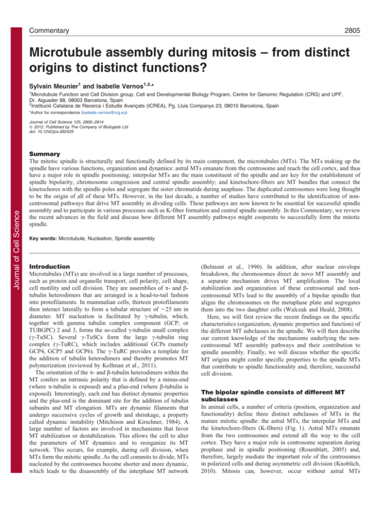
Commentary 2805 Microtubule assembly during mitosis – from distinct origins to distinct functions? Sylvain Meunier1 and Isabelle Vernos1,2,* 1 Microtubule Function and Cell Division group, Cell and Developmental Biology Program, Centre for Genomic Regulation (CRG) and UPF, Dr. Aiguader 88, 08003 Barcelona, Spain 2 Institució Catalana de Recerca i Estudis Avançats (ICREA), Pg. Lluis Companys 23, 08010 Barcelona, Spain *Author for correspondence (isabelle.vernos@crg.eu) Journal of Cell Science Journal of Cell Science 125, 2805–2814 ß 2012. Published by The Company of Biologists Ltd doi: 10.1242/jcs.092429 Summary The mitotic spindle is structurally and functionally defined by its main component, the microtubules (MTs). The MTs making up the spindle have various functions, organization and dynamics: astral MTs emanate from the centrosome and reach the cell cortex, and thus have a major role in spindle positioning; interpolar MTs are the main constituent of the spindle and are key for the establishment of spindle bipolarity, chromosome congression and central spindle assembly; and kinetochore-fibers are MT bundles that connect the kinetochores with the spindle poles and segregate the sister chromatids during anaphase. The duplicated centrosomes were long thought to be the origin of all of these MTs. However, in the last decade, a number of studies have contributed to the identification of noncentrosomal pathways that drive MT assembly in dividing cells. These pathways are now known to be essential for successful spindle assembly and to participate in various processes such as K-fiber formation and central spindle assembly. In this Commentary, we review the recent advances in the field and discuss how different MT assembly pathways might cooperate to successfully form the mitotic spindle. Key words: Microtubule, Nucleation, Spindle assembly Introduction Microtubules (MTs) are involved in a large number of processes, such as protein and organelle transport, cell polarity, cell shape, cell motility and cell division. They are assemblies of a- and btubulin heterodimers that are arranged in a head-to-tail fashion into protofilaments. In mammalian cells, thirteen protofilaments then interact laterally to form a tubular structure of ,25 nm in diameter. MT nucleation is facilitated by c-tubulin, which, together with gamma tubulin complex component (GCP; or TUBGPC) 2 and 3, forms the so-called c-tubulin small complex (c-TuSC). Several c-TuSCs form the large c-tubulin ring complex (c-TuRC), which includes additional GCPs (namely GCP4, GCP5 and GCP6). The c-TuRC provides a template for the addition of tubulin heterodimers and thereby promotes MT polymerization (reviewed by Kollman et al., 2011). The orientation of the a- and b-tubulin heterodimers within the MT confers an intrinsic polarity that is defined by a minus-end (where a-tubulin is exposed) and a plus-end (where b-tubulin is exposed). Interestingly, each end has distinct dynamic properties and the plus-end is the dominant site for the addition of tubulin subunits and MT elongation. MTs are dynamic filaments that undergo successive cycles of growth and shrinkage, a property called dynamic instability (Mitchison and Kirschner, 1984). A large number of factors are involved in mechanisms that favor MT stabilization or destabilization. This allows the cell to alter the parameters of MT dynamics and to reorganize its MT network. This occurs, for example, during cell division, when MTs form the mitotic spindle. As the cell commits to divide, MTs nucleated by the centrosomes become shorter and more dynamic, which leads to the disassembly of the interphase MT network (Belmont et al., 1990). In addition, after nuclear envelope breakdown, the chromosomes direct de novo MT assembly and a separate mechanism drives MT amplification. The local stabilization and organization of these centrosomal and noncentrosomal MTs lead to the assembly of a bipolar spindle that aligns the chromosomes on the metaphase plate and segregates them into the two daughter cells (Walczak and Heald, 2008). Here, we will first review the recent findings on the specific characteristics (organization, dynamic properties and function) of the different MT subclasses in the spindle. We will then describe our current knowledge of the mechanisms underlying the noncentrosomal MT assembly pathways and their contribution to spindle assembly. Finally, we will discuss whether the specific MT origins might confer specific properties to the spindle MTs that contribute to spindle functionality and, therefore, successful cell division. The bipolar spindle consists of different MT subclasses In animal cells, a number of criteria (position, organization and functionality) define three distinct subclasses of MTs in the mature mitotic spindle: the astral MTs, the interpolar MTs and the kinetochore-fibers (K-fibers) (Fig. 1). Astral MTs emanate from the two centrosomes and extend all the way to the cell cortex. They have a major role in centrosome separation during prophase and in spindle positioning (Rosenblatt, 2005) and, therefore, largely mediate the important role of the centrosomes in polarized cells and during asymmetric cell division (Knoblich, 2010). Mitosis can, however, occur without astral MTs 2806 Journal of Cell Science 125 (12) B + + + + A – + Interpolar MTs + t1/2= 30 seconds–1 minute C + + + + + + + + + + + – + K-fibers + + t1/2= 4–8 minutes D Journal of Cell Science + + + + – + Astral MTs t1/2= 30 seconds–1 minute (Khodjakov et al., 2000; Mahoney et al., 2006), and we will therefore focus on interpolar MTs and K-fibers. Interpolar MTs The interpolar MTs are the most dynamic class of spindle MTs and have an average half-life (t1/2) of less than 1 minute (Zhai et al., 1995). They are the most abundant class of MTs and comprise the main body of the mature spindle in many systems. Interpolar MTs emanate from the centrosome and extend towards the center of the spindle, where they interact in an antiparallel fashion with interpolar MTs emanating from the opposite spindle pole (Fig. 1). However, some interpolar MTs are shorter and do not extend half-way through the spindle (Mastronarde et al., 1993). In spindles assembled in Xenopus egg extracts, interpolar MTs are particularly abundant and comprise 95% of the spindle MTs (Ohi et al., 2007). They form a ‘polar’ array of parallel MTs near the spindle poles and a ‘barrel’ array of antiparallel, overlapping MTs close to the chromosomes (Yang et al., 2007). During anaphase, interpolar MTs are rearranged and become the major component of the central spindle, where they form an array of antiparallel MTs with bundled overlapping plus-ends (Glotzer, 2009). Interpolar MTs have multiple functions, including the establishment and maintenance of spindle bipolarity. In addition, they have a role in chromosome congression because they interact dynamically with the chromosomes through the chromokinesins [KID (also known as KIF22), kinesin family member (KIF) 4 and KLP2 (also known as KIF15) (Vanneste et al., 2011)] or through lateral interactions with the kinetochores (Cai et al., 2009; Magidson et al., 2011; Wignall and Villeneuve, 2009). However, the interpolar MTs are not directly responsible for chromosome segregation. This function is provided by the K-fibers. K-fibers K-fibers are bundles of 20–40 parallel MTs (McEwen et al., 1997; Rieder, 1981) whose main function is to attach the chromosomes to the two spindle poles and to segregate the sister Fig. 1. The three subclasses of spindle MTs. (A) Schematic metaphase spindle, showing its three different MT subclasses: interpolar MTs (orange), K-fibers (red) and astral MTs (green). The ‘+’ indicates the plus-ends of the MTs. (B–D) Representation of the three kinds of spindle MTs and their dynamic properties (see the text for more details). Astral MTs have dynamic properties that are similar to those of the interpolar MTs (Mitchison et al., 1986). Kfibers are less dynamic and undergo a constant polewards tubulin flux (arrows). chromatids into the daughter cells. K-fibers are much less dynamic than the interpolar MTs. Depending on the experimental system, their half-life is 4–8 minutes, which is closer to that of interphase MTs (t1/259–10 minutes) (Bakhoum et al., 2009; Zhai et al., 1995). Consistent with this, K-fibers are the last MT subclass to depolymerize following the exposure of cells to cold or nocodazole. The mechanism driving the assembly of the K-fibers is still not fully understood. The attachment of MTs to the kinetochore occurs progressively as the cell moves from prometaphase to anaphase, in a process called K-fiber maturation (McEwen et al., 1997). The characteristic bundling of the K-fiber MTs seems to be, in part, achieved by a protein complex that includes clathrin, TACC3 (for transforming, acidic coiled-coil containing protein 3, also known as maskin) and chTOG (also known as CKAP5 and, in Xenopus, XMAP215) (Booth et al., 2011). It is possible that some K-fiberassociated proteins that show MT-bundling activities in vitro might also be important players in K-fiber formation (Ribbeck et al., 2006; Silljé et al., 2006; Yokoyama et al., 2009). Further work is required to understand how MTs organize into the K-fiber bundles. The K-fiber plus-ends become attached to the outer kinetochore through the KMN complex (for KNL1–MIS12-complex–NDC80) network (Joglekar et al., 2010), which couples force generation to MT plus-end polymerization and depolymerization (Salmon et al., 2005). Erroneous K-fiber–kinetochore attachments are released for error correction through a mechanism involving the targeting and regulation of various kinetochore factors by the kinase Aurora B (Kelly and Funabiki, 2009). The mechanism controlling the polymerization at the K-fiber plus-ends is largely dependent on microtubule plus-end tracking proteins, including CLASP (for cytoplasmic linker associated protein, also known as orbit) (Maiato et al., 2005) and EB1 (also known as MAPRE1) (Tirnauer et al., 2002). The activities of kinesin-8 and -13 negatively regulate Kfiber plus-end stability (Joglekar et al., 2010; Manning et al., 2010). It has still not been fully resolved how the individual MT dynamics in the K-fibers result in directional kinetochore Microtubule assembly in mitosis (Khodjakov and Rieder, 1999). This process, called centrosome maturation, results in a dramatic increase in MT nucleation at the centrosomes during mitosis (Piehl et al., 2004). The presence of two centrosomes actively nucleating MTs from opposite sides of the nucleus in cells entering mitosis was central to the ‘search-and-capture’ model proposed by Kirschner and Mitchison in 1986 (Fig. 2). This model postulated that dynamic centrosomal MTs emanating from the two centrosomes grow and shrink, thereby exploring the cellular space until their plus-ends become captured by the paired sister kinetochores. This mechanism would naturally lead to the assembly of the bipolar spindle (Kirschner and Mitchison, 1986). In the past decade, both experimental and theoretical models have established that additional mechanisms are at work to help the search-andcapture process to connect all 92 kinetochores to the spindle poles in human cells (Magidson et al., 2011; Paul et al., 2009; Wollman et al., 2005). Early observations made in systems lacking centrosomes, such as plant cells (De Mey et al., 1982) and female germ cells undergoing meiosis in some animal species (Manandhar et al., 2005), have shown that acentrosomal cell divisions can also take place. More recently, different experimental approaches in cells containing centrosomes (Khodjakov et al., 2000; Mahoney et al., 2006), as well as in whole organisms (Basto et al., 2006), have lent additional support to the idea that spindle assembly and mitosis can occur in the absence of centrosomes (Fig. 2). This implies that additional non-centrosomal source(s) of MTs that drive spindle formation and chromosome segregation must exist in dividing cells, even when they naturally contain centrosomes. Karsenti and Kirschner observed in 1984 that the injection of bacteriophage lambda or E. coli DNA into Xenopus eggs is sufficient to trigger MT formation, thereby obtaining the first experimental evidence that chromatin has a role in MT assembly (Karsenti et al., 1984). The demonstration that DNA-coated beads incubated in Xenopus egg extract induce the assembly of a bipolar spindle provided further support to the idea that chromatin itself is sufficient to induce MT assembly and Journal of Cell Science movements that can abruptly switch from a polewards to an antipolewards direction (Skibbens et al., 1993; VandenBeldt et al., 2006). It has been shown, however, that the loss of tubulin subunits at the K-fiber minus-end occurs without spindle pole detachment and drives the polewards tubulin flux. This mechanism is sufficient for generating pulling forces at the kinetochores that stretch the centromeres in metaphase (Waters et al., 1996). In anaphase, dramatic changes in K-fiber dynamics at the plus-end, and also at the minus-end in some systems, promote the shortening of the K-fibers and chromosome segregation (Ganem and Compton, 2006; Kwok and Kapoor, 2007; Cheerambathur et al., 2007; Rogers et al., 2004). Various observations in mammalian and Drosophila cells suggest that the initial step of K-fiber formation occurs close to the kinetochores (Maiato et al., 2004; Tulu et al., 2006). A possible scenario is that the plus-ends of these MTs would be efficiently captured and stabilized by the kinetochore, and their minus-ends would connect with interpolar MTs before becoming focused at the spindle poles in a dynein- and nuclear mitotic apparatus protein 1 (NUMA1)-dependent manner (Khodjakov et al., 2003). These observations suggest that some MTs, and especially those forming the K-fibers, are not nucleated at the centrosomes. Moving towards non-centrosomal MT assembly pathways Since the initial description of the centrosome and its ‘radiating aster’ in the late 19th century (Paweletz, 2001), detailed structural studies have shown that centrosomes comprise two centrioles surrounded by pericentriolar material (PCM) (Chrétien et al., 1997; Ibrahim et al., 2009; Paintrand et al., 1992; Vorobjev and Chentsov, 1982). Centrosome biogenesis follows a cycle that is closely coordinated with the cell cycle, during which the single centrosome of the interphase cell duplicates (Bettencourt-Dias and Glover, 2007). At the onset of mitosis, the duplicated centrosomes recruit several components to the PCM, including ctubulin whose amount increases by more than three times First observation of MTs originating at the mitotic chromosomes (Witt et al., 1980) Mitotic spindle is a dynamic structure made of filaments called MTs (Inoue and Sato, 1967) 1967 1968 Identification of tubulin as the component of MTs (Mohri, 1968) 1984 Acentrosomal spindle assembly in mammalian cells (Khodjakov et al., 2000) DNA-coated beads induce spindle assembly (Heald et al., 1996) MT dynamic instability (Mitchison and Kirschner, 1984) 1980 Discovery of the Augmin pathway (Goshima et al., 2008) γ-Tubulin nucleates MTs (Wilde and Zheng, 1999) 1986 ‘Search-and-capture’ model (Kirschner and Mitchison, 1986) DNA triggers MT assembly (Karsenti and Kirschner, 1984) 2807 1996 1999 2000 Discovery of the Ran-GTP pathway (Carazo-Salas et al., 1999; Kalab et al., 1999; Zhang et al., 1999; Ohba et al., 1999) 2004 2008 Discovery of the CPC pathway (Sampath et al., 2004) Fig. 2. Timeline of the key discoveries on spindle formation. The timeline highlights the key findings that have led to the current model of spindle formation involving the different pathways of MT assembly discussed in the text. For previous historical discoveries, please see the excellent reviews by Paweletz (Paweletz, 2001) and Mitchison and Salmon (Mitchison and Salmon, 2001). 2808 Journal of Cell Science 125 (12) organization in M phase (Heald et al., 1996; Walczak et al., 1998). Furthermore, MT nucleation away from the centrosome has also been observed during mitosis in Drosophila and mammalian cells (Khodjakov et al., 2000; Mahoney et al., 2006; Tulu et al., 2006; Witt et al., 1980). Extensive studies over the past 10 years have provided mechanistic insights into the noncentrosomal MT assembly during cell division and have shown that two distinct chromosomal MT assembly pathways are involved: one is triggered by GTP-bound Ran and the other is controlled by the chromosomal passenger complex (CPC) (Fig. 2, timeline). Chromosomes as a source of MTs in dividing cells Journal of Cell Science The Ran-GTP pathway Ran is a small GTPase that drives nucleo-cytoplasmic transport during interphase, whereby the high concentration of the GTPbound form of Ran (Ran-GTP) in the nucleus allows the release of newly imported proteins from their binding to importins (Clarke and Zhang, 2008). In the late 1990s, several groups identified a mitosis-specific function for Ran in MT assembly. Their studies defined a new pathway that turned out to be essential for spindle assembly (Carazo-Salas et al., 2001; CarazoSalas et al., 1999; Kalab et al., 1999; Nachury et al., 2001; Ohba et al., 1999; Zhang et al., 1999). Spatially, the system is defined by the presence of the Ran guanine nucleotide exchange factor RCC1 (regulator of chromosome condensation 1) on chromatin (Karsenti and Vernos, 2001). This results in the formation of a Ran-GTP gradient that is centered around the chromosomes, which has been observed using fluorescence resonance energy transfer (FRET) approaches in Xenopus egg extract as well as in human somatic cells (Kaláb et al., 2006; Kalab et al., 2002). This Ran-GTP gradient ensures the local nucleation of MTs close to the chromatin and has a longer-range effect on MT stabilization (Caudron et al., 2005). The fine-tuned regulation of this system in space and time is still not fully understood and will require further investigation (Arnaoutov et al., 2005; Joseph et al., 2002; Torosantucci et al., 2008). The basic mechanism by which the Ran system functions both during interphase and in dividing cells is based on the differential affinity of the GDP- and GTP-bound forms of Ran for karyopherins (Clarke and Zhang, 2008). It has been postulated that, during cell division, Ran-GTP can trigger the release of putative spindle assembly factors (SAFs) from importins, thereby enabling the SAFs to perform their function in spindle assembly. The first protein that was shown to be regulated in this way is TPX2 (targeting protein for XKLP2) (Gruss et al., 2001; Wittmann et al., 2000). Once released from importin-a and importin-b, TPX2 triggers MT assembly around the chromatin and this activity is essential for spindle assembly (Gruss et al., 2001; Gruss et al., 2002; Tulu et al., 2006) (Fig. 3). Over the last decade, a number of Ran-GTP-regulated SAFs have been identified, and most of them have been shown to be involved in MT stabilization and/or organization (Table 1). In addition, other proteins that are not directly regulated by RanGTP, like ctubulin and ch-TOG (also known as CKAP5) (Groen et al., 2009; Wilde and Zheng, 1999), are also required for MT assembly through this pathway. However, the precise mechanism by which all these proteins promote MT assembly and organization is still not clearly understood. The CPC pathway Using a ‘mitosis with unreplicated genomes’ (MUG) system in which chromatin is, at least partially, excluded from the spindle, O’Connell and collaborators have shown that MT assembly also occurs at the kinetochores in a manner that is independent of chromatin, RCC1 and the Ran-GTP gradient (O’Connell et al., 2009). In fact, the molecular nature of this pathway has been described by the Funabiki group, and it is now known to involve the CPC, which comprises INCENP (inner centromere protein antigens 135/155 kDa), survivin (also known as BIRC5), borealin (also known as CDCA8) and the kinase Aurora B (Sampath et al., 2004; Tseng et al., 2010). The role of the CPC in MT stabilization and spindle assembly has been extensively characterized in Xenopus egg extracts (Kelly et al., 2007; Maresca et al., 2009; Sampath et al., 2004; Tseng et al., 2010). Beads coated with reconstituted CPC have been shown to promote MT assembly and the formation of bipolar spindles in Xenopus egg extract in the absence of Ran-GTP (Kelly et al., 2007; Maresca et al., 2009). The mechanism involves the local B A TPX2 CPC Ran-GTP αβ TPX2 RCC1 Ran-GDP C FAM29A HICE1 Fig. 3. MT assembly pathways. (A) Schematic prometaphase spindle, showing the three known sites of MT assembly during spindle assembly. MTs that originate from the centrosomes are in red, those originating from the chromosomes in orange and those emanating directly from pre-existing MTs are shown in green. (B,C) Representation of the main components of the two pathways of acentrosomal MT nucleation. (B) At the chromosomes, the presence of RCC1 bound to chromatin creates a Ran-GTP gradient around the chromosomes, allowing the release of SAFs such as TPX2 from importins (which inhibit SAF activity). TPX2, together with c-tubulin complexes (grey rings), then triggers MT nucleation. Chromosomal MTs are stabilized around the kinetochores by a mechanism that depends on the CPC (see text for details). (C) During MT amplification on pre-existing MTs, the Augmin complex (purple) is recruited to a preexisting MT (red), through its subunit HICE1. FAM29A is responsible for recruiting ctubulin (grey ring) to the complex and thereby triggering MT assembly. Microtubule assembly in mitosis 2809 Table 1. Ran-GTP-regulated SAFs Protein MT nucleation TPX2 MT stabilization HURP RAE1 ISWI1 CDK11 APC TACC3 MCRS1 Journal of Cell Science NUSAP1 Spindle organization NUMA1 Kid HSET Transport receptors Properties Mice models (effect of loss-of-function) Importin-a, importin-b (Gruss et al., 2001; Wittmann et al., 2000) Interacts with and activates Aurora A (Bayliss et al., 2003) Early embryonic lethality (AguirrePortolés et al., 2012) Importin-b (Silljé et al., 2006) MT-bundling activity (Koffa et al., 2006; Silljé et al., 2006; Wong and Fang, 2006) Ribonuncleoprotein complex involved in spindle assembly (Blower et al., 2005; Wong et al., 2006) MT-bundling activity (Yokoyama et al., 2009) Female infertility (Tsai et al., 2008) Importin-b (Blower et al., 2005; Wong et al., 2006) Importin-a, importin-b (Yokoyama et al., 2009) Importin-b (Yokoyama et al., 2008) Importin-b (Dikovskaya et al., 2010) Importin-b (Albee et al., 2006) Importin-b (Meunier and Vernos, 2011) Importin-a and/or importin-b, importin 7 (Ribbeck et al., 2006; Ribbeck et al., 2007) Importin-b (Nachury et al., 2001; Wiese et al., 2001) Importin-a, importin-b (Tahara et al., 2008) Importin-a, importin-b (Ems-McClung et al., 2004) Necessary for Ran-GTP-dependent MT stabilization(Yokoyama et al., 2008) MT-bundling activity (Dikovskaya et al., 2010) Phosphorylated by Aurora A (Barros et al., 2005; Peset et al., 2005) Binds and protects chromosomal MT minusends (Meunier and Vernos, 2011) Interacts with chromosomes and MTs (Ribbeck et al., 2006; Ribbeck et al., 2007) Pole focusing (Haren et al., 2009; Merdes et al., 2000; Merdes et al., 1996; Silk et al., 2009) Chromokinesin; additional MT-binding site (ATP independent) (Antonio et al., 2000; Funabiki and Murray, 2000; Tokai et al., 1996) Minus-end directed motor; additional MT-binding site (ATP independent) (Walczak et al., 1997) Early embryonic lethality (Babu et al., 2003) Early embryonic lethality (Stopka and Skoultchi, 2003) Early embryonic lethality (Li et al., 2004) Model of colorectal cancer (Oshima et al., 1996) Malformations of axial skeleton (Yao et al., 2007) n.d. Early embryonic lethality (Vanden Bosch et al., 2010) n.d. 50% embryonic lethality (Ohsugi et al., 2008) n.d. TPX2, the first Ran-GTP-regulated SAF to be identified, is essential for MT nucleation, some SAFs are involved in MT stabilization or bundling and others are more likely to be involved in spindle organization. n.d., not determined. activation of the kinase Aurora B (Sampath et al., 2004), which must be targeted to the MTs (through INCENP) in order to promote spindle assembly (Tseng et al., 2010). Taken together, these studies show that the CPC has a role in MT assembly that might be independent of the Ran-GTP pathway. It is not currently clear whether the CPC-dependent pathway is directly involved in MT nucleation or stabilization. There are no reports on the involvement of c-tubulin in the CPC-induced mechanism. However, Aurora B has been shown to negatively regulate the MT-catastrophe-promoting factors MCAK (for mitotic centromere-associated kinesin, also known as KIF2C) (Andrews et al., 2004; Lan et al., 2004; Ohi et al., 2004) and stathmin 1 (STMN1, also known as OP18) (Gadea and Ruderman, 2006). It is therefore probable that the CPC pathway promotes MT stabilization, rather than nucleation itself. The Ran-GTP- and CPC-dependent pathways probably work together, first nucleating MTs near chromatin and then stabilizing these nascent MTs preferentially near the kinetochores (Maresca et al., 2009; Needleman et al., 2010; Tulu et al., 2006) (Fig. 3). MT branching and amplification by the Augmin complex The observations reporting the localization of c-tubulin to spindle MTs (Khodjakov and Rieder, 1999; Lajoie-Mazenc et al., 1994) and the distribution of MT minus-ends throughout the whole spindle in Xenopus egg extracts (Burbank et al., 2006), could be explained by the transport of the MTs nucleated on chromosomes towards the poles. However, a mechanism of MT nucleation on preexisting MTs has been described in higher plants and fission yeast (Bratman and Chang, 2008), and the identification the Dgt (for dim c-tubulin) protein family (Goshima et al., 2007) has revealed a similar mechanism for MT amplification in the spindles of animal cells. These proteins define a complex called Augmin in Drosophila (Goshima et al., 2008) and HAUS (homologous to Augmin subunits) in human cells (Lawo et al., 2009; Uehara et al., 2009). The Augmin complex has been shown to recruit the c-TuRC to preexisting MTs and to promote MT nucleation, amplification or branching (Zhu et al., 2008; Zhu et al., 2009) (Fig. 3). The role of the different Augmin complex subunits has not been entirely resolved, but FAM29A (also known as Dgt6 and HAUS6) binds the c-TuRC, and another component, HICE1 (also known as HAUS8), associates with MTs (Tsai et al., 2011; Zhu et al., 2008). These two activities might account for targeting of the c-TuRC to a pre-existing MT. It is currently unknown whether this targeting can happen anywhere along a MT or whether there are some specific sites for MT branching and/or amplification. Moreover, it is also unknown whether all spindle MTs can act as docking sites with the same efficiency. The severity of the phenotypes observed when the function of this complex is impaired seem to be largely dependent on the system and more specifically on the presence or absence of centrosomes (Petry et al., 2011; Uehara et al., 2009; Wainman et al., 2009). In the absence of centrosomes, Augmin-dependent 2810 Journal of Cell Science 125 (12) Journal of Cell Science MT amplification is required, together with the chromosomal pathway, to build a bipolar spindle (Wainman et al., 2009). During anaphase, the Augmin complex is essential for central spindle assembly, for promoting MT nucleation and possibly for the very specific organization of the interpolar MTs composing the structure (Uehara and Goshima, 2010; Uehara et al., 2009). A unifying principle for MT nucleation in dividing cells A common requirement for the assembly of MTs through any of the three pathways – centrosomal, chromosomal and MT-dependent – is c-tubulin, which acts as a template for MT nucleation (Goshima et al., 2008; Mishra et al., 2010; Wilde and Zheng, 1999). The mitotic kinases cyclin-dependent kinase 1 (CDK1), polo-like kinase 1 (PLK1) and Aurora A have been shown to be involved in c-tubulin recruitment to the centrosome (Blagden and Glover, 2003). PLK1 and Aurora A activities are involved in c-TuRC recruitment by the Augmin complex (Johmura et al., 2011; Tsai et al., 2011) (Tables 2, 3). Although the mechanism of c-tubulin targeting in the chromosomal pathway remains poorly understood, Aurora A kinase is also required for MT nucleation and stabilization around chromatin (Bayliss et al., 2003; Sardon et al., 2008) (Table 3). The targeting of c-tubulin to each MT nucleation site is, at least in part, directed by the c-TuRCassociated protein NEDD1 (neural precursor cell expressed, developmentally down-regulated 1, also known as GCP7) (Lüders et al., 2006; Zhu et al., 2008) (Table 2). Interestingly, NEDD1 is hyperphosphorylated in mitosis, and its activity is differentially regulated by mitotic kinases at the centrosomes and in the acentrosomal MT amplification pathway (Johmura et al., 2011; Zhang et al., 2009). The selective targeting and activation of c-TuRCs by factors specific for each pathway might control MT assembly at different sites in the mitotic cell (Tables 2, 3). It is an interesting possibility that these different targeting complexes compete for a limited pool of c-TuSCs and c-TuRCs in the cell. The efficiency and/or regulation of c-tubulin targeting would therefore explain the relative contribution of the different MT assembly pathways, which varies dramatically depending on the organism and/or the cell type. In addition to the distinct mechanisms of c-tubulin targeting, the type of c-tubulin-containing complex involved in MT nucleation in each MT assembly pathway might be different. Although the c-TuRC is an active MT nucleator in higher eukaryotes, work in Drosophila has shown that the c-TuSC alone is essential and sufficient for MT nucleation at the centrosome. Indeed, none of the specific c-TuRC proteins (i.e. GCP4, GCP5 and GCP6) are required for c-tubulin recruitment to the centrosome or for MT nucleation in Drosophila cells (Vérollet et al., 2006) (Table 2). Interestingly, it has recently been shown that c-TuSCs are able to form a ring structure without the need of the other GCPs (Kollman et al., 2010). It is therefore tempting to speculate that the c-TuSCs could be sufficient to promote MT nucleation in metazoans, as it is the case in budding yeast (Kollman et al., 2011; Vinh et al., 2002). Taken together, these data suggest the possibility that different c-tubulin nucleation complexes might exist in the eukaryotic cell. If this is the case, it will be important to determine with precision which complexes function in the different MT assembly pathways and whether there are functional implications. Could different origins confer specificity to distinct spindle MTs? Having identified different means by which spindle MTs are assembled, the question remains as to what the relative contribution of the different MTs assembly pathways is to spindle assembly and function. The current view is that MTs that originate from the different pathways cooperate to form the spindle: the centrosome asters define the position of the two spindle poles and the axis of cell division; the chromosomal MTs facilitate the capture of the kinetochores and the formation of the K-fibers; and MT amplification by the Augmin complex contributes to the strengthening of the whole structure (Figs 1, 3). The combination of MTs from the three pathways could therefore allow the timely assembly of the bipolar spindle and allow mitosis to progress successfully. The recent identification of microspherule protein 1 (MCRS1) as a SAF (Meunier and Vernos, 2011) suggests that there is a possibility that the different MTs are not equivalent but differ in terms of dynamics and molecular composition. Table 2. MT nucleation complexes Protein Centrosomal pathway c-TuRC proteins c-tubulin (c-TuSC component) GCP2 (c-TuSC component) GCP3 (c-TuSC component) GCP4 GCP5 GCP6 3 3 3 7 (Vérollet et al., 2006) 7 (Vérollet et al., 2006) 7 (Vérollet et al., 2006) 3 ? ? ? ? ? 3 (Mishra et al., 2010) 3 (Mishra et al., 2010) 3 (Mishra et al., 2010) ? ? ? ? ? ? 7 7 7 7 7 7 3 (Lüders et al., 2006) 3 (Zhu et al., 2008) 3 (Lüders et al., 2006) c-TuRC-associated proteins MOZART1 (Hutchins et al., 2010) MOZART2A (Teixidó-Travesa et al., 2010) MOZART2B (Teixidó-Travesa et al., 2010) NEDD1 MT amplification pathway Chromosomal pathway The table shows the requirements of the components of the c-TuSC, c-TuRC and c-TuRC-associated proteins in the three MT assembly pathways (centrosomes, MTs and chromosomes). 7, not required for the assembly pathway; 3, required for the assembly pathway; ?, involvement not known. Microtubule assembly in mitosis 2811 Table 3. c-Tubulin-targeting mechanisms Centrosomes Targeting factors CEP192, ninein, centrosomin (Gomez-Ferreria et al., 2007; Raynaud-Messina and Merdes, 2007) Targeting regulators PLK1, Aurora A, CDK1 (Barr and Gergely, 2007) MT amplification Chromosomes Augmin complex, FAM29A (Bucciarelli et al., 2009) TPX2, RHAMM (also known as HMMR) (Groen et al., 2004) PLK1, Aurora A (Johmura et al., 2011; Tsai et al., 2011) Ran-GTP, Aurora A (Gruss et al., 2001; Tsai et al., 2003) Journal of Cell Science In the upper part of the table, the proteins involved specifically in c-tubulin targeting to the MT nucleation sites are indicated for each of the three pathways. In the lower part of the table, the main regulators of c-tubulin recruitment for each MT assembly pathway are shown. MCRS1 is a nuclear protein that associates specifically with the chromosomal Ran-GTP-dependent MTs. As such it is the first example of a protein showing specificity for this class of MTs. Interestingly, MCRS1 also shows full specificity for the minusends of the K-fibers. These data provide a molecular link between the chromosomal MTs and the K-fibers that goes beyond previous observations of the formation of acentrosomal MTs at the kinetochores (Khodjakov et al., 2003; Maiato et al., 2004). Interestingly, the distinct spindle MT subclasses have distinct dynamic properties, (Rizk et al., 2009), and functional studies have shown that MCRS1 could have a role in the regulation of Kfiber minus-end stability (Meunier and Vernos, 2011). The localization and function of MCRS1 therefore suggests that some specific proteins can be ‘loaded’ on the K-fibers by the Ran-GTP pathway to confer specific properties in terms of organization and dynamics to the K-fibers. Interestingly, other Ran-GTP-regulated proteins show activities consistent with this idea (Table 1). For instance, HURP (also known as DLGAP5), nucleolar and spindle associated protein 1 (NUSAP1) and ISWI (also known as SMRCA) have MT-bundling activity (Ribbeck et al., 2006; Silljé et al., 2006; Yokoyama et al., 2009) and preferentially bind to K-fibers (Silljé et al., 2006). Moreover, NUMA1 is involved in K-fiber minus-end transport towards the poles and is involved in the regulation of K-fiber stability (Haren et al., 2009; Khodjakov et al., 2003; Silk et al., 2009). TACC3 has also recently been shown to have a role in MT bridging in Kfibers (Booth et al., 2011; Cheeseman et al., 2011). Taken together, these data suggest a role for the Ran-GTP-dependent MT assembly pathway in promoting and regulating K-fiber formation. It is however important to note that the MTs nucleated by the chromosomal pathway are not all organized in K-fibers, as they can also form interpolar MTs. It is therefore probable that both the interaction of these MTs with the kinetochore and their plusend stabilization through the CPC also have a contribution in conferring specific properties to K-fibers. Perspectives Although three pathways for MT assembly in mitotic cells have been identified so far, it is possible that others exist. For instance, remnants of the nuclear envelope have been shown to function as MT nucleation sites in dividing Drosophila spermatocytes (Rebollo et al., 2004) but nothing is known about this process in mammalian cells. Golgi stacks have also been reported to efficiently promote MT assembly in interphase cells (Efimov et al., 2007; Rı́os et al., 2004; Rivero et al., 2009). Are these membrane-dependent mechanisms also involved in mitotic spindle assembly and how do they participate in successful cell division? Further studies will certainly clarify these questions and help us to obtain a global view of spindle assembly and function. As a result of decades of extensive research in the field of MT assembly during mitosis, we are now starting to reach a good understanding of spindle formation. However, an exciting challenge for the future will not only be to elucidate the exact role of each MT assembly pathway and the mechanism by which they work together to build the mitotic bipolar spindle but also to identify new putative pathways of MT assembly during mitosis. This is fundamental for our understanding of how, in only a few minutes, the cell is able to build a machinery as complex as the mitotic spindle. Acknowledgements We would like to thank M. Mendoza, R. Pinyol, T. Cavazza, M. Schütz and J. Scrofani for critical reading of the manuscript, and all the members of the Vernos lab for helpful discussions. We apologize to authors whose works could not be cited owing to space limitation. We thank the reviewers, whose comments greatly improved the manuscript. Funding The work of our laboratory is supported by grants from the Spanish Ministerio de Educación y Ciencia; the Ministerio de Ciencia e Innovaciœn [grant numbers BFU2006-04694, BFU2009-10202, CSD2006-00023]; and institutional funds. References Aguirre-Portolés, C., Bird, A. W., Hyman, A., Cañamero, M., Pérez de Castro, I. and Malumbres, M. (2012). Tpx2 controls spindle integrity, genome stability, and tumor development. Cancer Res. 72, 1518-1528. Albee, A. J., Tao, W. and Wiese, C. (2006). Phosphorylation of maskin by Aurora-A is regulated by RanGTP and importin beta. J. Biol. Chem. 281, 38293-38301. Andrews, P. D., Ovechkina, Y., Morrice, N., Wagenbach, M., Duncan, K., Wordeman, L. and Swedlow, J. R. (2004). Aurora B regulates MCAK at the mitotic centromere. Dev. Cell 6, 253-268. Antonio, C., Ferby, I., Wilhelm, H., Jones, M., Karsenti, E., Nebreda, A. R. and Vernos, I. (2000). Xkid, a chromokinesin required for chromosome alignment on the metaphase plate. Cell 102, 425-435. Arnaoutov, A., Azuma, Y., Ribbeck, K., Joseph, J., Boyarchuk, Y., Karpova, T., McNally, J. and Dasso, M. (2005). Crm1 is a mitotic effector of Ran-GTP in somatic cells. Nat. Cell Biol. 7, 626-632. Babu, J. R., Jeganathan, K. B., Baker, D. J., Wu, X., Kang-Decker, N. and van Deursen, J. M. (2003). Rae1 is an essential mitotic checkpoint regulator that cooperates with Bub3 to prevent chromosome missegregation. J. Cell Biol. 160, 341353. Bakhoum, S. F., Thompson, S. L., Manning, A. L. and Compton, D. A. (2009). Genome stability is ensured by temporal control of kinetochore-microtubule dynamics. Nat. Cell Biol. 11, 27-35. Barr, A. R. and Gergely, F. (2007). Aurora-A: the maker and breaker of spindle poles. J. Cell Sci. 120, 2987-2996. Barros, T. P., Kinoshita, K., Hyman, A. A. and Raff, J. W. (2005). Aurora A activates D-TACC-Msps complexes exclusively at centrosomes to stabilize centrosomal microtubules. J. Cell Biol. 170, 1039-1046. Journal of Cell Science 2812 Journal of Cell Science 125 (12) Basto, R., Lau, J., Vinogradova, T., Gardiol, A., Woods, C. G., Khodjakov, A. and Raff, J. W. (2006). Flies without centrioles. Cell 125, 1375-1386. Bayliss, R., Sardon, T., Vernos, I. and Conti, E. (2003). Structural basis of Aurora-A activation by TPX2 at the mitotic spindle. Mol. Cell 12, 851-862. Belmont, L. D., Hyman, A. A., Sawin, K. E. and Mitchison, T. J. (1990). Real-time visualization of cell cycle-dependent changes in microtubule dynamics in cytoplasmic extracts. Cell 62, 579-589. Bettencourt-Dias, M. and Glover, D. M. (2007). Centrosome biogenesis and function: centrosomics brings new understanding. Nat. Rev. Mol. Cell Biol. 8, 451-463. Blagden, S. P. and Glover, D. M. (2003). Polar expeditions – provisioning the centrosome for mitosis. Nat. Cell Biol. 5, 505-511. Blower, M. D., Nachury, M., Heald, R. and Weis, K. (2005). A Rae1-containing ribonucleoprotein complex is required for mitotic spindle assembly. Cell 121, 223234. Booth, D. G., Hood, F. E., Prior, I. A. and Royle, S. J. (2011). A TACC3/ch-TOG/ clathrin complex stabilises kinetochore fibres by inter-microtubule bridging. EMBO J. 30, 906-919. Bratman, S. V. and Chang, F. (2008). Mechanisms for maintaining microtubule bundles. Trends Cell Biol. 18, 580-586. Bucciarelli, E., Pellacani, C., Naim, V., Palena, A., Gatti, M. and Somma, M. P. (2009). Drosophila Dgt6 interacts with Ndc80, Msps/XMAP215, and gamma-tubulin to promote kinetochore-driven MT formation. Curr. Biol. 19, 1839-1845. Burbank, K. S., Groen, A. C., Perlman, Z. E., Fisher, D. S. and Mitchison, T. J. (2006). A new method reveals microtubule minus ends throughout the meiotic spindle. J. Cell Biol. 175, 369-375. Cai, S., O’Connell, C. B., Khodjakov, A. and Walczak, C. E. (2009). Chromosome congression in the absence of kinetochore fibres. Nat. Cell Biol. 11, 832-838. Carazo-Salas, R. E., Guarguaglini, G., Gruss, O. J., Segref, A., Karsenti, E. and Mattaj, I. W. (1999). Generation of GTP-bound Ran by RCC1 is required for chromatin-induced mitotic spindle formation. Nature 400, 178-181. Carazo-Salas, R. E., Gruss, O. J., Mattaj, I. W. and Karsenti, E. (2001). Ran-GTP coordinates regulation of microtubule nucleation and dynamics during mitotic-spindle assembly. Nat. Cell Biol. 3, 228-234. Caudron, M., Bunt, G., Bastiaens, P. and Karsenti, E. (2005). Spatial coordination of spindle assembly by chromosome-mediated signaling gradients. Science 309, 13731376. Cheerambathur, D. K., Civelekoglu-Scholey, G., Brust-Mascher, I., Sommi, P., Mogilner, A. and Scholey, J. M. (2007). Quantitative analysis of an anaphase B switch: predicted role for a microtubule catastrophe gradient. J. Cell Biol. 177, 9951004. Cheeseman, L. P., Booth, D. G., Hood, F. E., Prior, I. A. and Royle, S. J. (2011). Aurora A kinase activity is required for localization of TACC3/ch-TOG/clathrin intermicrotubule bridges. Commun. Integr. Biol. 4, 409-412. Chrétien, D., Buendia, B., Fuller, S. D. and Karsenti, E. (1997). Reconstruction of the centrosome cycle from cryoelectron micrographs. J. Struct. Biol. 120, 117-133. Clarke, P. R. and Zhang, C. (2008). Spatial and temporal coordination of mitosis by Ran GTPase. Nat. Rev. Mol. Cell Biol. 9, 464-477. De Mey, J., Lambert, A. M., Bajer, A. S., Moeremans, M. and De Brabander, M. (1982). Visualization of microtubules in interphase and mitotic plant cells of Haemanthus endosperm with the immuno-gold staining method. Proc. Natl. Acad. Sci. USA 79, 1898-1902. Dikovskaya, D., Li, Z., Newton, I. P., Davidson, I., Hutchins, J. R., Kalab, P., Clarke, P. R. and Näthke, I. S. (2010). Microtubule assembly by the Apc protein is regulated by importin-beta–RanGTP. J. Cell Sci. 123, 736-746. Efimov, A., Kharitonov, A., Efimova, N., Loncarek, J., Miller, P. M., Andreyeva, N., Gleeson, P., Galjart, N., Maia, A. R., McLeod, I. X. et al. (2007). Asymmetric CLASP-dependent nucleation of noncentrosomal microtubules at the trans-Golgi network. Dev. Cell 12, 917-930. Ems-McClung, S. C., Zheng, Y. and Walczak, C. E. (2004). Importin alpha/beta and Ran-GTP regulate XCTK2 microtubule binding through a bipartite nuclear localization signal. Mol. Biol. Cell 15, 46-57. Funabiki, H. and Murray, A. W. (2000). The Xenopus chromokinesin Xkid is essential for metaphase chromosome alignment and must be degraded to allow anaphase chromosome movement. Cell 102, 411-424. Gadea, B. B. and Ruderman, J. V. (2006). Aurora B is required for mitotic chromatininduced phosphorylation of Op18/Stathmin. Proc. Natl. Acad. Sci. USA 103, 44934498. Ganem, N. J. and Compton, D. A. (2006). Functional roles of poleward microtubule flux during mitosis. Cell Cycle 5, 481-485. Glotzer, M. (2009). The 3Ms of central spindle assembly: microtubules, motors and MAPs. Nat. Rev. Mol. Cell Biol. 10, 9-20. Gomez-Ferreria, M. A., Rath, U., Buster, D. W., Chanda, S. K., Caldwell, J. S., Rines, D. R. and Sharp, D. J. (2007). Human Cep192 is required for mitotic centrosome and spindle assembly. Curr. Biol. 17, 1960-1966. Goshima, G., Wollman, R., Goodwin, S. S., Zhang, N., Scholey, J. M., Vale, R. D. and Stuurman, N. (2007). Genes required for mitotic spindle assembly in Drosophila S2 cells. Science 316, 417-421. Goshima, G., Mayer, M., Zhang, N., Stuurman, N. and Vale, R. D. (2008). Augmin: a protein complex required for centrosome-independent microtubule generation within the spindle. J. Cell Biol. 181, 421-429. Groen, A. C., Cameron, L. A., Coughlin, M., Miyamoto, D. T., Mitchison, T. J. and Ohi, R. (2004). XRHAMM functions in ran-dependent microtubule nucleation and pole formation during anastral spindle assembly. Curr. Biol. 14, 1801-1811. Groen, A. C., Maresca, T. J., Gatlin, J. C., Salmon, E. D. and Mitchison, T. J. (2009). Functional overlap of microtubule assembly factors in chromatin-promoted spindle assembly. Mol. Biol. Cell 20, 2766-2773. Gruss, O. J., Carazo-Salas, R. E., Schatz, C. A., Guarguaglini, G., Kast, J., Wilm, M., Le Bot, N., Vernos, I., Karsenti, E. and Mattaj, I. W. (2001). Ran induces spindle assembly by reversing the inhibitory effect of importin alpha on TPX2 activity. Cell 104, 83-93. Gruss, O. J., Wittmann, M., Yokoyama, H., Pepperkok, R., Kufer, T., Silljé, H., Karsenti, E., Mattaj, I. W. and Vernos, I. (2002). Chromosome-induced microtubule assembly mediated by TPX2 is required for spindle formation in HeLa cells. Nat. Cell Biol. 4, 871-879. Haren, L., Gnadt, N., Wright, M. and Merdes, A. (2009). NuMA is required for proper spindle assembly and chromosome alignment in prometaphase. BMC Res. Notes 2, 64. Heald, R., Tournebize, R., Blank, T., Sandaltzopoulos, R., Becker, P., Hyman, A. and Karsenti, E. (1996). Self-organization of microtubules into bipolar spindles around artificial chromosomes in Xenopus egg extracts. Nature 382, 420-425. Hutchins, J. R., Toyoda, Y., Hegemann, B., Poser, I., Hériché, J. K., Sykora, M. M., Augsburg, M., Hudecz, O., Buschhorn, B. A., Bulkescher, J. et al. (2010). Systematic analysis of human protein complexes identifies chromosome segregation proteins. Science 328, 593-599. Ibrahim, R., Messaoudi, C., Chichon, F. J., Celati, C. and Marco, S. (2009). Electron tomography study of isolated human centrioles. Microsc. Res. Tech. 72, 42-48. Joglekar, A. P., Bloom, K. S. and Salmon, E. D. (2010). Mechanisms of force generation by end-on kinetochore-microtubule attachments. Curr. Opin. Cell Biol. 22, 57-67. Johmura, Y., Soung, N. K., Park, J. E., Yu, L. R., Zhou, M., Bang, J. K., Kim, B. Y., Veenstra, T. D., Erikson, R. L. and Lee, K. S. (2011). Regulation of microtubulebased microtubule nucleation by mammalian polo-like kinase 1. Proc. Natl. Acad. Sci. USA 108, 11446-11451. Joseph, J., Tan, S. H., Karpova, T. S., McNally, J. G. and Dasso, M. (2002). SUMO-1 targets RanGAP1 to kinetochores and mitotic spindles. J. Cell Biol. 156, 595-602. Kalab, P., Pu, R. T. and Dasso, M. (1999). The Ran GTPase regulates mitotic spindle assembly. Curr. Biol. 9, 481-484. Kalab, P., Weis, K. and Heald, R. (2002). Visualization of a Ran-GTP gradient in interphase and mitotic Xenopus egg extracts. Science 295, 2452-2456. Kaláb, P., Pralle, A., Isacoff, E. Y., Heald, R. and Weis, K. (2006). Analysis of a RanGTP-regulated gradient in mitotic somatic cells. Nature 440, 697-701. Karsenti, E. and Vernos, I. (2001). The mitotic spindle: a self-made machine. Science 294, 543-547. Karsenti, E., Newport, J. and Kirschner, M. (1984). Respective roles of centrosomes and chromatin in the conversion of microtubule arrays from interphase to metaphase. J. Cell Biol. 99 Suppl, 47-54. Kelly, A. E. and Funabiki, H. (2009). Correcting aberrant kinetochore microtubule attachments: an Aurora B-centric view. Curr. Opin. Cell Biol. 21, 51-58. Kelly, A. E., Sampath, S. C., Maniar, T. A., Woo, E. M., Chait, B. T. and Funabiki, H. (2007). Chromosomal enrichment and activation of the aurora B pathway are coupled to spatially regulate spindle assembly. Dev. Cell 12, 31-43. Khodjakov, A. and Rieder, C. L. (1999). The sudden recruitment of gamma-tubulin to the centrosome at the onset of mitosis and its dynamic exchange throughout the cell cycle, do not require microtubules. J. Cell Biol. 146, 585-596. Khodjakov, A., Cole, R. W., Oakley, B. R. and Rieder, C. L. (2000). Centrosomeindependent mitotic spindle formation in vertebrates. Curr. Biol. 10, 59-67. Khodjakov, A., Copenagle, L., Gordon, M. B., Compton, D. A. and Kapoor, T. M. (2003). Minus-end capture of preformed kinetochore fibers contributes to spindle morphogenesis. J. Cell Biol. 160, 671-683. Kirschner, M. and Mitchison, T. (1986). Beyond self-assembly: from microtubules to morphogenesis. Cell 45, 329-342. Knoblich, J. A. (2010). Asymmetric cell division: recent developments and their implications for tumour biology. Nat. Rev. Mol. Cell Biol. 11, 849-860. Koffa, M. D., Casanova, C. M., Santarella, R., Köcher, T., Wilm, M. and Mattaj, I. W. (2006). HURP is part of a Ran-dependent complex involved in spindle formation. Curr. Biol. 16, 743-754. Kollman, J. M., Polka, J. K., Zelter, A., Davis, T. N. and Agard, D. A. (2010). Microtubule nucleating gamma-TuSC assembles structures with 13-fold microtubulelike symmetry. Nature 466, 879-882. Kollman, J. M., Merdes, A., Mourey, L. and Agard, D. A. (2011). Microtubule nucleation by c-tubulin complexes. Nat. Rev. Mol. Cell Biol. 12, 709-721. Kwok, B. H. and Kapoor, T. M. (2007). Microtubule flux: drivers wanted. Curr. Opin. Cell Biol. 19, 36-42. Lajoie-Mazenc, I., Tollon, Y., Detraves, C., Julian, M., Moisand, A., GuethHallonet, C., Debec, A., Salles-Passador, I., Puget, A., Mazarguil, H. et al. (1994). Recruitment of antigenic gamma-tubulin during mitosis in animal cells: presence of gamma-tubulin in the mitotic spindle. J. Cell Sci. 107, 2825-2837. Lan, W., Zhang, X., Kline-Smith, S. L., Rosasco, S. E., Barrett-Wilt, G. A., Shabanowitz, J., Hunt, D. F., Walczak, C. E. and Stukenberg, P. T. (2004). Aurora B phosphorylates centromeric MCAK and regulates its localization and microtubule depolymerization activity. Curr. Biol. 14, 273-286. Lawo, S., Bashkurov, M., Mullin, M., Ferreria, M. G., Kittler, R., Habermann, B., Tagliaferro, A., Poser, I., Hutchins, J. R., Hegemann, B. et al. (2009). HAUS, the 8-subunit human Augmin complex, regulates centrosome and spindle integrity. Curr. Biol. 19, 816-826. Journal of Cell Science Microtubule assembly in mitosis Li, T., Inoue, A., Lahti, J. M. and Kidd, V. J. (2004). Failure to proliferate and mitotic arrest of CDK11(p110/p58)-null mutant mice at the blastocyst stage of embryonic cell development. Mol. Cell. Biol. 24, 3188-3197. Lüders, J., Patel, U. K. and Stearns, T. (2006). GCP-WD is a gamma-tubulin targeting factor required for centrosomal and chromatin-mediated microtubule nucleation. Nat. Cell Biol. 8, 137-147. Magidson, V., O’Connell, C. B., Lončarek, J., Paul, R., Mogilner, A. and Khodjakov, A. (2011). The spatial arrangement of chromosomes during prometaphase facilitates spindle assembly. Cell 146, 555-567. Mahoney, N. M., Goshima, G., Douglass, A. D. and Vale, R. D. (2006). Making microtubules and mitotic spindles in cells without functional centrosomes. Curr. Biol. 16, 564-569. Maiato, H., Rieder, C. L. and Khodjakov, A. (2004). Kinetochore-driven formation of kinetochore fibers contributes to spindle assembly during animal mitosis. J. Cell Biol. 167, 831-840. Maiato, H., Khodjakov, A. and Rieder, C. L. (2005). Drosophila CLASP is required for the incorporation of microtubule subunits into fluxing kinetochore fibres. Nat. Cell Biol. 7, 42-47. Manandhar, G., Schatten, H. and Sutovsky, P. (2005). Centrosome reduction during gametogenesis and its significance. Biol. Reprod. 72, 2-13. Manning, A. L., Bakhoum, S. F., Maffini, S., Correia-Melo, C., Maiato, H. and Compton, D. A. (2010). CLASP1, astrin and Kif2b form a molecular switch that regulates kinetochore-microtubule dynamics to promote mitotic progression and fidelity. EMBO J. 29, 3531-3543. Maresca, T. J., Groen, A. C., Gatlin, J. C., Ohi, R., Mitchison, T. J. and Salmon, E. D. (2009). Spindle assembly in the absence of a RanGTP gradient requires localized CPC activity. Curr. Biol. 19, 1210-1215. Mastronarde, D. N., McDonald, K. L., Ding, R. and McIntosh, J. R. (1993). Interpolar spindle microtubules in PTK cells. J. Cell Biol. 123, 1475-1489. McEwen, B. F., Heagle, A. B., Cassels, G. O., Buttle, K. F. and Rieder, C. L. (1997). Kinetochore fiber maturation in PtK1 cells and its implications for the mechanisms of chromosome congression and anaphase onset. J. Cell Biol. 137, 1567-1580. Merdes, A., Ramyar, K., Vechio, J. D. and Cleveland, D. W. (1996). A complex of NuMA and cytoplasmic dynein is essential for mitotic spindle assembly. Cell 87, 447458. Merdes, A., Heald, R., Samejima, K., Earnshaw, W. C. and Cleveland, D. W. (2000). Formation of spindle poles by dynein/dynactin-dependent transport of NuMA. J. Cell Biol. 149, 851-862. Meunier, S. and Vernos, I. (2011). K-fibre minus ends are stabilized by a RanGTPdependent mechanism essential for functional spindle assembly. Nat. Cell Biol. 13, 1406-1414. Mishra, R. K., Chakraborty, P., Arnaoutov, A., Fontoura, B. M. and Dasso, M. (2010). The Nup107-160 complex and gamma-TuRC regulate microtubule polymerization at kinetochores. Nat. Cell Biol. 12, 164-169. Mitchison, T. and Kirschner, M. (1984). Dynamic instability of microtubule growth. Nature 312, 237-242. Mitchison, T. J. and Salmon, E. D. (2001). Mitosis: a history of division. Nat. Cell Biol. 3, E17-E21. Mitchison, T., Evans, L., Schulze, E. and Kirschner, M. (1986). Sites of microtubule assembly and disassembly in the mitotic spindle. Cell 45, 515-527. Nachury, M. V., Maresca, T. J., Salmon, W. C., Waterman-Storer, C. M., Heald, R. and Weis, K. (2001). Importin beta is a mitotic target of the small GTPase Ran in spindle assembly. Cell 104, 95-106. Needleman, D. J., Groen, A., Ohi, R., Maresca, T., Mirny, L. and Mitchison, T. (2010). Fast microtubule dynamics in meiotic spindles measured by single molecule imaging: evidence that the spindle environment does not stabilize microtubules. Mol. Biol. Cell 21, 323-333. O’Connell, C. B., Loncarek, J., Kaláb, P. and Khodjakov, A. (2009). Relative contributions of chromatin and kinetochores to mitotic spindle assembly. J. Cell Biol. 187, 43-51. Ohba, T., Nakamura, M., Nishitani, H. and Nishimoto, T. (1999). Self-organization of microtubule asters induced in Xenopus egg extracts by GTP-bound Ran. Science 284, 1356-1358. Ohi, R., Sapra, T., Howard, J. and Mitchison, T. J. (2004). Differentiation of cytoplasmic and meiotic spindle assembly MCAK functions by Aurora B-dependent phosphorylation. Mol. Biol. Cell 15, 2895-2906. Ohi, R., Burbank, K., Liu, Q. and Mitchison, T. J. (2007). Nonredundant functions of Kinesin-13s during meiotic spindle assembly. Curr. Biol. 17, 953-959. Ohsugi, M., Adachi, K., Horai, R., Kakuta, S., Sudo, K., Kotaki, H., TokaiNishizumi, N., Sagara, H., Iwakura, Y. and Yamamoto, T. (2008). Kid-mediated chromosome compaction ensures proper nuclear envelope formation. Cell 132, 771782. Oshima, M., Dinchuk, J. E., Kargman, S. L., Oshima, H., Hancock, B., Kwong, E., Trzaskos, J. M., Evans, J. F. and Taketo, M. M. (1996). Suppression of intestinal polyposis in Apc delta716 knockout mice by inhibition of cyclooxygenase 2 (COX-2). Cell 87, 803-809. Paintrand, M., Moudjou, M., Delacroix, H. and Bornens, M. (1992). Centrosome organization and centriole architecture: their sensitivity to divalent cations. J. Struct. Biol. 108, 107-128. Paul, R., Wollman, R., Silkworth, W. T., Nardi, I. K., Cimini, D. and Mogilner, A. (2009). Computer simulations predict that chromosome movements and rotations accelerate mitotic spindle assembly without compromising accuracy. Proc. Natl. Acad. Sci. USA 106, 15708-15713. 2813 Paweletz, N. (2001). Walther Flemming: pioneer of mitosis research. Nat. Rev. Mol. Cell Biol. 2, 72-75. Peset, I., Seiler, J., Sardon, T., Bejarano, L. A., Rybina, S. and Vernos, I. (2005). Function and regulation of Maskin, a TACC family protein, in microtubule growth during mitosis. J. Cell Biol. 170, 1057-1066. Petry, S., Pugieux, C., Nédélec, F. J. and Vale, R. D. (2011). Augmin promotes meiotic spindle formation and bipolarity in Xenopus egg extracts. Proc. Natl. Acad. Sci. USA 108, 14473-14478. Piehl, M., Tulu, U. S., Wadsworth, P. and Cassimeris, L. (2004). Centrosome maturation: measurement of microtubule nucleation throughout the cell cycle by using GFP-tagged EB1. Proc. Natl. Acad. Sci. USA 101, 1584-1588. Raynaud-Messina, B. and Merdes, A. (2007). Gamma-tubulin complexes and microtubule organization. Curr. Opin. Cell Biol. 19, 24-30. Rebollo, E., Llamazares, S., Reina, J. and Gonzalez, C. (2004). Contribution of noncentrosomal microtubules to spindle assembly in Drosophila spermatocytes. PLoS Biol. 2, e8. Ribbeck, K., Groen, A. C., Santarella, R., Bohnsack, M. T., Raemaekers, T., Köcher, T., Gentzel, M., Görlich, D., Wilm, M., Carmeliet, G. et al. (2006). NuSAP, a mitotic RanGTP target that stabilizes and cross-links microtubules. Mol. Biol. Cell 17, 2646-2660. Ribbeck, K., Raemaekers, T., Carmeliet, G. and Mattaj, I. W. (2007). A role for NuSAP in linking microtubules to mitotic chromosomes. Curr. Biol. 17, 230-236. Rieder, C. L. (1981). The structure of the cold-stable kinetochore fiber in metaphase PtK1 cells. Chromosoma 84, 145-158. Rı́os, R. M., Sanchı́s, A., Tassin, A. M., Fedriani, C. and Bornens, M. (2004). GMAP210 recruits gamma-tubulin complexes to cis-Golgi membranes and is required for Golgi ribbon formation. Cell 118, 323-335. Rivero, S., Cardenas, J., Bornens, M. and Rios, R. M. (2009). Microtubule nucleation at the cis-side of the Golgi apparatus requires AKAP450 and GM130. EMBO J. 28, 1016-1028. Rizk, R. S., Bohannon, K. P., Wetzel, L. A., Powers, J., Shaw, S. L. and Walczak, C. E. (2009). MCAK and paclitaxel have differential effects on spindle microtubule organization and dynamics. Mol. Biol. Cell 20, 1639-1651. Rogers, G. C., Rogers, S. L., Schwimmer, T. A., Ems-McClung, S. C., Walczak, C. E., Vale, R. D., Scholey, J. M. and Sharp, D. J. (2004). Two mitotic kinesins cooperate to drive sister chromatid separation during anaphase. Nature 427, 364-370. Rosenblatt, J. (2005). Spindle assembly: asters part their separate ways. Nat. Cell Biol. 7, 219-222. Salmon, E. D., Cimini, D., Cameron, L. A. and DeLuca, J. G. (2005). Merotelic kinetochores in mammalian tissue cells. Philos. Trans. R. Soc. Lond. B Biol. Sci. 360, 553-568. Sampath, S. C., Ohi, R., Leismann, O., Salic, A., Pozniakovski, A. and Funabiki, H. (2004). The chromosomal passenger complex is required for chromatin-induced microtubule stabilization and spindle assembly. Cell 118, 187-202. Sardon, T., Peset, I., Petrova, B. and Vernos, I. (2008). Dissecting the role of Aurora A during spindle assembly. EMBO J. 27, 2567-2579. Silk, A. D., Holland, A. J. and Cleveland, D. W. (2009). Requirements for NuMA in maintenance and establishment of mammalian spindle poles. J. Cell Biol. 184, 677690. Silljé, H. H., Nagel, S., Körner, R. and Nigg, E. A. (2006). HURP is a Ran-importin beta-regulated protein that stabilizes kinetochore microtubules in the vicinity of chromosomes. Curr. Biol. 16, 731-742. Skibbens, R. V., Skeen, V. P. and Salmon, E. D. (1993). Directional instability of kinetochore motility during chromosome congression and segregation in mitotic newt lung cells: a push-pull mechanism. J. Cell Biol. 122, 859-875. Stopka, T. and Skoultchi, A. I. (2003). The ISWI ATPase Snf2h is required for early mouse development. Proc. Natl. Acad. Sci. USA 100, 14097-14102. Tahara, K., Takagi, M., Ohsugi, M., Sone, T., Nishiumi, F., Maeshima, K., Horiuchi, Y., Tokai-Nishizumi, N., Imamoto, F., Yamamoto, T. et al. (2008). Importin-beta and the small guanosine triphosphatase Ran mediate chromosome loading of the human chromokinesin Kid. J. Cell Biol. 180, 493-506. Teixidó-Travesa, N., Villén, J., Lacasa, C., Bertran, M. T., Archinti, M., Gygi, S. P., Caelles, C., Roig, J. and Lüders, J. (2010). The gammaTuRC revisited: a comparative analysis of interphase and mitotic human gammaTuRC redefines the set of core components and identifies the novel subunit GCP8. Mol. Biol. Cell 21, 39633972. Tirnauer, J. S., Canman, J. C., Salmon, E. D. and Mitchison, T. J. (2002). EB1 targets to kinetochores with attached, polymerizing microtubules. Mol. Biol. Cell 13, 4308-4316. Tokai, N., Fujimoto-Nishiyama, A., Toyoshima, Y., Yonemura, S., Tsukita, S., Inoue, J. and Yamamota, T. (1996). Kid, a novel kinesin-like DNA binding protein, is localized to chromosomes and the mitotic spindle. EMBO J. 15, 457-467. Torosantucci, L., De Luca, M., Guarguaglini, G., Lavia, P. and Degrassi, F. (2008). Localized RanGTP accumulation promotes microtubule nucleation at kinetochores in somatic mammalian cells. Mol. Biol. Cell 19, 1873-1882. Tsai, M. Y., Wiese, C., Cao, K., Martin, O., Donovan, P., Ruderman, J., Prigent, C. and Zheng, Y. (2003). A Ran signalling pathway mediated by the mitotic kinase Aurora A in spindle assembly. Nat. Cell Biol. 5, 242-248. Tsai, C. Y., Chou, C. K., Yang, C. W., Lai, Y. C., Liang, C. C., Chen, C. M. and Tsai, T. F. (2008). Hurp deficiency in mice leads to female infertility caused by an implantation defect. J. Biol. Chem. 283, 26302-26306. Tsai, C. Y., Ngo, B., Tapadia, A., Hsu, P. H., Wu, G. and Lee, W. H. (2011). AuroraA phosphorylates Augmin complex component Hice1 protein at an N-terminal serine/ Journal of Cell Science 2814 Journal of Cell Science 125 (12) threonine cluster to modulate its microtubule binding activity during spindle assembly. J. Biol. Chem. 286, 30097-30106. Tseng, B. S., Tan, L., Kapoor, T. M. and Funabiki, H. (2010). Dual detection of chromosomes and microtubules by the chromosomal passenger complex drives spindle assembly. Dev. Cell 18, 903-912. Tulu, U. S., Fagerstrom, C., Ferenz, N. P. and Wadsworth, P. (2006). Molecular requirements for kinetochore-associated microtubule formation in mammalian cells. Curr. Biol. 16, 536-541. Uehara, R. and Goshima, G. (2010). Functional central spindle assembly requires de novo microtubule generation in the interchromosomal region during anaphase. J. Cell Biol. 191, 259-267. Uehara, R., Nozawa, R. S., Tomioka, A., Petry, S., Vale, R. D., Obuse, C. and Goshima, G. (2009). The augmin complex plays a critical role in spindle microtubule generation for mitotic progression and cytokinesis in human cells. Proc. Natl. Acad. Sci. USA 106, 6998-7003. Vanden Bosch, A., Raemaekers, T., Denayer, S., Torrekens, S., Smets, N., Moermans, K., Dewerchin, M., Carmeliet, P. and Carmeliet, G. (2010). NuSAP is essential for chromatin-induced spindle formation during early embryogenesis. J. Cell Sci. 123, 3244-3255. VandenBeldt, K. J., Barnard, R. M., Hergert, P. J., Meng, X., Maiato, H. and McEwen, B. F. (2006). Kinetochores use a novel mechanism for coordinating the dynamics of individual microtubules. Curr. Biol. 16, 1217-1223. Vanneste, D., Ferreira, V. and Vernos, I. (2011). Chromokinesins: localization-dependent functions and regulation during cell division. Biochem. Soc. Trans. 39, 1154-1160. Vérollet, C., Colombié, N., Daubon, T., Bourbon, H. M., Wright, M. and RaynaudMessina, B. (2006). Drosophila melanogaster c-TuRC is dispensable for targeting ctubulin to the centrosome and microtubule nucleation. J. Cell Biol 172, 517-528. Vinh, D. B., Kern, J. W., Hancock, W. O., Howard, J. and Davis, T. N. (2002). Reconstitution and characterization of budding yeast gamma-tubulin complex. Mol. Biol. Cell 13, 1144-1157. Vorobjev, I. A. and Chentsov, Y. S. (1982). Centrioles in the cell cycle. I. Epithelial cells. J. Cell Biol. 93, 938-949. Wainman, A., Buster, D. W., Duncan, T., Metz, J., Ma, A., Sharp, D. and Wakefield, J. G. (2009). A new Augmin subunit, Msd1, demonstrates the importance of mitotic spindle-templated microtubule nucleation in the absence of functioning centrosomes. Genes Dev. 23, 1876-1881. Walczak, C. E. and Heald, R. (2008). Mechanisms of mitotic spindle assembly and function. Int. Rev. Cytol. 265, 111-158. Walczak, C. E., Verma, S. and Mitchison, T. J. (1997). XCTK2: a kinesin-related protein that promotes mitotic spindle assembly in Xenopus laevis egg extracts. J. Cell Biol. 136, 859-870. Walczak, C. E., Vernos, I., Mitchison, T. J., Karsenti, E. and Heald, R. (1998). A model for the proposed roles of different microtubule-based motor proteins in establishing spindle bipolarity. Curr. Biol. 8, 903-913. Waters, J. C., Mitchison, T. J., Rieder, C. L. and Salmon, E. D. (1996). The kinetochore microtubule minus-end disassembly associated with poleward flux produces a force that can do work. Mol. Biol. Cell 7, 1547-1558. Wiese, C., Wilde, A., Moore, M. S., Adam, S. A., Merdes, A. and Zheng, Y. (2001). Role of importin-beta in coupling Ran to downstream targets in microtubule assembly. Science 291, 653-656. Wignall, S. M. and Villeneuve, A. M. (2009). Lateral microtubule bundles promote chromosome alignment during acentrosomal oocyte meiosis. Nat. Cell Biol. 11, 839844. Wilde, A. and Zheng, Y. (1999). Stimulation of microtubule aster formation and spindle assembly by the small GTPase Ran. Science 284, 1359-1362. Witt, P. L., Ris, H. and Borisy, G. G. (1980). Origin of kinetochore microtubules in Chinese hamster ovary cells. Chromosoma 81, 483-505. Wittmann, T., Wilm, M., Karsenti, E. and Vernos, I. (2000). TPX2, A novel xenopus MAP involved in spindle pole organization. J. Cell Biol. 149, 1405-1418. Wollman, R., Cytrynbaum, E. N., Jones, J. T., Meyer, T., Scholey, J. M. and Mogilner, A. (2005). Efficient chromosome capture requires a bias in the ‘searchand-capture’ process during mitotic-spindle assembly. Curr. Biol. 15, 828-832. Wong, J. and Fang, G. (2006). HURP controls spindle dynamics to promote proper interkinetochore tension and efficient kinetochore capture. J. Cell Biol. 173, 879-891. Wong, R. W., Blobel, G. and Coutavas, E. (2006). Rae1 interaction with NuMA is required for bipolar spindle formation. Proc. Natl. Acad. Sci. USA 103, 19783-19787. Yang, G., Houghtaling, B. R., Gaetz, J., Liu, J. Z., Danuser, G. and Kapoor, T. M. (2007). Architectural dynamics of the meiotic spindle revealed by single-fluorophore imaging. Nat. Cell Biol. 9, 1233-1242. Yao, R., Natsume, Y. and Noda, T. (2007). TACC3 is required for the proper mitosis of sclerotome mesenchymal cells during formation of the axial skeleton. Cancer Sci. 98, 555-562. Yokoyama, H., Gruss, O. J., Rybina, S., Caudron, M., Schelder, M., Wilm, M., Mattaj, I. W. and Karsenti, E. (2008). Cdk11 is a RanGTP-dependent microtubule stabilization factor that regulates spindle assembly rate. J. Cell Biol. 180, 867-875. Yokoyama, H., Rybina, S., Santarella-Mellwig, R., Mattaj, I. W. and Karsenti, E. (2009). ISWI is a RanGTP-dependent MAP required for chromosome segregation. J. Cell Biol. 187, 813-829. Zhai, Y., Kronebusch, P. J. and Borisy, G. G. (1995). Kinetochore microtubule dynamics and the metaphase-anaphase transition. J. Cell Biol. 131, 721-734. Zhang, C., Hughes, M. and Clarke, P. R. (1999). Ran-GTP stabilises microtubule asters and inhibits nuclear assembly in Xenopus egg extracts. J. Cell Sci. 112, 24532461. Zhang, X., Chen, Q., Feng, J., Hou, J., Yang, F., Liu, J., Jiang, Q. and Zhang, C. (2009). Sequential phosphorylation of Nedd1 by Cdk1 and Plk1 is required for targeting of the cTuRC to the centrosome. J. Cell Sci. 122, 2240-2251. Zhu, H., Coppinger, J. A., Jang, C. Y., Yates, J. R., 3rd and Fang, G. (2008). FAM29A promotes microtubule amplification via recruitment of the NEDD1-ctubulin complex to the mitotic spindle. J. Cell Biol. 183, 835-848. Zhu, H., Fang, K. and Fang, G. (2009). Microtubule amplification in the assembly of mitotic spindle and the maturation of kinetochore fibers. Commun. Integr. Biol. 2, 208-210.


