Lipids, Membranes, and the First Cells
advertisement
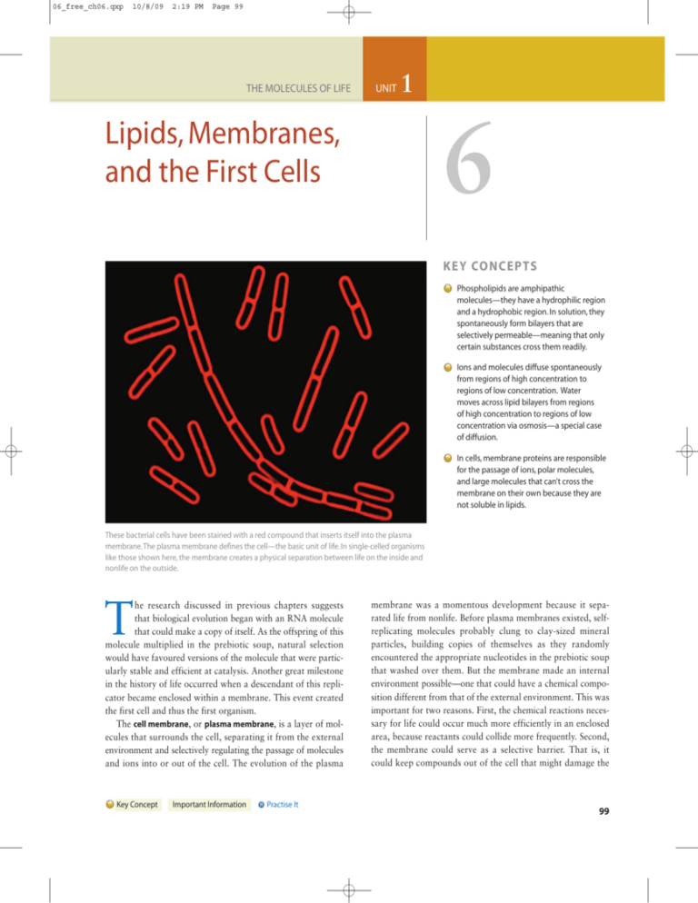
06_free_ch06.qxp 10/8/09 2:19 PM Page 99 THE MOLECULES OF LIFE UNIT 1 6 Lipids, Membranes, and the First Cells K E Y CO N C E P TS Phospholipids are amphipathic molecules—they have a hydrophilic region and a hydrophobic region. In solution, they spontaneously form bilayers that are selectively permeable—meaning that only certain substances cross them readily. Ions and molecules diffuse spontaneously from regions of high concentration to regions of low concentration. Water moves across lipid bilayers from regions of high concentration to regions of low concentration via osmosis—a special case of diffusion. In cells, membrane proteins are responsible for the passage of ions, polar molecules, and large molecules that can’t cross the membrane on their own because they are not soluble in lipids. These bacterial cells have been stained with a red compound that inserts itself into the plasma membrane.The plasma membrane defines the cell—the basic unit of life. In single-celled organisms like those shown here, the membrane creates a physical separation between life on the inside and nonlife on the outside. T he research discussed in previous chapters suggests that biological evolution began with an RNA molecule that could make a copy of itself. As the offspring of this molecule multiplied in the prebiotic soup, natural selection would have favoured versions of the molecule that were particularly stable and efficient at catalysis. Another great milestone in the history of life occurred when a descendant of this replicator became enclosed within a membrane. This event created the first cell and thus the first organism. The cell membrane, or plasma membrane, is a layer of molecules that surrounds the cell, separating it from the external environment and selectively regulating the passage of molecules and ions into or out of the cell. The evolution of the plasma Key Concept Important Information Practise It membrane was a momentous development because it separated life from nonlife. Before plasma membranes existed, selfreplicating molecules probably clung to clay-sized mineral particles, building copies of themselves as they randomly encountered the appropriate nucleotides in the prebiotic soup that washed over them. But the membrane made an internal environment possible—one that could have a chemical composition different from that of the external environment. This was important for two reasons. First, the chemical reactions necessary for life could occur much more efficiently in an enclosed area, because reactants could collide more frequently. Second, the membrane could serve as a selective barrier. That is, it could keep compounds out of the cell that might damage the 99 06_free_ch06.qxp 100 10/8/09 2:19 PM Page 100 Unit 1 The Molecules of Life replicator, but it might allow the entry of compounds required by the replicator. The membrane not only created the cell but also made it into an efficient and dynamic reaction vessel. The goal of this chapter is to investigate how membranes behave, with an emphasis on how they differentiate the internal environment from the external environment. Let’s begin by examining the structure and properties of the most abundant molecules in plasma membranes: the “oily” or “fatty” compounds called lipids. Then we can delve into analyzing the way lipids behave when they form membranes. Which ions and molecules can pass through a membrane that consists of lipids? Which cannot, and why? The chapter ends by exploring how proteins that become incorporated into a lipid membrane can control the flow of materials across the membrane. (a) In solution, lipids form water-filled vesicles. 50 nm (b) Red blood cells resemble vesicles. 6.1 Lipids Most biochemists are convinced that the building blocks of membranes, called lipids, existed in the prebiotic soup. This conclusion is based on the observation that several types of lipids have been produced in experiments designed to mimic the chemical and energetic conditions that prevailed early in Earth’s history. For example, the spark-discharge experiments reviewed in Chapter 3 succeeded in producing at least two types of lipids. An observation made by A. D. Bangham illustrates why this result is interesting. In the late 1950s, Bangham performed experiments to determine how lipids behave when they are immersed in water. But until the electron microscope was invented, he had no idea what these lipid–water mixtures looked like. Once transmission electron microscopes became available, Bangham was able to produce high-magnification, high-resolution images of his lipid–water mixtures. (Transmission electron microscopy is introduced in BioSkills 8.) The images that resulted, called micrographs, were astonishing. As Figure 6.1a shows, the lipids had spontaneously formed enclosed compartments filled with water. Bangham called these membrane-bound structures vesicles and noted that they resembled cells (Figure 6.1b). Bangham had not done anything special to the lipid–water mixtures; he had merely shaken them by hand. The experiment raises a series of questions: How could these structures have formed? Is it possible that vesicles like these existed in the prebiotic soup? If so, could they have surrounded a self-replicating molecule and become the first plasma membrane? Let’s begin answering these questions by investigating what lipids are and how they behave. What Is a Lipid? Earlier chapters analyzed the structures of the organic molecules called amino acids, nucleotides, and monosaccharides 50 μm FIGURE 6.1 Lipids Can Form Cell-like Vesicles When in Water. (a) Transmission electron micrograph showing a cross section through the tiny, bag-like compartments that formed when a researcher shook a mixture of lipids and water. (b) Scanning electron micrograph showing red blood cells from humans. Note the scale bars. and explored how these monomers polymerize to form macromolecules. Here let’s focus on another major type of mid-sized molecule found in living organisms: lipids. Lipid is a catch-all term for carbon-containing compounds that are found in organisms and are largely nonpolar and hydrophobic—meaning that they do not dissolve readily in water. (Recall from Chapter 2 that water is a polar solvent.) Lipids do dissolve, however, in liquids consisting of nonpolar organic compounds. To understand why lipids do not dissolve in water, examine the five-carbon compound called isoprene illustrated in Figure 6.2a; notice that it consists of a group of carbon atoms bonded to hydrogen atoms. Molecules that contain only carbon and hydrogen, such as isoprene or octane (see Chapter 2) are known as hydrocarbons. Hydrocarbons are nonpolar, because electrons are shared equally in carbon–hydrogen bonds. This property makes hydrocarbons hydrophobic. Thus, the reason lipids do not dissolve in water is that they have a significant hydrocarbon component. Figure 6.2b is a type of compound called a fatty acid, which consists of a hydrocarbon chain bonded to a carboxyl (COOH) functional group. Isoprene 06_free_ch06.qxp 10/8/09 2:19 PM Page 101 Chapter 6 Lipids, Membranes, and the First Cells (a) Isoprene O C H2C Carboxyl group A Look at Three Types of Lipids Found in Cells H2C CH3 Unlike amino acids, nucleotides, and carbohydrates, lipids are defined by a physical property—their solubility—instead of their chemical structure. As a result, the structure of lipids varies widely. To drive this point home, consider the structures of the most important types of lipids found in cells: fats, steroids, and phospholipids. CH2 C H 2C C H and fatty acids are key building blocks of the lipids found in organisms. (b) Fatty acid HO CH2 CH2 H2C CH2 H2C CH2 H2C Hydrocarbon chain • CH2 CH2 H2C CH2 H3C FIGURE 6.2 Hydrocarbon Groups Make Lipids Hydrophobic. (a) Isoprenes are hydrocarbons. Isoprene subunits can be linked end to end to form long hydrocarbon chains. (b) Fatty acids typically contain a total of 14–20 carbon atoms, most found in their long hydrocarbon tails. EXERCISE Circle the hydrophobic portion of a fatty acid. Glycerol H H H H C C C OH OH OH H2O HO O C H Fats are composed of three fatty acids that are linked to a three-carbon molecule called glycerol. Because of this struc- ture, fats are also called triacylglycerols or triglycerides. As Figure 6.3a shows, fats form when a dehydration reaction occurs between a hydroxyl group of glycerol and the carboxyl group of a fatty acid. The glycerol and fatty-acid molecules become joined by an ester linkage, which is analogous to the peptide bonds, phosphodiester bonds, and glycosidic linkages in proteins, nucleic acids, and carbohydrates, respectively. Fats are not polymers, however, and fatty acids are not monomers. As Figure 6.3b shows, fatty acids are not linked together to form a macromolecule in the way that amino acids, nucleotides, and monosaccharides H2C (a) Fats form via dehydration reactions. 101 (b) Fats consist of glycerol linked by ester linkages to three fatty acids. H H H H C C C O O O C O C O C H O Ester linkages Dehydration reaction Fatty acid FIGURE 6.3 Fats Are One Type of Lipid Found in Cells. (a) When glycerol and a fatty acid react, a water molecule leaves. (b) The covalent bond that results from this reaction is termed an ester linkage.The fat shown here as a structural formula and a space-filling model is tristearin, the most common type of fat in beef. 06_free_ch06.qxp 2:19 PM Page 102 Unit 1 The Molecules of Life (a) A steroid Formula Space-filling Polar (hydrophilic) Schematic HO d roi Ste CH3 gs Nonpolar (hydrophobic) ri n CH3 H C CH3 Isoprene chain 102 10/8/09 H2C CH2 H2C HC CH3 H3C (b) A phospholipid H3C CH3 N+ CH3 CH2 H2C O Polar head (hydrophilic) H O P H H O C C C O O H O– Choline H OC O H2C CH2 CH2 H2C H 2C CH2 CH2 H 2C H2C CH2 CH2 H2C H 2C CH CH2 H2C CH CH2 H C 2 H2C CH2 CH2 H C 2 H2C CH2 CH2 H C 2 H3C CH2 H 2C CH3 Phosphate Glycerol C Fatty acid Nonpolar tail (hydrophobic) Fatty acid H2C FIGURE 6.4 Amphipathic Lipids Contain Hydrophilic and Hydrophobic Elements. (a) All steroids have a distinctive four-ring structure. (b) All phospholipids consist of a glycerol that is linked to a phosphate group and to either two chains of isoprene or two fatty acids. QUESTION What makes cholesterol—the steroid shown in part (a)—different from other steroids? QUESTION If these molecules were in solution, where would water molecules interact with them? are. After studying the structure in Figure 6.3b, you should be able to explain why fats store a great deal of chemical energy, and why they are hydrophobic. • Steroids are a family of lipids distinguished by the four-ring structure shown in solid orange in Figure 6.4a. The various steroids differ from one another by the functional groups or 06_free_ch06.qxp 10/8/09 2:19 PM Page 103 Chapter 6 Lipids, Membranes, and the First Cells side groups attached to those rings. The molecule pictured in Figure 6.4a is cholesterol, which is distinguished by a hydrocarbon “tail” formed of isoprene subunits. Cholesterol is an important component of plasma membranes in many organisms. In mammals, it is also used as the starting point for the synthesis of several of the signalling molecules called hormones. Estrogen, progesterone, and testosterone are examples of hormones derived from cholesterol. These molecules are responsible for regulating sexual development and activity in humans. • Phospholipids consist of a glycerol that is linked to a phos- phate group (PO422) and to either two chains of isoprene or two fatty acids. In some cases, the phosphate group is bonded to another small organic molecule, such as the choline shown on the phospholipid in Figure 6.4b. Phospholipids with isoprene tails are found in the domain Archaea introduced in Chapter 1; phospholipids composed of fatty acids are found in the domains Bacteria and Eukarya. In all three domains of life, phospholipids are critically important components of the plasma membrane. To summarize, the lipids found in organisms have a wide array of structures and functions. In addition to storing chemical energy and serving as signals between cells, lipids act as pigments that capture or respond to sunlight, form waterproof coatings on leaves and skin, and act as vitamins used in an array of cellular processes. The most important lipid function, however, is their role in the plasma membrane. Let’s take a closer look at the specific types of lipids found in membranes. The Structures of Membrane Lipids Not all lipids can form the artificial membranes that Bangham and his colleagues observed. In fact, just two types of lipids are usually found in plasma membranes. Membrane-forming lipids have a polar, hydrophilic region in addition to the nonpolar, hydrophobic region found in all lipids. To better understand this structure, take another look at the phospholipid illustrated in Figure 6.4b. Notice that the molecule has a “head” region containing highly polar covalent bonds as well as positive and negative charges. The charges and polar bonds in the head region interact with water molecules when a phospholipid is placed in solution. In contrast, the long isoprene or fatty-acid tails of a phospholipid are nonpolar. Water molecules cannot form hydrogen bonds with the hydrocarbon tail, so they do not interact with this part of the molecule. Compounds that contain both hydrophilic and hydrophobic elements are amphipathic (“dual-sympathy”). Phospholipids are amphipathic. As Figure 6.4a shows, cholesterol is also amphipathic. It has both hydrophilic and hydrophobic regions. The amphipathic nature of phospholipids is far and away their most important feature biologically. It is responsible for their presence in plasma membranes. 103 Check Your Understanding If you understand that… ● Fats, steroids, and phospholipids differ in structure and function: Fats store chemical energy; amphipathic steroids are important components of cell membranes; phospholipids are amphipathic and are usually the most abundant component of cell membranes. You should be able to… 1) Draw a generalized version of a fat, a steroid, and a phospholipid. 2) Use these diagrams to explain why cholesterol and phospholipids are amphipathic. 3) Explain how the structure of a fat correlates with its function in the cell. 6.2 Phospholipid Bilayers Phospholipids do not dissolve when they are placed in water. Water molecules interact with the hydrophilic heads of the phospholipids, but not with their hydrophobic tails. Instead of dissolving in water, then, phospholipids may form one of two types of structures: micelles or lipid bilayers. Micelles (Figure 6.5a) are tiny droplets created when the hydrophilic heads of phospholipids face the water and the hydrophobic tails are forced together, away from the water. Lipids with compact tails tend to form micelles. Because their double-chain tails are often too bulky to fit in the interior of a micelle, most phospholipids tend to form bilayers. Phospholipid bilayers, or simply, lipid bilayers, are created when two sheets of phospholipid molecules align. As Figure 6.5b shows, the hydrophilic heads in each layer face a surrounding solution while the hydrophobic tails face one another inside the bilayer. In this way, the hydrophilic heads interact with water while the hydrophobic tails interact with each other. Micelles tend to form from phospholipids with relatively short tails; bilayers tend to form from phospholipids with longer tails. Once you understand the structure of micelles and phospholipid bilayers, the most important point to recognize about them is that they form spontaneously. No input of energy is required. This concept can be difficult to grasp, because the ormation of these structures clearly decreases entropy. Micelles and lipid bilayers are much more highly organized than phospholipids floating free in the solution. The key is to recognize that micelles and lipid bilayers are much more stable energetically than are independent molecules in solution. Stated another way, micelles and lipid bilayers have much lower potential energy than do independent phospholipids in solution. Independent phospholipids are unstable in water because their hydrophobic tails disrupt hydrogen bonds that otherwise 06_free_ch06.qxp 104 10/8/09 2:19 PM Page 104 Unit 1 The Molecules of Life (a) Lipid micelles (b) Lipid bilayers Water No water Water Hydrophilic heads interact with water Hydrocarbon surrounded by water molecules Hydrophobic tails interact with each other FIGURE 6.6 Hydrocarbons Disrupt Hydrogen Bonds between Water Molecules. Hydrocarbons are unstable in water because they disrupt hydrogen bonding between water molecules. EXERCISE Label the area where no hydrogen bonding is occurring between water molecules. QUESTION Hydrogen bonds pull water molecules closer together. Which way are the water molecules in this figure being pulled, relative to the hydrocarbon? Hydrophilic heads interact with water FIGURE 6.5 Phospholipids Form Bilayers in Solution. In (a) a micelle or (b) a lipid bilayer, the hydrophilic heads of lipids face out, toward water; the hydrophobic tails face in, away from water. Plasma membranes consist in part of lipid bilayers. would form between water molecules ( Figure 6.6; see also Figure 2.13b). As a result, amphipathic molecules are much more stable in aqueous solution when their hydrophobic tails avoid water and instead participate in the hydrophobic (van der Waals) interactions introduced in Chapter 3. In this case, the loss of potential energy outweighs the decrease in entropy. Overall, the free energy of the system decreases. Lipid bilayer formation is exergonic and spontaneous. If you understand this reasoning, you should be able to add water molecules that are hydrogen-bonded to each hydrophilic head in Figure 6.5, and explain the logic behind your drawing. Artificial Membranes as an Experimental System When lipid bilayers are agitated by shaking, the layers break and re-form as small, spherical structures. This is what happened in Bangham’s experiment. The resulting vesicles had water on the inside as well as the outside because the hydrophilic heads of the lipids faced outward on each side of the bilayer. Researchers have produced these types of vesicles by using dozens of different types of phospholipids. Artificial membrane- bound vesicles like these are called liposomes. The ability to create them supports an important conclusion: If phospholipid molecules accumulated during chemical evolution early in Earth’s history, they almost certainly formed water-filled vesicles. To better understand the properties of vesicles and plasma membranes, researchers began creating and experimenting with liposomes and other types of artificial bilayers. Some of the first questions they posed concerned the permeability of lipid bilayers. The permeability of a structure is its tendency to allow a given substance to pass across it. Once a membrane forms a water-filled vesicle, can other molecules or ions pass in or out? If so, is this permeability selective in any way? The permeability of membranes is a critical issue, because if certain molecules or ions pass through a lipid bilayer more readily than others, the internal environment of a vesicle can become different from the outside. This difference between exterior and interior environments is a key characteristic of cells. Figure 6.7 shows the two types of artificial membranes that are used to study the permeability of lipid bilayers. Figure 6.7a shows liposomes, roughly spherical vesicles. Figure 6.7b illustrates planar bilayers, which are lipid bilayers constructed across a hole in a glass or plastic wall separating two aqueous (watery) solutions. Using liposomes and planar bilayers, researchers can study what happens when a known ion or molecule is added to one side of a lipid bilayer (Figure 6.7c). Does the ion or molecule cross the membrane and show up on the other side? If so, how 06_free_ch06.qxp 10/8/09 2:19 PM Page 105 Chapter 6 Lipids, Membranes, and the First Cells (a) Liposomes: Artificial membrane-bound vesicles Water Water 50 nm (b) Planar bilayers: Artificial membranes 105 factor changes from one experimental treatment to the next. Control, in turn, is why experiments are such an effective means of exploring scientific questions. You might recall from Chapter 1 that good experimental design allows researchers to alter one factor at a time and determine what effect, if any, each has on the process being studied. Equally important for experimental purposes, liposomes and planar bilayers provide a clear way to determine whether a given change in conditions has an effect. By sampling the solutions on both sides of the membrane before and after the treatment and then analyzing the concentration of ions and molecules in the samples, researchers have an effective way to determine whether the treatment had any consequences. Using such systems, what have biologists learned about membrane permeability? Selective Permeability of Lipid Bilayers Water Water Lipid bilayer (c) Artificial-membrane experiments How rapidly can different solutes cross the membrane (if at all) when ... Solute (ion or molecule) ? 1. Different types of phospholipids are used to make the membrane? 2. Proteins or other molecules are added to the membrane? FIGURE 6.7 Liposomes and Planar Bilayers Are Important Experimental Systems. (a) Electron micrograph of liposomes in cross section (left) and a cross-sectional diagram of the lipid bilayer in a liposome. (b) The construction of planar bilayers across a hole in a glass wall separating two water-filled compartments (left), and a close-up sketch of the bilayer. (c) A wide variety of experiments are possible with liposomes and planar bilayers; a few are suggested here. rapidly does the movement take place? What happens when a different type of phospholipid is used to make the artificial membrane? Does the membrane’s permeability change when proteins or other types of molecules become part of it? Biologists describe such an experimental system as elegant and powerful because it gives them precise control over which When researchers put molecules or ions on one side of a liposome or planar bilayer and measure the rate at which the molecules arrive on the other side, a clear pattern emerges: Lipid bilayers are highly selective. Selective permeability means that some substances cross a membrane more easily than other substances can. Small, nonpolar molecules move across bilayers quickly. In contrast, large molecules and charged substances cross the membrane slowly, if at all. According to the data in Figure 6.8, small, nonpolar molecules such as oxygen (O2) move across selectively permeable membranes more than a billion times faster than do chloride ions (Cl2). Very small and uncharged molecules such as water (H 2 O) can also cross membranes relatively rapidly, even if they are polar. Small, polar molecules such as glycerol and urea have intermediate permeability. The leading hypothesis to explain this pattern is that charged compounds and large, polar molecules can’t pass through the nonpolar, hydrophobic tails of a lipid bilayer. Because of their electrical charge, ions are more stable in solution where they form hydrogen bonds with water than they are in the interior of membranes, which is electrically neutral. If you understand this hypothesis, you should be able to predict whether amino acids and nucleotides will cross a membrane readily. To test the hypothesis, researchers have manipulated the size and structure of the tails in liposomes or planar bilayers. Does the Type of Lipid in a Membrane Affect Its Permeability? Theoretically, two aspects of a hydrocarbon chain could affect the way the chain behaves in a lipid bilayer: (1) the number of double bonds it contains and (2) its length. Recall from Chapter 2 that when carbon atoms form a double bond, the attached atoms are found in a plane instead of a (threedimensional) tetrahedron. The carbon atoms involved are 06_free_ch06.qxp 106 10/8/09 2:19 PM Page 106 Unit 1 The Molecules of Life (a) Permeability scale (cm/s) (b) Size and charge affect the rate of diffusion across a membrane. Phospholipid bilayer 100 High permeability Small, nonpolar molecules O2, CO2, N2 Small, uncharged polar molecules H2O, urea, glycerol Large, uncharged polar molecules Glucose, sucrose O2 ,CO 2 –2 10 H2O 10–4 Glycerol, urea 10–6 Glucose 10–8 10–10 Cl – K+ Na+ –12 10 Low permeability Ions Cl – , K+ , Na+ FIGURE 6.8 Selective Permeability of Lipid Bilayers. (a) The numbers represent “permeability coefficients,” or the rate (cm/s) at which an ion or molecule crosses a lipid bilayer. (b) The relative permeabilities of various molecules and ions, based on data like those presented in part (a). QUESTION About how fast does water cross the lipid bilayer? also locked into place. They cannot rotate freely, as they do in carbon–carbon single bonds. As a result, a double bond between carbon atoms produces a “kink” in an otherwise straight hydrocarbon chain (Figure 6.9). CH2 Double bonds cause kinks in phospholipid tails H2C CH2 H2C C H2C H2C H2C CH2 H C H CH2 CH3 Unsaturated fatty acid Saturated fatty acid FIGURE 6.9 Unsaturated Hydrocarbons Contain Carbon–Carbon Double Bonds. A double bond in a hydrocarbon chain produces a “kink.”The icon on the right indicates that one of the hydrocarbon tails in a phospholipid is unsaturated and therefore kinked. EXERCISE Draw the structural formula and a schematic diagram for an unsaturated fatty acid containing two double bonds. When a double bond exists between two carbon atoms in a hydrocarbon chain, the chain is said to be unsaturated. Conversely, hydrocarbon chains without double bonds are said to be saturated. This choice of terms is logical, because if a hydrocarbon chain does not contain a double bond, it is saturated with the maximum number of hydrogen atoms that can attach to the carbon skeleton. If it is unsaturated, then fewer than the maximum number of hydrogen atoms are attached. Because they contain more C–H bonds, which have much more free energy than C@C bonds, saturated fats have much more chemical energy than unsaturated fats do. People who are dieting are often encouraged to eat fewer saturated fats. Foods that contain lipids with many double bonds are said to be polyunsaturated and are advertised as healthier than foods with more-saturated fats. Why do double bonds affect the permeability of membranes? When hydrophobic tails are packed into a lipid bilayer, the kinks created by double bonds produce spaces among the tightly packed tails. These spaces reduce the strength of hydrophobic interactions among the tails. Because the interior of the membrane is “glued together” less tightly, the structure should become more fluid and more permeable (Figure 6.10). Hydrophobic interactions also become stronger as saturated hydrocarbon tails increase in length. Membranes dominated by phospholipids with long, saturated hydrocarbon tails should be stiffer and less permeable because the interactions among the tails are stronger. 06_free_ch06.qxp 10/8/09 2:19 PM Page 107 Chapter 6 Lipids, Membranes, and the First Cells Lipid bilayer with no unsaturated fatty acids Lower permeability Lipid bilayer with many unsaturated fatty acids Higher permeability FIGURE 6.10 Fatty-Acid Structure Changes the Permeability of Membranes. Lipid bilayers containing many unsaturated fatty acids have more gaps and should be more permeable than are bilayers with few unsaturated fatty acids. A biologist would predict, then, that bilayers made of lipids with long, straight, saturated fatty-acid tails should be much less permeable than membranes made of lipids with short, kinked, unsaturated fatty-acid tails. Experiments on liposomes have shown exactly this pattern. Phospholipids with long, saturated tails form membranes that are much less permeable than membranes consisting of phospholipids with shorter, unsaturated tails. The central point here is that the degree of hydrophobic interactions dictates the behaviour of these molecules. This is another example in which the structure of a molecule— specifically, the number of double bonds in the hydrocarbon chain and its overall length—correlates with its properties and function. These data are also consistent with the basic observation that highly saturated fats are solid at room temperature (Figure 6.11a). (a) Saturated lipids O HO Lipids that have extremely long hydrocarbon tails, as waxes do, form stiff solids at room temperature due to the extensive hydrophobic interactions that occur (Figure 6.11b). Birds, sea otters, and many other organisms synthesize waxes and spread them on their exterior surface as a waterproofing; plant cells secrete a waxy layer that covers the surface of leaves and stems and keeps water from evaporating. In contrast, highly unsaturated fats are liquid at room temperature (Figure 6.11c). Liquid triacylglycerides are called oils. Besides exploring the role of hydrocarbon chain length and degree of saturation on membrane permeability, biologists have investigated the effect of adding cholesterol molecules. Because the steroid rings in cholesterol are bulky, adding cholesterol to a membrane should increase the density of the hydrophobic section. As predicted, researchers found that adding cholesterol molecules to liposomes dramatically reduced the permeability of the liposomes. The data behind this claim are presented in Figure 6.12. The graph in this figure makes another important point, however: Temperature has a strong influence on the behaviour of lipid bilayers. Why Does Temperature Affect the Fluidity and Permeability of Membranes? At about 25°C—or “room temperature”—the phospholipids found in plasma membranes are liquid, and bilayers have the consistency of olive oil. This fluidity, as well as the membrane’s permeability, decreases as temperature decreases. As temperatures drop, individual molecules in the bilayer move more slowly. As a result, the hydrophobic tails in the interior of membranes pack together more tightly. At very low temperatures, (b) Saturated lipids with long hydrocarbon tails Butter (c) Unsaturated lipids Beeswax O O C O HO C C FIGURE 6.11 The Fluidity of Lipids Depends on the Characteristics of Their Hydrocarbon Chains. The fluidity of a lipid depends on the length and saturation of its hydrocarbon chain. (a) Butter consists primarily of saturated lipids. (b) Waxes are lipids with extremely long hydrocarbon chains. (c) Oils are dominated by “polyunsaturates”—lipids with hydrocarbon chains that contain multiple double bonds. QUESTION Why are waxes so effective for waterproofing floors? 107 Safflower oil 06_free_ch06.qxp 108 10/8/09 2:19 PM Page 108 Unit 1 The Molecules of Life Experiment Question: Does adding cholesterol to a membrane affect its permeability? Hypothesis: Cholesterol reduces permeability because it fills spaces in phospholipid bilayers. Null hypothesis: Cholesterol has no effect on permeability. Experimental setup: Phospholipids Cholesterol 1. Create liposomes with no cholesterol, 20% cholesterol, and 50% cholesterol. Liposome 2. Record how quickly glycerol moves across each type of membrane at different temperatures. FIGURE 6.13 Phospholipids Move within Membranes. Membranes are dynamic—in part because phospholipid molecules move within each layer in the structure. Glycerol Prediction: Liposomes with higher cholesterol levels will have reduced permeability. Prediction of null hypothesis: All liposomes will have the same permeability. Permeability of membrane to glycerol Results: No cholesterol 20% of lipids = cholesterol 50% of lipids = cholesterol 0 Phospholipids are in constant lateral motion, but rarely flip to the other side of the bilayer 10 20 Temperature (°C) (mm)/second at room temperature. At these speeds, phospholipids could travel the length of a small bacterial cell in a second. These experiments on lipid and ion movement demonstrate that membranes are dynamic. Phospholipid molecules whiz around each layer while water and small, nonpolar molecules shoot in and out of the membrane. How quickly molecules move within and across membranes is a function of temperature and the structure of the hydrocarbon tails in the bilayer. Given these insights into the permeability and fluidity of lipid bilayers, an important question remains: Why do certain molecules move across membranes spontaneously? 30 Conclusion: Adding cholesterol to membranes decreases their permeability to glycerol. The permeability of all membranes analyzed in this experiment increases with increasing temperature. FIGURE 6.12 The Permeability of a Membrane Depends on Its Composition. lipid bilayers begin to solidify. As the graph in Figure 6.12 indicates, low temperatures can make membranes impervious to molecules that would normally cross them readily. The fluid nature of membranes also allows individual lipid molecules to move laterally within each layer, a little like a person moving about in a dense crowd (Figure 6.13). By tagging individual phospholipids and following their movement, researchers have clocked average speeds of 2 micrometres Check Your Understanding If you understand that… ● In solution, phospholipids form bilayers that are selectively permeable—meaning that some substances cross them much more readily than others do. ● Permeability is a function of temperature, the amount of cholesterol in the membrane, and the length and degree of saturation of the hydrocarbon tails in membrane phospholipids. You should be able to… Fill in a chart with rows called “Temperature,” “Cholesterol,” “Length of hydrocarbon tails,” and “Saturation of hydrocarbon tails” and columns named “Factor,” “Effect on permeability,” and “Reason.” 06_free_ch06.qxp 10/8/09 2:19 PM Page 109 Chapter 6 Lipids, Membranes, and the First Cells CANADIAN ISSUES 6.1 Lipids in Our Diet: Cholesterol, Unsaturated Oils, Saturated Fats, and Trans Fats Most of the foods we eat contain one or more types of lipids. Of all of these, cholesterol is the most vilified, which is somewhat unfair. Too much in the diet does result in atherosclerosis when the unneeded cholesterol begins to coat the sides of blood vessels; as discussed in Chapter 44, this can lead to heart attacks and strokes. But cholesterol is also essential. Our bodies use it to maintain membrane fluidity and to synthesize important molecules such as sex hormones and vitamin D. We also eat fats and oils.These are essential in our diet as well because there are some we require but are unable to synthesize; they provide chemical energy, and they help us absorb vitamins from our gut. Fats and oils are made up of three fatty acids joined to a glycerol. As can be seen in Figure 6.9 on page 106, saturated fatty acids are straight, while unsaturated fatty acids have one bend for each carbon–carbon double bond. Unsaturated fatty acids take more energy to synthesize but they do remain liquid at lower temperatures. Plants and animals that store chemical energy as lipids use a mixture of saturated and unsaturated fatty acids appropriate for the temperature within their cells. Mammals can use saturated fatty acids to make fats for long-term energy storage because their cells are warm. Plants and cold blooded animals such as fish must use unsaturated fatty acids to make oils because fats would solidify in their tissues. From this discussion, it would seem that there would be three types of fats and oils in our diets: saturated, monounsaturated, and polyunsaturated. Polyunsaturated lipids are the most healthful for us, and are found in such foods as fish, sunflower oil, and walnuts. Next are the monounsaturated lipids, from sources including olive oil and peanuts.The least healthy are the saturated lipids from coconut oil, dairy products, beef, and pork. As is Stearic acid (saturated) Oleic acid (unsaturated) commonly known, too much saturated fat in the diet leads to atherosclerosis. In fact, there is a fourth category, known as the trans fats.Their fatty acids contain carbon–carbon double bonds, but different from the ones previously discussed. The double bond in Figure 6.9 is a cis bond. Note how the hydrogens are on one side and the molecule continues in both directions from the other side.This is what puts the kink into the fatty acid. Trans double bonds, on the other hand, do not result in a kink (Figure A). Because trans bonds in fatty acids take energy to make but don’t make a kink in the molecule, they are rare in nature. Dairy products and animal fats such as those in beef and pork contain a small amount. Even though trans fats are rare in nature, until quite recently they were common in our diet. To explain why, it is necessary to know that it is possible to treat oils to make them saturated. This process is called hydrogenation because it converts unsaturated carbon–carbon double bonds (–CH=CH–) into carbon–carbon single bonds (–CH2–CH2–). This is how vegetable oil is turned into margarine, for example. Margarine is a cheap alternative to butter and, because it does not contain cholesterol, healthier too. A byproduct of hydrogenation is the generation of trans fats. At the molecular level, some of the cis double bonds were converted into trans double bonds rather than single bonds. Because trans fats are also solid at room temperature, they are unnoticed in the final product and were common in partially hydrogenated vegetable oils and the foods made with them.The most prevalent source was greasy foods served at fast food restaurants (Figure B). Since the discovery that trans fats are even more likely to cause atherosclerosis than are saturated fats, Health Canada has been active in reducing and eliminating them from foods. Canada was HH HO− O HH HO− H O H The cis double bond kinks the molecule H HO− Elaidic acid (unsaturated) O Figure A A comparison between 18-carbon-long fatty acids with no double bonds, a cis double bond, and a trans double bond. H The trans double bond does not kink the molecule continued 109 06_free_ch06.qxp 110 10/8/09 2:19 PM Page 110 Unit 1 The Molecules of Life CANADIAN ISSUES 6.1 (continued) the first country to require that pre-packaged foods include the amount of trans fat in the Nutrition Facts labelling. In 2006, Health Canada and the Heart and Stroke Foundation recommended that the proportion of fat that is trans fat should be less than 2 percent in vegetable oils and margarine and less than 5 percent in all other foods. It was also suggested that trans fats be replaced with unsaturated rather than saturated fats. The food and restaurant industry was given two years to meet these recommendations voluntarily or they would become regulations. To monitor compliance, Health Canada has surveyed food from restaurants and grocery stores and is continuing to do so. Foods found to have a high proportion of trans fats were tested in 2006 and then again in 2008, and for the most part, the amount of trans fats has decreased substantially. For example, a sample of McDonald’s french fries purchased in October 2006 contained 18.8 percent fat; of this, 8.8 percent was trans fat and 48.7 percent was saturated fat. A second sample of fries purchased in April 2008 contained almost as much fat but only 1.0 percent was trans fat and 12.6 percent was saturated fat. While Health Canada and the food industry’s goal of phasing out trans fats is becoming a reality, 6.3 Why Molecules Move across Lipid Bilayers: Diffusion and Osmosis A thought experiment can help explain why molecules and ions are able to move across membranes spontaneously. Suppose you rack up a set of blue billiard balls on a pool table containing many white balls and then begin to vibrate the table. Because of the vibration, the balls will move about randomly. They will also bump into one another. After these collisions, some blue balls will move outward—away from their original position. In fact, the overall (or net) movement of blue balls will be outward. This occurs because the random motion of the blue balls disrupts their original, nonrandom position—as they move at random, they are more likely to move away from each other than to stay together. Eventually, the blue billiard balls will be distributed randomly across the table. The entropy of the blue billiard balls has increased. Recall from Chapter 2 that entropy is a measure of the randomness or disorder in a system. The second law of thermodynamics states that in a closed system, entropy always increases. This hypothetical example illustrates why molecules or ions located on one side of a lipid bilayer move to the other side spontaneously. The dissolved molecules and ions, or solutes, have thermal energy and are in constant, random motion. Movement of molecules and ions that results from their kinetic energy is known as diffusion. Because solutes change position randomly due to diffusion, they tend to move from a region of high concentration to a region of low concentration. A difference in solute concentrations creates a concentration gradient. Figure B Until recently, fast food contained a large proportion of trans fats. it is important not to overlook the health risks from consuming excess amounts of cholesterol and saturated fats even though they are natural lipids. Molecules and ions still move randomly in all directions when a concentration gradient exists, but there is a net movement from regions of high concentration to regions of low concentration. Diffusion along a concentration gradient is a spontaneous process because it results in an increase in entropy. Once the molecules or ions are randomly distributed throughout a solution, equilibrium is established. For example, consider two aqueous solutions separated by a lipid bilayer. Figure 6.14 shows how molecules that pass through the bilayer diffuse to the other side. At equilibrium, molecules continue to move back and forth across the membrane, but at equal rates—simply because each molecule or ion is equally likely to move in any direction. This means that there is no longer a net movement of molecules across the membrane. What about water itself? As the data in Figure 6.8 (page 106) showed, water moves across lipid bilayers relatively quickly. Like other substances that diffuse, water moves along its concentration gradient—from higher to lower concentration. The movement of water is a special case of diffusion that is given its own name: osmosis. Osmosis occurs only when solutions are separated by a membrane that is permeable to some molecules but not others—that is, a selectively permeable membrane. The best way to think about water moving in response to a concentration gradient is to focus on the concentration of solutes in the solution. Let’s suppose the concentration of a particular solute is higher on one side of a selectively permeable membrane than it is on the other side (Figure 6.15, step 1). Further, suppose that this solute cannot diffuse through the membrane to establish equilibrium. What happens? Water will 06_free_ch06.qxp 10/8/09 2:19 PM Page 111 Chapter 6 Lipids, Membranes, and the First Cells DIFFUSION ACROSS A LIPID BILAYER Lipid bilayer 1. Start with different solutes on opposite sides of a lipid bilayer. Both molecules diffuse freely across bilayer. 2. Solutes diffuse across the membrane— each undergoes a net movement along its own concentration gradient. 111 OSMOSIS Lipid bilayer Osmosis 1. Start with more solute on one side of the lipid bilayer than the other, using molecules that cannot cross the selectively permeable membrane. 2. Water undergoes a net movement from the region of low concentration of solute (high concentration of water) to the region of high concentration of solute (low concentration of water). FIGURE 6.15 Osmosis. 3. Equilibrium is established. Solutes continue to move back and forth across the membrane but at equal rates. FIGURE 6.14 Diffusion across a Selectively Permeable Membrane. EXERCISE If a solute’s rate of diffusion increases linearly with its concentration difference across the membrane, write an equation for the rate of diffusion across a membrane. move from the side with a lower concentration of solute to the side with a higher concentration of solute (step 2). It dilutes the higher concentration and equalizes the concentrations on both sides. This movement is spontaneous. It is driven by the increase in entropy achieved when solute concentrations are equal on both sides of the membrane. Another way to think about osmosis is to realize that water is at higher concentration on the left side of the beaker in Figure 6.15 than it is on the right side of the beaker. As water diffuses, then, there will be net movement of water molecules from the left side to the right side: from a region of high concentration to a region of low concentration. QUESTION Suppose you doubled the number of molecules on the right side of the membrane (at the start). At equilibrium, would the water level on the right side be higher or lower than what is shown here? The movement of water by osmosis is important because it can swell or shrink a membrane-bound vesicle. Consider the liposomes illustrated in Figure 6.16. If the solution outside the membrane has a higher concentration of solutes than the interior has, and the solutes are not able to pass through the lipid bilayer, then water will move out of the vesicle into the solution outside. As a result, the vesicle will shrink and the membrane shrivel. Such a solution is said to be hypertonic (“excess-tone”) relative to the inside of the vesicle. The word root hyper refers to the outside solution containing more solutes than the solution on the other side of the membrane. Conversely, if the solution outside the membrane has a lower concentration of solutes than the interior has, water will move into the vesicle via osmosis. The incoming water will cause the vesicle to swell or even burst. Such a solution is termed hypotonic (“lower-tone”) relative to the inside of the vesicle. Here the word root hypo refers to the outside solution containing fewer solutes than the inside solution has. If solute concentrations are equal on either side of the membrane, the liposome will maintain its size. When the outside solution does not affect the membrane’s shape, that solution is called isotonic (“equal-tone”). Note that the terms hypertonic, hypotonic, and isotonic are relative—they can be used only to express the relationship 06_free_ch06.qxp 112 10/8/09 2:19 PM Page 112 Unit 1 The Molecules of Life Start with: Hypertonic solution Hypotonic solution Isotonic solution Lipid bilayer Result: Arrows represent the direction of net water movement via osmosis Net flow of water out of cell; cell shrinks Net flow of water into cell; cell swells or even bursts No change FIGURE 6.16 Osmosis Can Shrink or Burst Membrane-Bound Vesicles. QUESTION Some species of bacteria can live in extremely salty environments, such as saltwater-evaporation ponds. Is this habitat likely to be hypertonic, hypotonic, or isotonic relative to the interior of the cells? between a given solution and another solution. If you understand this concept, you should be able to draw liposomes in Figure 6.16 that change the relative “tonicity” of the surrounding solution. Specifically, draw (1) a liposome on the left such that the surrounding solution is hypotonic relative to the solution inside the liposome, and (2) a liposome in the centre where the surrounding solution is hypertonic relative to the solution inside the liposome. Web Animation descendants would continue to occupy cell-like structures that grew and divided. Now let’s investigate the next great event in the evolution of life: the formation of a true cell. How can lipid bilayers become a barrier capable of creating and maintaining a specialized internal environment that is conducive to life? How could an effective plasma membrane—one that admits ions and molecules needed by the replicator while excluding ions and molecules that might damage it—evolve in the first cell? at www.masteringbio.com Diffusion and Osmosis To summarize, diffusion and osmosis move solutes and water across lipid bilayers. What does all this have to do with the first membranes floating in the prebiotic soup? Osmosis and diffusion tend to reduce differences in chemical composition between the inside and outside of membrane-bound structures. If liposome-like structures were present in the prebiotic soup, it’s unlikely that their interiors offered a radically different environment from the surrounding solution. In all likelihood, the primary importance of the first lipid bilayers was simply to provide a container for self-replicating molecules. Experiments have shown that ribonucleotides can diffuse across lipid bilayers. Further, it is clear that cell-like vesicles grow as additional lipids are added and then divide if sheared by shaking, bubbling, or wave action. Based on these observations, it is reasonable to hypothesize that once a self-replicating ribozyme had become surrounded by a lipid bilayer, this simple life-form and its Check Your Understanding If you understand that… ● Diffusion is the movement of ions or molecules in solution from regions of high concentration to regions of low concentration. ● Osmosis is the movement of water across a selectively permeable membrane, from a region of low solute concentration to a region of high solute concentration. You should be able to… Make a concept map (see BioSkills 6) that includes the concepts of water movement, solute movement, solution, osmosis, diffusion, semipermeable membrane, hypertonic, hypotonic, and isotonic. 06_free_ch06.qxp 10/8/09 2:19 PM Page 113 Chapter 6 Lipids, Membranes, and the First Cells 113 CANADIAN RESEARCH 6.1 Liposomal Nanomedicines Because of their amphipathic nature, phospholipids will spontaneously arrange themselves into spheres if placed in water. As is shown in Figure 6.7a on page 105, these vesicles are called liposomes if they are made in vitro. Pieter Cullis at the University of British Columbia is one of the pioneers in using liposomes to deliver medicines to where they are needed within patients.This is the new field of liposomal nanomedicines, or LNs. To make these LN particles, phospholipids and the therapeutic agent are mixed together. If the concentration of each is optimal, the lipids will arrange themselves into either a bilayer surrounding a fluid-filled space containing the agents, or a monolayer surrounding a hydrophobic space containing the agents. A common use for this system is to deliver cancer cell–killing drugs into tumours (Figure A), and several cancer treatments based upon LNs are being used in Canada. The LNs are made in vitro and then injected into the patient’s circulatory system, but how do they end up at the tumours? In tests on rodents, Dr. Cullis and his colleagues injected LNs into rodents that had tumours. They found that the LNs accumulated in the tumours but not in healthy tissue. If the blood vessels are intact, the LNs remain in the circulatory system, but if the blood vessels are damaged, as they are in a tumour, the LNs enter the tissue and become trapped. Once the liposomes have entered the tumour, the final step is for the drugs they contain to enter the cancerous cells. Some LNs are designed to slowly leak the therapeutic agent, which is then absorbed into the cancer cells. Other LNs are made to fuse with the plasma membrane of the cancerous cells. In this case, the fusion of the liposomal membrane with the plasma membrane releases the liposome’s internal contents into the cell. Liposomal nanoparticles have two main advantages to injecting medicine directly into a person’s body. First, they allow the medicine to accumulate in the desired location rather than in healthy tissues. Second, they protect the therapeutic agents from being broken down or modified when they are in the circulatory system. While relatively simple in concept, LNs are challenging to design. They must be large enough to contain a sufficient amount of therapeutic agent and yet small enough to leave the blood vessel and enter the damaged tissue. They must also be stable enough to travel in the circulatory system for the hours it takes for chance to deliver them to the tumour sites. Dr. Cullis and his research group have tested various combinations of lipids for use in LNs. Just as with animal cell membranes, they found that including cholesterol prevented leakage from the liposomes and made them more durable. By testing different combinations of unmodi- 6.4 Membrane Proteins What sort of molecule could become incorporated into a lipid bilayer and affect the bilayer’s permeability? The title of this Phospholipids 1. Make LNs. Drugs or 2. Introduce the LNs into the organism’s or patient’s circulatory system. LNs exit the blood vessel where it is damaged LNs in blood vessel Tumour 3. Transfer of drugs into cancer cells. or LN Cancer cell The drugs leak out of the LN The LN has fused with the cell membrane Figure A Liposomal nanomedicines can be used to deliver cancer cell–killing drugs into tumours. fied and modified phospholipids, they were also able to improve on the performance of the LNs. LNs represent an imaginative way to make use of a naturally occurring phenomenon—the selfassembly of phospholipids into spheres—to influence the movement of medicines within our bodies. Reference: Fenske, Chonn, and Cullis (2008). Liposomal nanomedicines: An emerging field. Toxicology Pathology 36:21–29. section gives the answer away. Proteins that are amphipathic can be inserted into lipid bilayers. Proteins can be amphipathic because they are made up of amino acids and because amino acids have side chains, or 06_free_ch06.qxp 114 10/8/09 2:19 PM Page 114 Unit 1 The Molecules of Life R-groups, that range from highly nonpolar to highly polar. (Some are even charged; see Figure 3.3 and Table 3.2 on pages 49 and 50.) It’s conceivable, then, that a protein could have a series of nonpolar amino acids in the middle of its primary structure, but polar or charged amino acids on both ends of its primary structure, as illustrated in Figure 6.17a. The nonpolar amino acids would be stable in the interior of a lipid bilayer, while the polar or charged amino acids would be stable alongside the polar heads and surrounding water (Figure 6.17b). Further, because the secondary and tertiary structures of proteins are almost limitless in their variety and complexity, it is possible for proteins to form tubes and thus function as some sort of channel or pore across a lipid bilayer. Based on these considerations, it is not surprising that when researchers began analyzing the chemical composition of plasma membranes in eukaryotes they found that proteins were just as common, in terms of mass, as phospholipids. How were these two types of molecules arranged? In 1935 Hugh Davson and James Danielli proposed that plasma membranes were structured like a sandwich, with hydrophilic proteins coating both sides of a pure lipid bilayer (Figure 6.18a). Early electron micrographs of plasma membranes seemed to be con- (a) Proteins can be amphipathic. Glu Phe Met Ile Ala (a) Sandwich model The polar and charged amino acids are hydrophilic Ile Ile Gly Val sistent with the sandwich model, and for decades it was widely accepted. The realization that membrane proteins could be amphipathic led S. Jon Singer and Garth Nicolson to suggest an alternative hypothesis, however. In 1972, they proposed that at least some proteins span the membrane instead of being found only outside the lipid bilayer. Their hypothesis was called the fluid-mosaic model. As Figure 6.18b shows, Singer and Nicolson suggested that membranes are a mosaic of phospholipids and different types of proteins. The overall structure was proposed to be dynamic and fluid. The controversy over the nature of the plasma membrane was resolved in the early 1970s with the development of an innovative technique for visualizing the surface of plasma membranes. The method is called freeze-fracture electron microscopy, because the steps involve freezing and fracturing the membrane before examining it with a scanning electron microscope, which produces images of an object’s surface (see BioSkills 8). As Figure 6.19 shows, the technique allows researchers to split plasma membranes and view the middle of the structure. The scanning electron micrographs that result show pits and mounds studding the inner surfaces of the lipid Ile Gly Thr Membrane proteins on cell exterior Ile Phospholipid bilayer Ser Ile The nonpolar amino acids are hydrophobic Membrane proteins on cell interior (b) Amphipathic proteins can integrate into lipid bilayers. Outside cell (b) Fluid-mosaic model Glu Thr Cell exterior Thr Phospholipid bilayer Ser Inside cell Cell interior Membrane proteins FIGURE 6.17 Proteins Can Be Amphipathic. QUESTION Researchers can analyze the primary structure of a membrane protein and predict which portions are embedded in the membrane and which are exposed to the cell’s interior or exterior. How is this possible? QUESTION What type of secondary structure is shown in part (b)? FIGURE 6.18 Past and Current Models of Membrane Structure. (a) The protein–lipid–lipid–protein sandwich model was the first hypothesis for the arrangement of lipids and proteins in plasma membranes. (b) The fluid-mosaic model was a radical departure from the sandwich hypothesis. 06_free_ch06.qxp 10/8/09 2:19 PM Page 115 Chapter 6 Lipids, Membranes, and the First Cells VISUALIZING MEMBRANE PROTEINS Lipid bilayer 1. Strike frozen cell with a knife. Cell Knife Membrane exterior 2. Fracture splits the lipid bilayer. Prepare cell surface for scanning electron microscopy. Membrane interior Membrane interior Exterior of membrane 3. Observe pits and mounds in the membrane interior. 0.1 μm 115 bilayer. Researchers interpret these structures as the locations of membrane proteins. As step 4 in Figure 6.19 shows, the pits and mounds are hypothesized to represent proteins that span the lipid bilayer. These observations conflicted with the sandwich model but were consistent with the fluid-mosaic model. Based on these and subsequent observations, the fluid-mosaic model is now widely accepted. Figure 6.20 summarizes the current hypothesis for where proteins and lipids are found in a plasma membrane. Note that some proteins span the membrane and have segments facing both the interior and exterior surfaces. Proteins such as these are called integral membrane proteins, or transmembrane proteins. Other proteins, called peripheral membrane proteins, are found only on one side of the membrane. Often, peripheral membrane proteins are attached to an integral membrane protein. In most cases, specific peripheral proteins are found only in the inside of the plasma membrane and thus inside the cell, while others are found only on the outside of the plasma membrane and thus facing the surrounding environment. The location of peripheral proteins is one of several reasons that the exterior surface of the plasma membrane is very different from the interior surface. It’s also important to realize that the position of these proteins is not static. Like the phospholipids in the bilayer, membrane proteins are in constant motion, diffusing through the oily film. What do all these proteins do? Later chapters will explore how certain membrane proteins act as enzymes or are involved in cell-to-cell signalling or making physical connections between cells. Here, let’s focus on how integral membrane proteins are involved in the transport of selected ions and molecules across the plasma membrane. Outside cell Mounds and pits in the middle of lipid bilayer Peripheral membrane protein 4. Interpret image as support for fluid-mosaic model of membrane structure. Integral membrane protein Exterior of membrane Inside cell Peripheral membrane protein FIGURE 6.19 Freeze-Fracture Preparations Allow Biologists to View Membrane Proteins. FIGURE 6.20 Integral and Peripheral Membrane Proteins. Integral membrane proteins are also called transmembrane proteins because they span the membrane. Peripheral membrane proteins are often attached to integral membrane proteins. QUESTION What would the micrograph in step 3 look like if the sandwich model of membrane structure were correct? QUESTION Are the external and internal faces of a plasma membrane the same or different? Explain. 06_free_ch06.qxp 116 10/8/09 2:19 PM Page 116 Unit 1 The Molecules Life Systems for Studying Membrane Proteins The discovery of integral membrane proteins was consistent with the hypothesis that proteins affect membrane permeability. The evidence was not considered conclusive enough, though, because it was also plausible to claim that integral membrane proteins were structural components that influenced membrane strength or flexibility. To test whether proteins actually do affect membrane permeability, researchers needed some way to isolate and purify membrane proteins. Figure 6.21 outlines one method that researchers developed to separate proteins from membranes. The key to the technique is the use of detergents. A detergent is a small, amphipathic molecule. As step 1 of Figure 6.21 shows, the hydrophobic tails of detergents clump in solution, forming micelles. When detergents are added to the solution surrounding a lipid bilayer, the hydrophobic tails of the detergent molecules interact with the hydrophobic tails of the lipids. In doing so, the detergent tends to disrupt the bilayer and break it apart (step 2). If the membrane contains proteins, the hydrophobic tails of the detergent molecules also interact with the hydrophobic parts of the membrane proteins. The detergent molecules displace the membrane phospholipids and end up forming water-soluble, detergent–protein complexes (step 3). To isolate and purify these membrane proteins once they are in solution, researchers use the technique called gel elec- ISOLATING MEMBRANE PROTEINS 1. Detergents are small, amphipathic molecules that tend to form micelles in water. 2. Detergents break up plasma membranes; they coat hydrophobic portions of membrane proteins and phospholipids. Isolated protein 3. Treating a plasma membrane with a detergent is an effective way to isolate membrane proteins so they can be purified and studied in detail. FIGURE 6.21 Detergents Can Be Used to Get Membrane Proteins into Solution. trophoresis, introduced in BioSkills 6. When detergent–protein complexes are loaded into a gel and a voltage is applied, the larger protein complexes migrate more slowly than smaller proteins. As a result, the various proteins isolated from a plasma membrane separate from each other. To obtain a pure sample of a particular protein, the appropriate band is cut out of the gel. The gel material is then dissolved to retrieve the protein. Once this protein is inserted into a planar bilayer or liposome, dozens of different experiments are possible. How Do Membrane Proteins Affect Ions and Molecules? In the 55 years since intensive experimentation on membrane proteins began, researchers have identified three broad classes of transport proteins—channels, transporters, and pumps— that affect membrane permeability. What do these molecules do? Can plasma membranes that contain these proteins create an internal environment more conducive to life than the external environment is? Facilitated Diffusion via Channel Proteins One of the first membrane peptides to be investigated in detail is called gramicidin. Gramicidin is produced by a bacterium called Bacillus brevis and is used as a weapon: B. brevis cells release the protein just before a resistant coating forms around their cell wall and membrane. The gramicidin wipes out competitors, giving B. brevis cells more room to grow when they emerge from the resistant phase. Gramicidin is also used medicinally in humans as an antibiotic. After observing that experimental cells treated with gramicidin seemed to lose large numbers of ions, researchers became interested in understanding how the molecule works. Could this protein alter the flow of ions across plasma membranes? Biologists answered this question by inserting purified gramicidin into planar bilayers. The experiment they performed was based on an important fact about ion movement across membranes: Not only do ions move from regions of high concentration to regions of low concentration via diffusion, but they also flow from areas of like charge to areas of unlike charge. In Figure 6.22, for example, a large concentration gradient favours the movement of sodium ions from the outside of the cell to the inside. But in addition, the inside of this cell has a net negative charge while the outside has a net positive charge. As a result, there is also a charge gradient that favours the movement of sodium ions from the outside to the inside of the cell. Based on this example, it should be clear that ions move in response to a combined concentration and electrical gradient, or what biologists call an electrochemical gradient. If you understand this concept, you should be able to add an arrow to Figure 6.22, indicating the electrochemical gradient for chloride ions assuming that chloride concentrations are equal on both sides of the membrane. 06_free_ch06.qxp 10/8/09 2:19 PM Page 117 Chapter 6 Lipids, Membranes, and the First Cells Na+ Na+ Na+ High concentration of Na+ Net + charge Na+ Cl – Na+ 117 Experiment Cl – Na+ Outside cell Question: Does gramicidin affect the flow of ions across a membrane? Hypothesis: Gramicidin increases the flow of cations across a Electrochemical gradient for sodium ions (Na+) membrane. Null hypothesis: Gramicidin has no effect on membrane permeability. Experimental setup: Inside cell Cl – Na+ Net – charge Low concentration of Na+ Membrane without gramicidin Cl – FIGURE 6.22 Electrochemical Gradients. When ions build up on one side of a membrane, they establish a combined concentration and electrical gradient. Membrane with gramicidin + + + + + + + + + + + + + + 1. Create planar bilayers with and without gramicidin. + + + + + + EXERCISE By adding sodium ions to this figure, illustrate a situation where there is no electrochemical gradient favouring the movement of either Na1 or Cl2. Ion flow? Ion flow? 3. Record electrical currents to measure ion flow across the planar bilayers. Prediction: Ion flow will be higher in membrane with gramicidin. Prediction of null hypothesis: Ion flow will be the same in both membranes. Results: Size of electric current To determine whether gramicidin affected the membrane’s permeability to ions, the researchers measured the flow of electric current across the membrane. Because ions carry a charge, the movement of ions produces an electric current. This property provides an elegant and accurate test for assessing the bilayer’s permeability to ions—one that is simpler and more sensitive than taking samples from either side of the membrane and determining the concentrations of solutes present. If gramicidin facilitates ion movement, then an investigator should be able to detect an electric current across planar bilayers that contain gramicidin. The result? The graph in Figure 6.23 shows that when gramicidin was absent, no electric current passed through the membrane. But when gramicidin was inserted into the membrane, current began to flow. Based on this observation, biologists proposed that gramicidin is an ion channel. An ion channel is a peptide or protein that makes lipid bilayers permeable to ions. (Recall from Chapter 3 that peptides are proteins containing fewer than 50 amino acids.) Follow-up work corroborated that gramicidin is selective. It allows only positively charged ions, or cations, to pass. Gramicidin does not allow negatively charged ions, or anions, to pass through the membrane. It was also established that gramicidin is most permeable to hydrogen ions or protons (H 1 ) , and somewhat less permeable to other cations, such as potassium (K 1 ) and sodium (Na 1 ). Researchers gained additional insight into the way gramicidin works when they determined its amino acid sequence (that is, primary structure) and tertiary structure. Figure 6.24 provides a view from the outside of a cell to the inside through gramicidin. The key observation is that the molecule forms a hole. The portions of amino acids that line this hole are hydrophilic, while regions on the exterior (in contact with the membrane phospholipids) are hydrophobic. The molecule’s structure correlates with its function. 2. Add cations to one side of the planar bilayer to create an electrochemical gradient. Where gramicidin is present, electric current increases Rate of ion flow flattens out Initial rapid increase in ion flow Where gramicidin is not present, no current Concentration of ions Conclusion: Gramicidin facilitates diffusion of cations along an electrochemical gradient. Gramicidin is an ion channel. FIGURE 6.23 Measuring Ion Flow through the Channel Gramicidin. Experiment for testing the hypothesis that gramicidin is an ion channel. QUESTION Why does the curve in the Results section flatten out? Subsequent research has shown that cells have many different types of channel proteins in their membranes, each featuring a structure that allows it to admit a particular type of ion or small molecule. For example, Peter Agre and co-workers recently discovered channels called aquaporins (“water-pores”) that allow water to cross the plasma membrane over 10 times faster than it does in the absence of aquaporins. Figure 6.25a shows a cutaway 06_free_ch06.qxp 118 10/8/09 2:19 PM Page 118 Unit 1 The Molecules of Life (a) Water pores allow only water to pass through. (a) Top view of gramicidin Hydrophobic exterior Hydrophilic interior Outside cell Hydrophobic exterior H2O Hydrophilic interior (b) Potassium channels allow only potassium ions to pass through. Inside cell (b) Side view of gramicidin Potassium ions can enter the channel, but cannot pass into the cell Outside cell K+ K+ K+ Inside cell Closed When a change in electrical charge occurs outside the membrane, the protein changes shape and allows the ions to pass through FIGURE 6.24 The Structure of a Channel Protein. Gramicidin is an ␣-helix consisting of only 15 amino acids. (a) In top view, the molecule forms a hole or pore. (b) In side view, a green helix traces the peptidebonded backbone of the polypeptide. R-groups hang off the backbone to the outside.The interior of the channel is hydrophilic; the exterior is hydrophobic. + + + + + + + + + + + + + + K+ K+ K+ EXERCISE In (a) and (b), add symbols indicating the locations of phospholipids relative to gramicidin in a plasma membrane. view from the side of an aquaporin, indicating how it fits in a plasma membrane. Like gramicidin, the channel has a pore that is lined with polar regions of amino acids—in this case, functional groups that interact with water. Hydrophobic groups form the outside of the structure and interact with the lipid bilayer. Unlike gramicidin, aquaporins are extremely selective. They admit water but not other small molecules or ions. Selectivity turns out to be a prominent characteristic of most channel proteins. The vast majority of these proteins admit only a single type of ion. In many cases, researchers are now able to identify exactly which amino acids are responsible for making the pore selective. Open FIGURE 6.25 Most Membrane Channels Are Highly Selective and Highly Regulated (a) A cutaway view looking at the side of an aquaporin—a membrane channel that admits only water. Water moves through its pore via osmosis over 10 times faster than it can move through the lipid bilayer. (b) A model of a K1 channel in the open and closed configurations. Recent research has also shown that the aquaporins and ion channels are gated channels—meaning that they open or close in response to the binding of a particular molecule or to a change in the electrical charge on the outside of the membrane. 06_free_ch06.qxp 10/8/09 2:19 PM Page 119 Chapter 6 Lipids, Membranes, and the First Cells For example, Figure 6.25b shows a potassium channel in the open and closed configuration. When the electrical charge on the membrane becomes positive on the outside relative to the inside, the protein’s structure changes in a way that opens the channel and allows potassium ions to cross. The important point here is that in almost all cases, the flow of ions and small molecules through membrane channels is carefully controlled. In all cases, the movement of substances through channels is passive—meaning it does not require an expenditure of energy. Passive transport is powered by diffusion along an electrochemical gradient. Channel proteins enable ions or polar molecules to move across lipid bilayers efficiently. If you understand the nature of membrane channels, you should be able to (1) draw the structure of a channel that admits calcium ions (Ca21) when a signalling molecule binds to it, (2) label hydrophilic and hydrophobic portions of the channel, (3) add ions to the outside and inside of a membrane containing the channel to explain why an electrochemical gradient favours entry of Ca21, (4) sketch the channel in the open versus closed configuration, and (5) suggest a hypothesis to explain why it might be important for the channel to be selective. To summarize, membrane proteins such as gramicidin, aquaporins, and potassium channels circumvent the lipid bilayer’s impermeability to small, charged compounds. They are responsible for facilitated diffusion: the passive transport of substances that otherwise would not cross a membrane readily. The presence of channels reduces differences between the interior and exterior. Water molecules and ions are not the only substances that move across membranes through membrane proteins, however. Larger molecules can, too. Facilitated Diffusion via Carrier Proteins Even though facilitated diffusion does not require an expenditure of energy, it is facilitated—aided—by the presence of a specialized membrane protein. Facilitated diffusion can occur through channels or through carrier proteins, also called transporters, that change shape during the process. Perhaps the best-studied transporter is specialized for moving glucose into cells. Next to ribose, the six-carbon sugar glucose is the most important sugar found in organisms. Virtually all cells alive today use glucose as a building block for important macromolecules and as a source of stored chemical energy. But as Figure 6.8 on page 106 showed, lipid bilayers are only moderately permeable to glucose. It is reasonable to expect, then, that plasma membranes have some mechanism for increasing their permeability to this sugar. This prediction was supported when researchers compared the permeability of glucose across planar bilayers with its permeability across membranes from cells. The plasma membrane in this study came from human red blood cells, which are among the simplest cells known. A mature red blood cell consists of a membrane, about 300 million hemoglobin molecules, and not much else (Figure 6.26, step 1). When these cells are placed in a hypotonic solution (step 2), water rushes into them by osmosis. As water flows inward, the cells swell. Eventually they burst, releasing the hemoglobin molecules and other cell contents. This leaves researchers with pure preparations of plasma membranes called red blood cell “ghosts” (step 3). Experiments have shown that these membranes are much more permeable to glucose than are pure lipid bilayers. Why? After isolating and analyzing many proteins from red blood cell ghosts, researchers found one protein that specifically increases membrane permeability to glucose. When this purified protein was added to liposomes, the artificial membrane transported glucose at the same rate as a membrane from a living cell. This experiment convinced biologists that a membrane protein was indeed responsible for transporting glucose across plasma membranes. Follow-up work showed that this glucosetransporting protein, a carrier that is now called GLUT-1, facilitates transport of the “right-handed” optical isomer of glucose but not the left-handed form. Cells use only the right-handed form of glucose, and GLUT-1’s binding site is specific for the HOW RESEARCHERS MAKE RED BLOOD CELL “GHOSTS” 1. Normal blood cells in isotonic solution. 119 2. In hypotonic solution, cells swell as water enters via osmosis. Eventually the cells burst. 3. After the cell contents have spilled out, all that remains are cell “ghosts,” which consist entirely of plasma membranes. FIGURE 6.26 Red Blood Cell “Ghosts.” Red blood cell ghosts are simple membranes that can be purified and studied in detail. 06_free_ch06.qxp 120 10/8/09 2:19 PM Page 120 Unit 1 The Molecules of Life A HYPOTHESIS FOR HOW GLUT-1 FACILITATES GLUCOSE DIFFUSION Outside cell O O O O O O O O O O O O O Glucose O GLUT-1 O O Inside cell 1. GLUT-1 is a transmembrane transport protein, shown with its binding site facing outside the cell. 2. Glucose binds to GLUT-1 from outside the cell. 3. A conformational change results, transporting glucose to the interior. 4. Glucose is released inside the cell. FIGURE 6.27 A Hypothesis to Explain How Membrane Transport Proteins Work. This model suggests that the GLUT-1 transporter acts like an enzyme. It binds a substrate (in this case, a glucose molecule), undergoes a conformation change, and releases the substrate. QUESTION GLUT’s binding site has the same affinity for glucose in both of its conformations. Explain how this trait allows glucose to diffuse along its concentration gradient. right-handed form. To make sense of these observations, biologists hypothesize that GLUT proteins with binding sites that interact with the right-handed form of glucose were favoured by natural selection. Stated another way, cells with proteins like GLUT-1 thrived much better than cells without a glucosetransport protein or with proteins that transported the lefthanded form. Exactly how GLUT-1 works is a focus of ongoing research. Biologists who are working on the problem have noted that because glucose transport by GLUT-1 is so specific, it is logical to predict that the mechanism resembles the action of enzymes. One hypothesis is illustrated in Figure 6.27. The idea is that glucose binds to GLUT-1 on the exterior of the membrane and that this binding induces a conformational change in the protein which transports glucose to the interior of the cell. Recall from Chapter 3 that enzymes frequently change shape when they bind substrates and that such conformational changes are often a critical step in the catalysis of chemical reactions. Importing molecules into cells via carrier proteins is still powered by diffusion, however. When glucose enters a cell via GLUT-1, it does so because it is following its concentration gradient. If the concentration of glucose is the same on both sides of the plasma membrane, then no net movement of glucose occurs even if the membrane contains GLUT-1. A large array of molecules moves across plasma membranes via facilitated diffusion through specific carrier proteins. HOW THE SODIUM–POTASSIUM PUMP (Na+/K+-ATPase) WORKS K+ Outside cell K+ K+ K+ K+ Na+ K+ K+ K+ Na+ Na+ Na+ Na+ Na+ Na+ Na+ Na+ Na+ Na+ Na+ Inside cell 1. Three binding sites within the protein have a high affinity for sodium ions. P P P P ATP 2. Three sodium ions from the inside of the cell bind to these sites. Phosphate group P P P 3. A phosphate group from ATP binds to the protein. In response, the protein changes shape. FIGURE 6.28 Active Transport Depends on an Input of Chemical Energy. EXERCISE Circle the two steps where addition or deletion of a phosphate group causes the protein to change conformation. Label each “Shape change.” ADP 4. The sodium ions leave the protein and diffuse to the exterior of the cell. 06_free_ch06.qxp 10/8/09 2:19 PM Page 121 121 Chapter 6 Lipids, Membranes, and the First Cells Active Transport by Pumps Whether diffusion is facilitated by channel proteins or by carrier proteins, it is a passive process that makes the cell interior and exterior more similar. But it is also possible for today’s cells to import molecules or ions against their electrochemical gradient. Accomplishing this task requires energy, however, because the cell must counteract the entropy loss that occurs when molecules or ions are concentrated. It makes sense, then, that transport against an electrochemical gradient is called active transport. In cells, the energy required to move substances against their electrochemical gradient is usually provided by a phosphate group (HPO42−) from adenosine triphosphate, or ATP. ATP contains three phosphate groups. When one of these phosphate groups leaves ATP and binds to a protein, two negative charges are added to the protein. These charges repel other charges on the protein’s amino acids. The protein’s potential energy increases in response, and its conformation (shape) usually changes. As Chapter 9 will detail, proteins usually move when a phosphate group binds to them or when a phosphate group drops off. When a phosphate group leaves ATP, the resulting molecule is adenosine diphosphate (ADP), which has two phosphate groups. Figure 6.28 shows how ions or molecules can move against an electrochemical gradient when membrane proteins called pumps change shape. The figure highlights the first pump that was discovered and characterized: a protein called the sodium–potassium pump, or more formally, Na1/K1-ATPase. The Na1/K1 part of this expression refers to the ions that are transported; ATP indicates that adenosine triphosphate is used; and –ase implies that the molecule acts like an enzyme. Unlike the situation with GLUT-1, the mechanism of action in Na1/K1-ATPase is now well known. When the protein is in the conformation shown in step 1 of Figure 6.28, binding sites K+ Na+ Na+ Na+ Na+ with a high affinity for sodium ions are available. As step 2 shows, three sodium ions from the inside of the cell bind to these sites. A phosphate group from ATP then binds to the pump (step 3). When the phosphate group attaches, the pump’s shape changes in a way that reduces its affinity for sodium ions. As a result, the ions leave the protein and diffuse to the exterior of the cell (step 4). In this conformation, though, the protein has binding sites with a high affinity for potassium ions (step 5). As step 6 shows, two potassium ions from outside the cell bind to the pump. When they do, the phosphate group drops off the protein and its shape changes in response—back to the original shape (step 7). In this conformation, the pump has low affinity for potassium ions. As step 8 shows, the potassium ions leave the protein and diffuse to the interior of the cell. The cycle then repeats. This movement of ions can occur even if an electrochemical gradient exists that favours the outflow of potassium and the inflow of sodium. By exchanging three sodium ions for every two potassium ions, the outside of the membrane becomes positively charged relative to the inside. In this way, the sodium– potassium pump sets up an electrical gradient as well as a chemical gradient across the membrane. Similar pumps are specialized for moving protons (H1), calcium ions (Ca21), or other ions or molecules. In this way, cells are capable of concentrating certain substances or setting up electrochemical gradients. It is difficult to overemphasize the importance of these gradients. For example, the electrochemical gradients produced by proton pumps allow plants to take up nutrients from the soil; the gradients established by the Na1/K1-ATPase and calcium pumps allow your nerve cells to transmit electrical signals throughout your body. You will encounter active transport, membrane pumps, and electrochemical gradients throughout this text. Na+ Na+ Na+ Na+ Na+ Na+ Na+ Na+ K+ K+ K+ K+ P K+ P K+ P 5. In this conformation, the protein has binding sites with a high affinity for potassium ions. 6. Two potassium ions bind to the pump. 7. The phosphate group drops off the protein. In response, the protein changes back to its original shape. K+ 8. The potassium ions leave the protein and diffuse to the interior of the cell. These 8 steps repeat. 06_free_ch06.qxp 122 10/8/09 2:19 PM Page 122 Unit 1 The Molecules of Life Web Animation ATP to move pumps and channels and other molecules and machines where they’re needed? Answering these and related questions is the focus of Unit 2. at www.masteringbio.com Membrane Transport Proteins Taken together, the lipid bilayer and the proteins involved in passive transport and active transport enable cells to create an internal environment that is much different from the external one. Membrane proteins allow ions and molecules to cross the plasma membrane, even though they are not lipid soluble. (Figure 6.29). When membrane proteins first evolved, then, the early cells acquired the ability to create an internal environment that was conducive to life—meaning that such an environment contained the substances required for manufacturing ATP and copying ribozymes. Cells with particularly efficient and selective membrane proteins would be favoured by natural selection and would come to dominate the population. Cellular life had begun. Some 3.5 billion years later, cells continue to evolve. What do today’s cells look like, and how do they produce and store the chemical energy that makes life possible? How do they use Diffusion H2O H2O H2O Outside cell Inside cell Description: CO2 If you understand that… ● Membrane proteins allow ions and molecules that ordinarily do not readily cross lipid bilayers to enter or exit cells. ● Substances may move along an electrochemical gradient via facilitated diffusion through channel proteins or transport proteins, or they may move against an electrochemical gradient in response to work done by pumps. You should be able to… 1) Sketch a phospholipid bilayer. 2) Indicate how ions and large molecules cross it via each major type of membrane transport protein. Facilitated diffusion Active transport O CO2 CO2 Check Your Understanding Na+ CO2 H2O H 2O O K+ K+ K+ Na+ K+ K+ K+ Na+ K+ K+ K+ K+ H 2O CO2 H2O Passive movement of small, uncharged molecules along an electrochemical gradient, through a membrane H2O Passive movement of ... Protein(s) involved: FIGURE 6.29 Mechanisms of Membrane Transport: A Summary. EXERCISE Complete the chart. Na+ Na+ O K+ Na+ Na+ Na+ Active movement of ... K+ K+ 06_free_ch06.qxp 10/8/09 2:19 PM Page 123 Chapter 6 Lipids, Membranes, and the First Cells 123 Chapter Review SUMMARY OF KEY CONCEPTS Phospholipids are amphipathic molecules—they have a hydrophilic region and a hydrophobic region. In solution, they spontaneously form bilayers that are selectively permeable—meaning that only certain substances cross them readily. The plasma membrane is a structure that forms a physical barrier between life and nonlife. The basic structure of plasma membranes is created by a phospholipid bilayer. Phospholipids have a polar head and a nonpolar tail. The nonpolar tail consists of a lipid—usually a fatty acid or an isoprene. Lipids do not dissolve in water. Small, nonpolar molecules tend to move across membranes readily; ions and other charged compounds cross rarely, if at all. The permeability and fluidity of lipid bilayers depend on temperature and on the types of phospholipids present. For example, because phospholipids that contain long, saturated fatty acids form a dense and highly hydrophobic membrane interior, they tend to be less permeable than phospholipids containing shorter, unsaturated fatty acids. You should be able to draw phospholipid bilayers that are highly permeable and fluid versus highly impermeable and lacking in fluidity. Ions and molecules diffuse spontaneously from regions of high concentration to regions of low concentration. Water moves across lipid bilayers from regions of high concentration to regions of low concentration via osmosis—a special case of diffusion. Diffusion is movement of ions or molecules due to their kinetic energy. Solutes move via diffusion from a region of high concentration to a region of low concentration. This is a spontaneous process driven by an increase in entropy. Water also moves across membranes spontaneously if a molecule or an ion that cannot cross the membrane is found in different concentrations on the two sides. In osmosis, water moves from the region with a lower concentration of solutes to the region of higher solute concentration. Osmosis is a passive process driven by an increase in entropy. You should be able to draw a beaker with solutions on either side separated by a plasma membrane, and then predict what will happen after addition of a solute to one side if the solute (1) crosses the membrane readily, versus (2) is incapable of crossing the membrane. Web Animation at www.masteringbio.com Diffusion and Osmosis In cells, membrane proteins are responsible for the passage of ions, polar molecules, and large molecules that can’t cross the membrane on their own because they are not soluble in lipids. The permeability of lipid bilayers can be altered significantly by membrane transport proteins, which are scattered throughout the plasma membrane. Channel proteins, for example, are molecules that provide holes in the membrane and facilitate the diffusion of specific ions into or out of the cell. Transport proteins are enzyme-like proteins that allow specific molecules to diffuse into the cell. In addition to these forms of facilitated diffusion, membrane proteins that act as energy-demanding pumps actively move ions or molecules against their electrochemical gradient. In combination, the selective permeability of phospholipid bilayers and the specificity of transport proteins make it possible to create an environment inside a cell that is radically different from the exterior. You should be able to draw and label the membrane of a cell that pumps hydrogen ions to the exterior, has channels that admit calcium ions along an electrochemical gradient, and has carriers that admit lactose (a sugar) molecules along a concentration gradient. Your drawing should include arrows and labels indicating the direction of solute movement and the direction of the appropriate electrochemical gradients. Web Animation at www.masteringbio.com Membrane Transport Proteins QUESTIONS Test Your Knowledge 1. What does the term hydrophilic mean when it is translated literally? a. “oil loving” b. “water loving” c. “oil fearing” d. “water fearing” 3. If a solution surrounding a cell is hypertonic relative to the inside of the cell, how will water move? a. It will move into the cell via osmosis. b. It will move out of the cell via osmosis. c. It will not move, because equilibrium exists. d. It will evaporate from the cell surface more rapidly. 2. If a solution surrounding a cell is hypotonic relative to the inside of the cell, how will water move? a. It will move into the cell via osmosis. b. It will move out of the cell via osmosis. c. It will not move, because equilibrium exists. d. It will evaporate from the cell surface more rapidly. 4. When does a concentration gradient exist? a. when membranes rupture b. when solute concentrations are high c. when solute concentrations are low d. when solute concentrations differ on the two sides of a membrane 06_free_ch06.qxp 2:49 PM Page 124 Unit 1 The Molecules of Life 5. Which of the following must be true for osmosis to occur? a. Water must be at room temperature or above. b. Solutions with the same concentration of solutes must be separated by a selectively permeable membrane. c. Solutions with different concentrations of solutes must be separated by a selectively permeable membrane. d. Water must be under pressure. 6. Why are the lipid bilayers in cells called “selectively permeable”? a. They are not all that permeable. b. Their permeability changes with their molecular composition. c. Their permeability is temperature dependent. d. They are permeable to some substances but not others. Test Your Knowledge answers: 1. b; 2. a; 3. b; 4. d; 5. c; 6. d 124 10/9/09 Test Your Understanding 1. Cooking oil is composed of lipids that consist of long hydrocarbon chains. Would you expect these lipids to form membranes spontaneously? Why or why not? Describe, on a molecular level, how you would expect these lipids to interact with water. Answers are available at www.masteringbio.com K+ H2O CO2 H2O Na + CO2 H2O Na + CO2 Cl – K+ CO2 H2O 2. Explain why phospholipids form a bilayer in solution. 3. Ethanol, the active ingredient in alcoholic beverages, is a small, polar, uncharged molecule. Would you predict that this molecule crosses plasma membranes quickly or slowly? Explain your reasoning. 4. Why can osmosis occur only if solutions are separated by a selectively permeable membrane? What happens in solutions that are not separated by a selectively permeable membrane? 5. The text claims that the portion of membrane proteins that spans the hydrophobic tails of phospholipids is itself hydrophobic (see Figure 6.17b on page 114). Why is this logical? Look back at Figure 3.3 and Table 3.2 (pages 49 and 50), and make a list of amino acids you might expect to find in these regions of transmembrane proteins. H2O Gramicidin molecule Cl – H2O Cl – Na + Cl – H2O Cl – Cl – K+ Cl – 6. Examine the membrane in the accompanying figure. Label the molecules and ions that will pass through the membrane as a result of osmosis, diffusion, and facilitated diffusion. Draw arrows to indicate where each of the molecules and ions will travel. Applying Concepts to New Situations 1. When phospholipids are arranged in a bilayer, it is theoretically possible for individual molecules in the bilayer to flip-flop. That is, a phospholipid could turn 180° and become part of the membrane’s other surface. Sketch this process. From what you know about the behaviour of polar heads and nonpolar tails, predict whether flipflops are frequent or rare. Then design an experiment, using a planar bilayer made up partly of fatty acids that contain a dye molecule on their hydrophilic head, to test your prediction. 2. Unicellular organisms that live in extremely cold habitats have an unusually high proportion of unsaturated fatty acids in their plasma membranes. Some of these membranes even contain polyunsaturated fatty acids, which have more than one double bond in each hydrocarbon chain. Draw a picture of this type of membrane and comment on the hypothesis that membranes with unsaturated fatty-acid tails function better at cold temperatures than do membranes with saturated fatty-acid tails. Make a prediction about the structure of fatty acids found in organisms that live in extremely hot environments. 3. When biomedical researchers design drugs that must enter cells to be effective, they sometimes add methyl (CH3) groups to make the Answers are available at www.masteringbio.com drug molecules more likely to pass through plasma membranes. Conversely, when researchers design drugs that act on the exterior of plasma membranes, they sometimes add a charged group to decrease the likelihood that the drugs will pass through membranes and enter cells. Explain why these strategies are effective. 4. Advertisements frequently claim that laundry and dishwashing detergents “cut grease.” What the ad writers mean is that the detergents surround oil droplets on clothing or dishes, making the droplets water soluble. When this happens, the oil droplets can be washed away. Explain how this happens on a molecular level. www.masteringbio.com is also your resource for • Answers to text, table, and figure caption questions and exercises • Answers to Check Your Understanding boxes • Online study guides and quizzes • Additional study tools including the E-Book for Biological Science, textbook art, animations, and videos.

