THE FOOT POSTURE INDEX©
Easy quantification of
standing foot posture
Six item version
FPI-6
USER GUIDE AND MANUAL
M
AUGUST 2005
© Anthony Redmond 1998
All rights reserved,
May not be copied without permission
www.leeds.ac.uk/medicine/FASTER/FPI/
1
Foot Posture Index - User guide and manual
Acknowledgments
The FPI was developed with funding from the following agencies
The CMT Association of the USA
The Australasian Podiatry Council, Australian Podiatry Education and Research Fund
The Podiatry Education and Research Account of the NSW Podiatrists’ Registration Board.
In-kind support was also provided by the Arthritis Research Campaign
Sincere thanks are due to the following institutions and individuals for their assistance in the development
and testing of the FPI
University of Sydney, Australia
University of Western Sydney, Australia
University of South Australia
University of Huddersfield, United Kingdom
University of Leeds, United Kingdom
Royal Alexandra Hospital for Children, Sydney,
Australia
Prof Robert Ouvrier
Dr Jack Crosbie
Dr Jennifer Peat
Dr Joshua Burns
Rolf Scharfbillig
Angela Evans
Alex Copper
Anne-Maree Keenan
Dr Jim Woodburn
Liz Barr
All staff and students at the University of Western Sydney, School of Exercise and Health
Sciences.
All of the other clinicians in the many disciplines who have contributed with their time,
suggestions and expertise in the development of the FPI to date.
About the author
Dr Anthony Redmond is Arthritis Research Campaign lecturer in the Academic Unit of Musculoskeletal
Disease at the University of Leeds. He has worked in clinical podiatry and foot-related research for the
majority of his career, mostly in multidisciplinary gait and lower limb clinics. The FPI was conceived as a
part answer to the recurring clinical problem of assessing gait and foot posture variables reliably in the
clinical setting. Work first started on the various iterations of the FPI in 1996, with a more formal
approach to the development of the FPI as part of his PhD candidature in the faculty of medicine at the
University of Sydney. Various iterations have appeared in the literature† but only this six-item version has
completed all validation studies satisfactorily. We now recommend that the use of any previous versions
be discontinued.
The validation process is described in full in:
Redmond AC. Foot Posture in Neuromuscular Disease (PhD Thesis) University of Sydney, 2004.
Redmond AC., Crosbie J., Ouvrier RA. Development and validation of a novel rating system for scoring
foot posture: the Foot Posture Index. Clinical Biomechanics (In Press)
FPI manuals and datasheets
The FPI concept and data sheets have been released into the public domain. The datasheets may be
copied freely for clinical or research purposes although they should not be altered or adapted without the
express permission of the copyright holder. All rights are reserved for this manual/user guide and it
should not should be copied or redistributed in any form without the author’s express consent.
Further information can be found on-line at www.leeds.ac.uk/medicine/FASTER/FPI
A.R. August 2005
†
Redmond A, Burns J, Crosbie J, Ouvrier R, Peat J. An initial appraisal of the validity of a criterion based, observational clinical rating system for foot
posture. Journal of Orthopedic and Sports Physical Therapy 2001;31(3):160.
Payne C, Oates M, Noakes H. Static stance response to different types of foot orthoses. J Am Pod Med Assoc 2003;93(6):492- 8.
Evans AM, Copper AW, Scharfbillig RW, Scutter SD, Williams MT. The reliability of the foot posture index and traditional measures of foot position. J
Am Pod Med Assoc 2003;93:203-13.
Yates B, White S . The incidence and risk factors in the development of medial tibial stress syndrome among naval recruits. Am J Sports Med 2004: 32
(3): 772-780
2
Foot Posture Index - User guide and manual
Introduction
The Foot Posture Index (FPI) is a diagnostic clinical tool aimed at
quantifying the degree to which a foot can be considered to be in a
pronated, supinated or neutral position.
It is intended to be a simple method of scoring the various features
of foot posture into a single quantifiable result, which in turn gives
an indication of the overall foot posture. The foot posture index
rates weightbearing posture according to a series of predefined
criteria. The FPI started life as an eight-item draft version, which
during a thorough validation process was eventually refined to the
six-item version detailed in this manual.
All observations are made with the subject standing in a relaxed
angle and base of gait, double limb support, static stance position.
This relaxed double limb support position has been reported to
approximate the position about which the foot functions during the
gait cycle.
Derivation of the
foot posture
index
The FPI was derived from a search of the literature yielding details
of clinical assessment in more than 140 papers. From these 140
papers, 36 distinct clinical measures were identified. In identifying
indicators potentially appropriate for use in the FPI, emphasis was
placed on indicators that met the following criteria:
a)
b)
c)
d)
e)
Measures must be easy to conduct
Measures must be time-efficient to perform
Using the measures must not depend on costly technology
The results of the measure must be simple to understand
Assessment yields quantifiable data (at a minimum of ordinal
level)
In addition it was considered essential for the combination of the
chosen measures to, between them, measure foot posture in all of
the three body planes and to also provide information on rearfoot,
midfoot and forefoot segments.
Eight measures were incorporated into a working draft of the FPI
and this was refined to six items after a series of validation studies.
Scoring foot
posture
The user attaches a score to a series of observations that are
routinely
used
by
experienced
practitioners.
Features
commensurate with an approximately neutral foot posture are
graded as zero, while pronated postures are given a positive value,
and supinated features a negative value.
3
Foot Posture Index - User guide and manual
When the scores are combined, the aggregate value gives an
estimate of the overall foot posture. High positive aggregate
values indicate a pronated posture, significantly negative
aggregate values indicate a supinated overall foot posture, while
for a neutral foot the final FPI aggregate score should lie
somewhere around zero. While the measures are conducted in
double limb support each foot should be scored independently.
FPI scoring
criteria
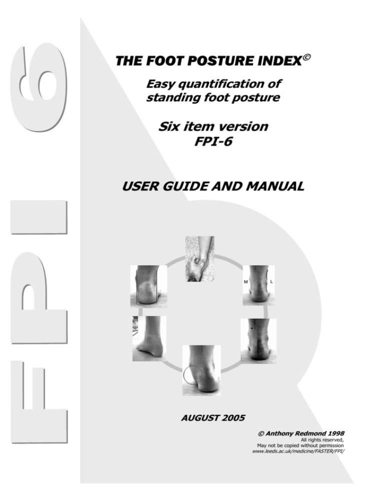
The six clinical criteria employed in the FPI-6 are:
1. Talar head palpation
2. Supra and infra lateral malleolar curvature
3. Calcaneal frontal plane position
4. Prominence in the region of the talonavicular joint
5. Congruence of the medial longitudinal arch
6. Abduction/adduction of the forefoot on the rearfoot
Using the
specified
criteria
Full explanations of each of the FPI constituent parts are
detailed subsequently, and the derivation of each is referenced
and detailed in Appendix 1. Each of the component tests or
observations are simply graded 0 for neutral, with a minimum
score of –2 for clear signs of supination, and + 2 for positive
signs of pronation. Unless the criteria outlined for each of the
features are clearly met then the more conservative score should
be awarded. It is also to be emphasised that the gradings need
to be awarded on the basis of the criteria outlined below.
Variation resulting from observations based on ‘clinical feel’ or
past experience alone will result in unacceptable inter-observer
error.
Preparing the
patient
The patient should stand in their relaxed stance position with
double limb support. The patient should be instructed to stand
still, with their arms by the side and looking straight ahead. It
may be helpful to ask the patient to take several steps, marching
on the spot, prior to settling into a comfortable stance position.
During the assessment, it is important to ensure that the patient
does not swivel to try to see what is happening for themself, as
this will significantly affect the foot posture. The patient will
need to stand still for approximately two minutes in total, in
order for the assessment to be conducted. The assessor needs
to be able to move around the patient during the assessment
and to have uninterrupted access to the posterior aspect of the
leg and foot.
4
Foot Posture Index - User guide and manual
1. Talar Head
Palpation
(Palpation for
talo-navicular
congruence)
This is the only scoring criterion that relies on palpation rather
than observation. The head of the talus is palpated on the
medial and lateral side of the anterior aspect of the ankle,
according to the standard method described variously by Root,
Elveru and many others. Scores are awarded for the observation
of the position as follows.
Diagram showing the position of the fingers when palpating of the
head of the talus. The circles indicate the precise point of palpation
on the medial and lateral side.
Clinical note: This is
not an attempt to
determine the so-called
subtalar neutral position.
For the FPI measure the
subtalar joint is not
manipulated into the
position where the head
of the talus is in maximal
congruence with the
navicular. For the FPI
measure the head of the
talus is simply palpated
in the relaxed stance
position and the talar
head orientation
reported.
It may however be
useful in some cases to
move the foot into
inversion and eversion
while palpating for the
talar head as this can aid
in determining wether
the head is still palpable
in individuals on the
border between 1 &2 or
–1&-2.
Score
-2
-1
0
1
2
Talar head
palpable
on lateral
side/but
not on
medial
side
Talar head
palpable on
lateral
side/slightly
palpable on
medial side
Talar head
equally
palpable on
lateral and
medial side
Talar head
slightly
palpable
on lateral
side/
palpable
on medial
side
Talar head
not
palpable
on lateral
side/ but
palpable
on medial
side
5
Foot Posture Index - User guide and manual
2. Supra and
infra lateral
malleolar
curvature
(Observation
and comparison
of the curves
above and
below the
lateral ankle
malleoli)
In the neutral foot it has been suggested that the curves should
be approximately equal. In the pronated foot the curve BELOW
the malleolus will be more acute than the curve above due to the
abduction of the foot, and eversion of the calcaneus. The opposite
is true in the supinated foot.
Supinated (-2)
Score
Clinical note 1: For
estimating malleolar
curvature, it may be
helpful to use a straight
edge for reference. This
can be a set square,
ruler or even a pen
according to availability.
Neutral (0)
Pronated (+2)
-2
-1
0
1
2
Curve
below the
malleolus
either
straight or
convex
Curve below
the
malleolus
concave, but
flatter/ more
shallow than
the curve
above the
malleolus
Both infra
and supra
malleolar
curves
roughly
equal
Curve
below
malleolus
more
concave
than curve
above
malleolus
Curve
below
malleolus
markedly
more
concave
than curve
above
malleolus
Clinical note 2: Where
oedema or obesity
obscures the curvature
this measures should be
either scored at zero or
removed from the
assessment and
indicated as such.
6
Foot Posture Index - User guide and manual
3. Calcaneal
frontal plane
position
(Inversion /
eversion of the
calcaneus)
This is an observational equivalent of the measurements often
employed in quantifying the relaxed and neutral calcaneal stance
positions. With the patient standing in the relaxed stance position,
the posterior aspect of the calcaneus is visualised with the observer
in line with the long axis of the foot.
Angular measurements are not required for the FPI, the foot is
graded according to visual appraisal of the frontal plane position.
Supinated (-2)
Score
-2
More than
an
estimated
5° inverted
(varus)
Neutral (0)
-1
Between
vertical and
an
estimated
5° inverted
(varus)
0
Vertical
Pronated (+2)
1
Between
vertical and
an
estimated
5° everted
(valgus)
2
More than
an
estimated
5° everted
(valgus)
7
Foot Posture Index - User guide and manual
4. Bulging in
the region of
the talonavicular
joint (TNJ)
In the neutral foot the area of skin immediately superficial to the
TNJ will be flat. The TNJ becomes more prominent if the head of the
talus is adducted in rearfoot pronation. Bulging in this area is thus
associated with a pronating foot. In the supinated foot this area may
be indented.
Supinated (-2)
Score
Clinical note:
Bulging of the TNJ
area is a common
finding in pronated
feet. However, true
convexity of the area
is usually only seen
with highly supinated
postures. Unless
there is a definite
indentation,
assigning negative
scores to this
observation should
be undertaken
judiciously.
Neutral (0)
Pronated (+2)
-2
-1
0
1
2
Area of
TNJ
markedly
concave
Area of TNJ
slightly, but
definitely
concave
Area of
TNJ flat
Area of
TNJ
bulging
slightly
Area of
TNJ
bulging
markedly
8
Foot Posture Index - User guide and manual
5. Height and
congruence of
the medial
longitudinal
arch
While arch height is a strong indicator of foot function, the shape of
the arch can also be equally important. In a neutral foot the
curvature of the arch should be relatively uniform, similar to a
segment of the circumference of a circle. When a foot is supinated
the curve of the MLA becomes more acute at the posterior end of
the arch. In the excessively pronated foot the MLA becomes
flattened in the centre as the midtarsal and Lisfranc’s joints open up.
Neutral (0)
This observation should be made
taking both the arch height and
the
arch
congruence
into
consideration.
Supinated foot (-2)
Clinical note: While
simple arch height
will usually be the
more readily
apparent of the two
components of this
measure, arch
congruence is
probably more subtle
and informative.
Careful observation
of the arch
congruence should
be the main element
of this measure with
arch height factored
in secondarily.
Score
-2
Arch high
and acutely
angled
towards the
posterior end
of the medial
arch
Pronated foot (+2)
-1
Arch
moderately
high and
slightly
acute
posteriorly
0
Arch
height
normal
and
concentric
ally
curved
1
Arch
lowered
with some
flattening
in the
central
portion
2
Arch very
low with
severe
flattening
in the
central
portion –
arch
making
ground
contact
9
Foot Posture Index - User guide and manual
6. Abduction/
adduction of
the forefoot on
the rearfoot.
(Too many toes
sign)
When viewed from directly behind, and in-line with the long axis of
the heel (not the long axis of the whole foot), the neutral foot will
allow the observer to see the forefoot equally on the medial and
lateral sides. In the supinated foot the forefoot will adduct on the
rearfoot resulting more of the forefoot being visible on the medial
side. Conversely pronation of the foot causes the forefoot to abduct
resulting in more of the forefoot being visible on the lateral side.
Supinated (-2)
Clinical note: This
measure should be
treated with caution
where there is a fixed
adduction deformity of
the forefoot on the
rearfoot in the nonweightbearing state.
Normally it is possible
to see the toes by the
observer raising their
angle of view slightly.
If the toes are
obscured by other
structures the mtp
joints or more proximal
structures can be used
as a guide.
Score
-2
No lateral
toes visible.
Medial toes
clearly
visible
Neutral(0)
-1
Medial toes
clearly
more
visible than
lateral
0
Medial
and
lateral
toes
equally
visible
Pronated (+2)
1
Lateral
toes
clearly
more
visible
than
medial
2
No medial
toes
visible.
Lateral toes
clearly
visible
10
Foot Posture Index - User guide and manual
FPI total
score
The final FPI score will be a whole number between –12 and +12.
In most cases there will be a consistent pattern of scores and the
clinical picture will be immediately clear. However in some patients
there will be a dominance of motion occurring in one of the three
body planes or a difference between the function of the forefoot and
rearfoot.
The foot segments and the body plane measured by each of the
observations are indicated on the FPI data sheet. This allows the FPI
to provide substantially more information than existing single
segment/single plane assessment techniques. While the information
needs careful clinical interpretation based on the clinician’s
knowledge of anatomy and function, the information yielded by the
FPI assessment allows such interpretation to be better informed by
data.
Examples
Example 1. Abnormal frontal plane observations predominate
in a patient, with transverse and sagittal plane measures
reading near neutral.
Talar head palpation
+1
Malleolar curves
+1
Inv/eversion calcaneus +1
TNJ prominence
0
Congruence of MLA
0
Abd/adduction of FF
+1
_______________________
TOTAL
+4
11
Foot Posture Index - User guide and manual
Example 2. The rearfoot factors may be near less marked in a
patient while the midfoot/forefoot observations indicate substantial
instability in the midfoot.
Talar head palpation
+1
Malleolar curves
+1
Inv/eversion calcaneus +1
TNJ prominence
+2
Congruence of MLA
+2
Abd/adduction of FF
+1
______________________
TOTAL
+8
In both of these cases the clinician interprets the results to put the
foot posture into its clinically relevant context. The clinician may
decide to use the FPI as a general overview of the foot function
(just using the total score) or conversely he or she may prefer to
keep the planar or segmental information disaggregated in order to
retain the differentiation of the individual components of the score.
Either way the clinician has more information available, upon which
to base a decision.
Getting to know
the FPI
The FPI is designed to be simple to use and for the set criteria to
limit variability in scoring. Nevertheless, it is worth developing some
exercise with using the measure before applying the scores in
earnest.
We recommend that the novice user rates approximately 30
individuals with as broad a range of foot types as possible before
using the FPI formally in clinic.
12
Foot Posture Index - User guide and manual
Validation of the
FPI
The validation of the FPI was conducted in several stages.
Item validity
FPI scores were compared initially to concurrently derived Valgus
Index (VI) scores. Ratings of the eight components making up the
draft FPI were undertaken for each of 131 subjects (91 male and
40 female aged 18-65 (Mean=33.7 years) while they stood on a
‘pedograph’, ink and paper mat.
In ordinal regression modelling the
FPI-8 total scores predicted 59% of
the variance in VI values (Cox and
Snell R2=0.590, B=0.551, P<0.001,
N=131)
The inter-item reliability (Cronbach’s
α) was 0.834, indicating good interitem reliability overall. The individual
coefficients were >0.65 for six of the
eight FPI components. The
components measuring Helbing’s sign
(0.36) and the congruence of the
lateral border (0.20) of the foot
showed poor inter-item reliability.
Principal components analysis yielded two separate factors. The
first included seven of the initial eight FPI items. A second factor,
explaining 12% of the variance, was mainly a function of the
congruence of the lateral border of the foot suggesting that a
separate subgroup with variation in foot position independent of
the lateral foot contour might be evident.
A Fastrak™ electromagnetic tracking (EMT) system
was then used to reconstruct a three-dimensional
lower limb model for the right leg of 20 healthy
volunteers in each of three positions (pronated,
neutral, supinated). The FPI scoring criteria (again
except lateral border shape) predicted between
63% and 80% of the variance in their EMT derived
equivalents.
Item reduction
The items Lateral border congruence and Helbing’s
sign had not demonstrated adequate validity and
were removed to produce the final six-item
instrument.
13
Foot Posture Index - User guide and manual
Validation of the
FPI
FPI-6 Instrument validity
Once the FPI had been reduced to its final six-item form the
validity was evaluated further. Six item FPI scores were compared
with contemporaneous EMT data obtained during quiet standing
and during normal walking. The FPI-6 scores predicted 64% of the
variation in the static ankle/subtalar position during quiet double
limb standing (adjusted R2=0.64, F=73.529, P<0.001, N=14). The
same FPI-6 scores predicted 41% of the variance in ankle/subtalar
position at midstance (R2 = 0.41, F=31.786, P<0.001, N=15).
Reliability
Reliability is a function of the user and patient group being
investigated rather than a characteristic of the instrument. The
independently reported inter-tester reliability of the original eight
item FPI has ranged from 0.62 to 0.91, depending on population,
and intra-tester reliability ranges from 0.81 to 0.91
See
Redmond AC. Foot Posture in Neuromuscular Disease (PhD Thesis) University of
Sydney, 2004.
Burns J., Keenan A., Redmond AC. Foot type and lower limb overuse injury in
triathletes. J Am Pod Med Assoc 2005, 95:3; 235-241.
Payne C, Oates M, Noakes H. Static stance response to different types of foot
orthoses. J Am Pod Med Assoc 2003;93(6):492- 8.
Evans AM, Copper AW, Scharfbillig RW, Scutter SD, Williams MT. The reliability
of the foot posture index and traditional measures of foot position. J Am Pod
Med Assoc 2003;93:203-13.
Yates B, White S . The incidence and risk factors in the development of medial
tibial stress syndrome among naval recruits. Am J Sports Med 2004: 32 (3): 772780
Psychometric
properties
The psychometric properties
including uni-dimensionality and
item-functioning have been evaluated
and demonstrated good fit to the
Rasch model. The robustness of its
psychometric properties (High person
separation, no differential item
functioning and good item fit), combined with the number of levels
in the scoring scale (25) means that the FPI can be used in studies
employing parametric statistical analysis.
See
Keenan AM, Redmond AC, Horton M, Conaghan PC, Tennant A. "The Foot
Posture Index: Rasch analysis of a novel, foot specific outcome measure".
Health Outcomes 2005: making a difference. Book of Proceedings. 11th Annual
National Conference, 17-18 August 2005, Canberra, Australia.
14
Foot Posture Index - User guide and manual
References and
further reading
Talar head
palpation
1.
2.
3.
4.
5.
6.
7.
8.
9.
10.
Supra and infra
lateral malleolar
curvature.
(Sanner compared
medial and lateral
malleoli)
1.
2.
Astrom M, Arvidson T. Alignment and joint motion in the normal foot.
Journal of Orthopaedic & Sports Physical Therapy 1995;22(5):216-22.
Bevans JS. Biomechanics and plantar ulcers in diabetes. The Foot
1992;2:166-172.
Diamond JE, Mueller MJ, Delitto A, Sinacore DR. Reliability of a diabetic
foot evaluation. Physical Therapy 1989;69(10):797-802.
Elveru RA, Rothstein JM, Lamb RL, Riddle DL. Methods for taking
subtalar joint measurements. A clinical report. Physical Therapy
1988;68(5):678-82.
McPoil TG, Cornwall MW. Relationship between three static angles of
the rearfoot and the pattern of rearfoot motion during walking. Journal
of Orthopaedic & Sports Physical Therapy 1996;23(6):370-5.
McPoil TG, Schuit D, Knecht HG. Comparison of three methods used to
obtain a neutral plaster foot impression. Physical Therapy
1989;69(6):448-52.
Pierrynowski MR, Smith SB. Rear foot inversion/eversion during gait
relative to the subtalar joint neutral position. Foot & Ankle
International 1996;17(7):406-12.
Pierrynowski MR, Smith SB, Mlynarczyk JH. Proficiency of foot care
specialists to place the rearfoot at subtalar neutral. Journal of the
American Podiatric Medical Association 1996;86(5):217-23.
Picciano AM, Rowlands MS, Worrell T. Reliability of open and closed
kinetic chain subtalar joint neutral positions and navicular drop test.
Journal of Orthopaedic & Sports Physical Therapy 1993;18(4):553-8.
Sell KE, Verity TM, Worrell TW, Pease BJ, Wigglesworth J. Two
measurement techniques for assessing subtalar joint position: a
reliability study. Journal of Orthopaedic & Sports Physical Therapy
1994;19(3):162-7.
Merriman LM, Tollafield DR, editors. Assessment of the Lower Limb.
Edinburgh: Churchill Livingstone; 1995.
Sanner WH. Clinical methods for predicting the effectiveness of
functional foot orthoses. Clinics in Podiatric Medicine & Surgery
1994;11(2):279-95.
15
Foot Posture Index - User guide and manual
Calcaneal
frontal plane
position
1.
Astrom M, Arvidson T. Alignment and joint motion in the normal foot.
Journal of Orthopaedic & Sports Physical Therapy 1995;22(5):216-22.
2.
Bevans JS. Biomechanics and plantar ulcers in diabetes. The Foot
1992;2:166-172.
3.
Coplan JA. Rotational motion of the knee: A comparison of normal and
pronating subjects. Journal of Orthopaedic & Sports Physical Therapy
1989;10(9):366-369.
4.
Dahle LK, Mueller M, Delitto A, Diamond JE. Visual assessment of foot type
and relationship of foot type to lower extremity injury. Journal of
Orthopaedic & Sports Physical Therapy 1991;14(2):70-4.
5.
Diamond JE, Mueller MJ, Delitto A, Sinacore DR. Reliability of a diabetic
foot evaluation. Physical Therapy 1989;69(10):797-802.
6.
Donatelli R, Wooden M, Ekedahl SR, Wilkes JS, Cooper J, Bush AJ.
Relationship between static and dynamic foot postures in professional
baseball players. Journal of Orthopaedics and Sports Physical Therapy
1999;29(6):316-330.
7.
Jahss MH. Evaluation of the cavus foot for orthopedic treatment. Clinical
Orthopaedics & Related Research 1983(181):52-63.
8.
Leppilahti J, Korpelainen R, Karpakka J, Kvist M, Orava S. Ruptures of the
Achilles Tendon - Relationship to Inequality in Length of Legs and to
Patterns in the Foot and Ankle. Foot & Ankle International
1998;19(10):683-687.
9.
Lepow GM, Valenza PL. Flatfoot overview. Clinics in Podiatric Medicine &
Surgery 1989;6(3):477-89.
10.
McPoil TG, Cornwall MW. Relationship between three static angles of the
rearfoot and the pattern of rearfoot motion during walking. Journal of
Orthopaedic & Sports Physical Therapy 1996;23(6):370-5.
11.
Merriman LM, Tollafield DR, editors. Assessment of the Lower Limb.
Edinburgh: Churchill Livingstone; 1995.
12.
Nester CJ. Rearfoot complex: A review of its interdependent components,
axis orientation and functional model. Foot 1997;7(2):86-96.
13.
Novick A, Kelley DL. Position and movement changes of the foot with
orthotic intervention during the loading response of gait. Journal of
Orthopaedic & Sports Physical Therapy 1990;11(7):301-312.
14.
Picciano AM, Rowlands MS, Worrell T. Reliability of open and closed kinetic
chain subtalar joint neutral positions and navicular drop test. Journal of
Orthopaedic & Sports Physical Therapy 1993;18(4):553-8.
15.
Sanner WH. Clinical methods for predicting the effectiveness of functional
foot orthoses. Clinics in Podiatric Medicine & Surgery 1994;11(2):279-95.
16.
Sell KE, Verity TM, Worrell TW, Pease BJ, Wigglesworth J. Two
measurement techniques for assessing subtalar joint position: a reliability
study. Journal of Orthopaedic & Sports Physical Therapy 1994;19(3):162-7.
17.
Sobel E, Levitz S, Caselli M, Brentnall Z, Tran MQ. Natural history of the
rearfoot angle: preliminary values in 150 children. Foot & Ankle
International 1996;20(2):119-125.
18.
Song J, Hillstrom HJ, Secord D, Levitt J. Foot type biomechanics.
comparison of planus and rectus foot types. Journal of the American
Podiatric Medical Association 1996;86(1):16-23.
19.
Weiner-Ogilvie S, Rome K. The reliability of three techniques for measuring
foot position. Journal of the American Podiatric Medical Association
1998;88(8):381-6.
20.
Wen DY, Puffer JC, Schmalzried TP. Lower extremity alignment and risk of
overuse injuries in runners. Medicine & Science in Sports & Exercise
1997;29(10):1291-8.
21.
Yamamoto H, Muneta T, Ishibashi T, Furuya K. Posteromedial release of
congenital club foot in children over five years of age. Journal of Bone &
Joint Surgery - British Volume 1994;76(4):555-8.
16
Foot Posture Index - User guide and manual
Prominence in
the region of the
talonavicular
joint
1.
2.
3.
4.
Height and
congruence of
the medial
longitudinal
arch
1.
2.
3.
4.
5.
6.
7.
8.
9.
10.
11.
12.
13.
Dahle LK, Mueller M, Delitto A, Diamond JE. Visual assessment of foot type
and relationship of foot type to lower extremity injury. Journal of
Orthopaedic & Sports Physical Therapy 1991;14(2):70-4.
Fraser RK, Menelaus MB, Williams PF, Cole WG. The Miller procedure for
mobile flat feet. Journal of Bone & Joint Surgery - British Volume
1995;77(3):396-9.
Gould N. Evaluation of hyperpronation and pes planus in adults. Clinical
Orthopaedics & Related Research 1983(181):37-45.
Merriman LM, Tollafield DR, editors. Assessment of the Lower Limb.
Edinburgh: Churchill Livingstone; 1995.
Cowan DN, Jones BH, Robinson JR. Foot morphologic characteristics and
risk of exercise-related injury. Archives of Family Medicine 1993;2(7):7737.
Dahle LK, Mueller M, Delitto A, Diamond JE. Visual assessment of foot type
and relationship of foot type to lower extremity injury. Journal of
Orthopaedic & Sports Physical Therapy 1991;14(2):70-4.
Fraser RK, Menelaus MB, Williams PF, Cole WG. The Miller procedure for
mobile flat feet. Journal of Bone & Joint Surgery - British Volume
1995;77(3): 396-9.
Jahss MH. Evaluation of the cavus foot for orthopedic treatment. Clinical
Orthopaedics & Related Research 1983(181):52-63.
Lepow GM, Valenza PL. Flatfoot overview. Clinics in Podiatric Medicine &
Surgery 1989;6(3):477-89.
Merriman LM, Tollafield DR, editors. Assessment of the Lower Limb.
Edinburgh: Churchill Livingstone; 1995.
Nester CJ. Rearfoot complex: A review of its interdependent components,
axis orientation and functional model. Foot 1997;7(2):86-96.
Saltzman CL, Nawoczenski DA, Talbot KD. Measurement of the medial
longitudinal arch. Archives of Physical Medicine & Rehabilitation
1995;76(1):45-9.
Sell KE, Verity TM, Worrell TW, Pease BJ, Wigglesworth J. Two
measurement techniques for assessing subtalar joint position: a reliability
study. Journal of Orthopaedic & Sports Physical Therapy 1994;19(3):1627.
Song J, Hillstrom HJ, Secord D, Levitt J. Foot type biomechanics.
comparison of planus and rectus foot types. Journal of the American
Podiatric Medical Association 1996;86(1):16-23.
Weiner-Ogilvie S, Rome K. The reliability of three techniques for measuring
foot position. Journal of the American Podiatric Medical Association
1998;88(8):381-6.
Spinner SM, Chussid F, Long DH. Criteria for combined procedure selection
in the surgical correction of the acquired flatfoot. Clinics in Podiatric
Medicine & Surgery 1989;6(3):561-75.
Wen DY, Puffer JC, Schmalzried TP. Lower extremity alignment and risk of
overuse injuries in runners. Medicine & Science in Sports & Exercise
1997;29(10):1291-8.
17
Foot Posture Index - User guide and manual
Abduction/
adduction of
the forefoot on
the rearfoot.
1.
2.
3.
4.
5.
6.
7.
8.
9.
10.
11.
12.
13.
Dahle LK, Mueller M, Delitto A, Diamond JE. Visual assessment of foot type
and relationship of foot type to lower extremity injury. Journal of
Orthopaedic & Sports Physical Therapy 1991;14(2):70-4.
Fraser RK, Menelaus MB, Williams PF, Cole WG. The Miller procedure for
mobile flat feet. Journal of Bone & Joint Surgery - British Volume
1995;77(3):396-9.
Freychat P, Belli A, Carret JP, Lacour JR. Relationship between rearfoot
and forefoot orientation and ground reaction forces during running.
Medicine & Science in Sports & Exercise 1996;28(2):225-32.
Jahss MH. Evaluation of the cavus foot for orthopedic treatment. Clinical
Orthopaedics & Related Research 1983(181):52-63.
Johnson KA. Tibialis posterior tendon rupture. Clinical Orthopaedics &
Related Research 1983(177):140-7.
Kouchi M, Tsutsumi E. Relation between the medial axis of the foot outline
and 3-D foot shape. Ergonmics 1996;39(6):853-861.
Lepow GM, Valenza PL. Flatfoot overview. Clinics in Podiatric Medicine &
Surgery 1989;6(3):477-89.
Merriman LM, Tollafield DR, editors. Assessment of the Lower Limb.
Edinburgh: Churchill Livingstone; 1995.
Nester CJ. Rearfoot complex: A review of its interdependent components,
axis orientation and functional model. Foot 1997;7(2):86-96.
Ross AS, Jones LJ. Non-weightbearing negative cast evaluation. Journal of
the American Podiatry Association 1982;72(12):634-8.
Sanner WH. Clinical methods for predicting the effectiveness of functional
foot orthoses. Clinics in Podiatric Medicine & Surgery 1994;11(2):279-95.
Spinner SM, Chussid F, Long DH. Criteria for combined procedure selection
in the surgical correction of the acquired flatfoot. Clinics in Podiatric
Medicine & Surgery 1989;6(3):561-75.
Yamamoto H, Muneta T, Ishibashi T, Furuya K. Posteromedial release of
congenital club foot in children over five years of age. Journal of Bone &
Joint Surgery - British Volume 1994;76(4):555-8.
18
Foot Posture Index Datasheet
Patient name
ID number
FACTOR
PLANE
SCORE 1
SCORE 2
SCORE 3
Date_______________
Date_______________
Date_______________
Comment___________
Comment___________
Comment___________
Forefoot
Rearfoot
Left
-2 to +2
Talar head palpation
Transverse
Curves above and below the lateral malleolus
Frontal/
transverse
Inversion/eversion of the calcaneus
Frontal
Prominence in the region of the TNJ
Transverse
Right
-2 to +2
Left
-2 to +2
Right
-2 to +2
Left
-2 to +2
Right
-2 to +2
Sagittal
Congruence of the medial longitudinal arch
Transverse
Abd/adduction forefoot on rearfoot
TOTAL
Anthony Redmond 1998
(May be copied for clinical use and adapted
with the permission of the copyright holder)
www.leeds.ac.uk/medicine/FASTER/FPI
Reference values
Normal = 0 to +5
Pronated = +6 to +9, Highly pronated 10+
Supinated = -1 to –4, Highly supinated –5 to -12
Foot Posture Index Datasheet
Patient name
ID number
FACTOR
PLANE
SCORE 1
SCORE 2
SCORE 3
Date_______________
Date_______________
Date_______________
Comment___________
Comment___________
Comment___________
Forefoot
Rearfoot
Left
-2 to +2
Talar head palpation
Transverse
Curves above and below the lateral malleolus
Frontal/
transverse
Inversion/eversion of the calcaneus
Frontal
Prominence in the region of the TNJ
Transverse
Congruence of the medial longitudinal arch
Abd/adduction forefoot on rearfoot
Right
-2 to +2
Left
-2 to +2
Right
-2 to +2
Left
-2 to +2
Right
-2 to +2
Sagittal
Transverse
TOTAL
Reference values
Normal = 0 to +5
Pronated = +6 to +9, Highly pronated 10+
Supinated = -1 to –4, Highly supinated –5 to -12
Anthony Redmond 1998
(May be copied for clinical use and adapted
with the permission of the copyright holder)
www.leeds.ac.uk/medicine/FASTER/FPI

