Sedimentation characterization and resuspendability
advertisement
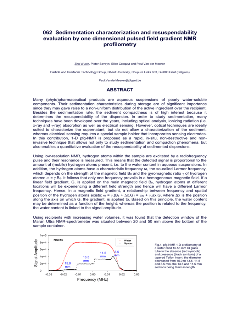
062 Sedimentation characterization and resuspendability evaluation by one dimensional pulsed field gradient NMR profilometry Zhu Wuxin, Pieter Saveyn, Ellen Cocquyt and Paul Van der Meeren Particle and Interfacial Technology Group, Ghent University, Coupure Links 653, B-9000 Gent (Belgium) Paul.VanderMeeren@Ugent.be ABSTRACT Many (phyto)pharmaceutical products are aqueous suspensions of poorly water-soluble components. Their sedimentation characteristics during storage are of significant importance since they may gave raise to a non-uniform distribution of the active ingredient over the recipient. Besides the sedimentation rate, the sediment compactness is of high interest because it determines the resuspendability of the dispersion. In order to study sedimentation, many techniques have been developed over the years, including optical analysis, ionizing radiation (i.e. x-ray and -ray) absorption as well as electrical sensing. However, optical techniques are ideally suited to characterize the supernatant, but do not allow a characterization of the sediment, whereas electrical sensing requires a special sample holder that incorporates sensing electrodes. In this contribution, 1-D pfg-NMR is proposed as a rapid, in-situ, non-destructive and noninvasive technique that allows not only to study sedimentation and compaction phenomena, but also enables a quantitative evaluation of the resuspendability of sedimented dispersions. Using low-resolution NMR, hydrogen atoms within the sample are excitated by a radiofrequency pulse and their resonance is measured. This means that the detected signal is proportional to the amount of (mobile) hydrogen atoms present, i.e. to the water content in aqueous suspensions. In addition, the hydrogen atoms have a characteristic frequency ω, the so-called Larmor frequency, which depends on the strength of the magnetic field B0 and the gyromagnetic ratio of hydrogen atoms: = .B0. It follows that only one frequency prevails in a homogeneous magnetic field. If a linear field gradient, G, is applied on the main magnetic field B0, hydrogen atoms at different locations will be experiencing a different field strength and hence will have a different Larmor frequency. Hence, in a magnetic field gradient, a relationship between frequency and spatial position of the hydrogen atoms exists: = .(Bo + x.G) = o + .x.G, where Δx is the position along the axis on which G, the gradient, is applied to. Based on this principle, the water content may be determined as a function of the height: whereas the position is related to the frequency, the water content is linked to the signal amplitude. Using recipients with increasing water volumes, it was found that the detection window of the Maran Ultra NMR-spectrometer was situated between 20 and 50 mm above the bottom of the sample container. Signal Amplitude 1e+5 8e+4 NS=16 Water Spacer 6e+4 4e+4 15.0 mm 2e+4 11.5 mm 13.5 mm Fig.1. pfg-NMR 1-D profilometry of a water-filled 15.56 mm ID glass tube in the absence (red symbols) and presence (black symbols) of a tapered Teflon insert: the diameter decreased from 15.0 to 13.5, 11.5 and 8.5 mm, the 13.5 and 11.5 mm sections being 9 mm in length. 8.5 mm 0 -0.03 -0.02 -0.01 0.00 0.01 Frequency (MHz) r content (%) 100 NS=16 80 60 40 Spacer value Calculated value 1 Calculated value 2 Calculated value 3 Calculated value 4 0.02 0.03 Considering these limitations, the spatial resolution was determined using a Teflon insert of varying diameter inside a water filled test tube (fig.1). From the three discontinuities in the profile of the tube with the insert, it is obvious that signal frequency is indeed proportional to the vertical position and that the spatial resolution is better than 0.5 mm. Figure 2 illustrates the usefulness of the 1-D profilometry approach for resuspendability evaluation. The black trace shows that the sample during storage has been sedimenting, giving raise to a (sediment) part of low signal amplitude (and hence low water content) at low frequencies and a (supernatant) part of high signal amplitude at high frequency. From the ratio of the amplitudes, it follows that the water content of the sediment is about 35%. As the resuspending intensity is increased, the water content of the supernatant is decreased (by dispersed particles), whereas the thickness of the sediment decreases and its water content increased. The fact that the area under the profile remains remarkably constant is a further indication of the accuracy of the method. Signal Amplitude 8000 6000 4000 No shaking (Area 6.1e7) 30 s shaking (Area 6.17e7) 45 s shaking (Area 6.19e7) 60 s shaking (Area 6.27e7) Vigorous shaking (Area 5.58e7) 10 s vortexing (Area 5.36e7) 2000 0 -0.010 -0.008 -0.006 -0.004 -0.002 0.000 0.002 0.004 Frequency (MHz) Fig.2. pfg-NMR 1-D profilometry of a sedimented aqueous drug suspension as a function of the respuspending intensity These preliminary results reveal that pfg-NMR enables the in-situ, non-destructive, noninvasive and accurate determination of one-dimensional (vertical) water profiles, even of turbid or viscous samples, which makes the method ideally suited to follow demixing (such as sedimentation or creaming), as well as mixing (resuspendability) phenomena.
