King Abdulaziz University
advertisement
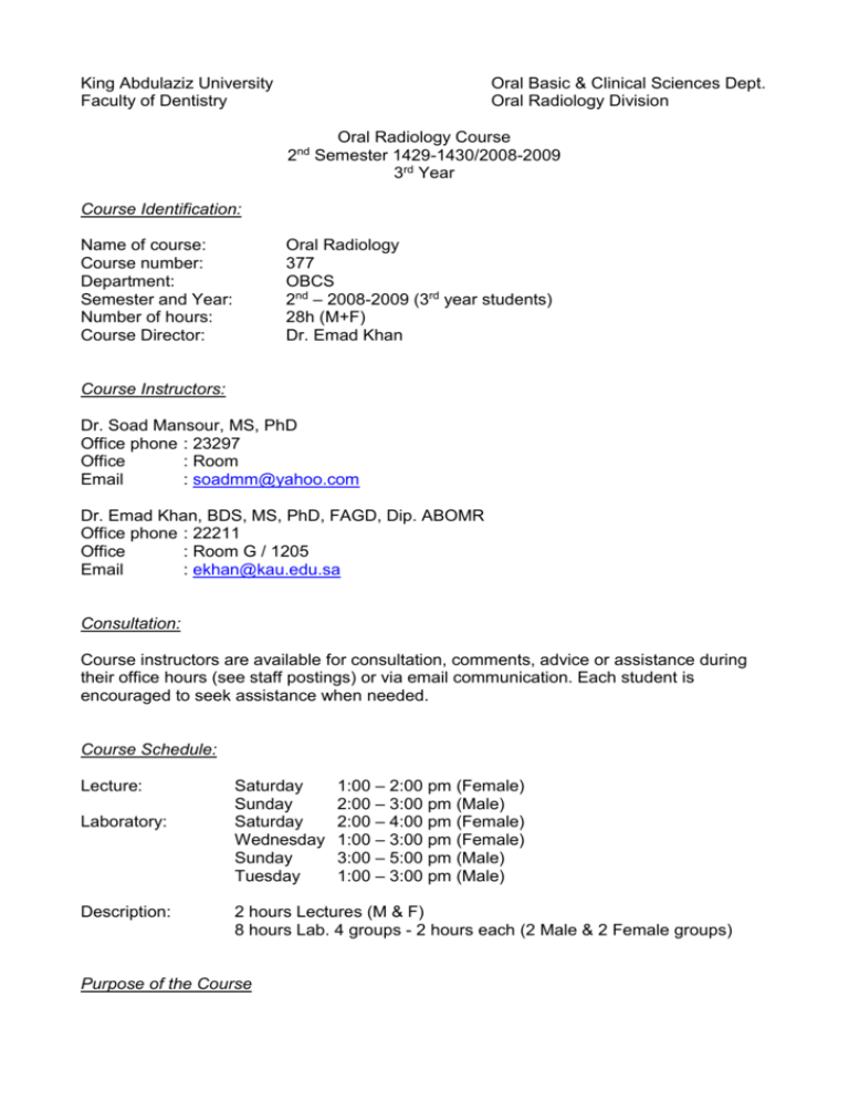
King Abdulaziz University Faculty of Dentistry Oral Basic & Clinical Sciences Dept. Oral Radiology Division Oral Radiology Course 2nd Semester 1429-1430/2008-2009 3rd Year Course Identification: Name of course: Course number: Department: Semester and Year: Number of hours: Course Director: Oral Radiology 377 OBCS 2nd – 2008-2009 (3rd year students) 28h (M+F) Dr. Emad Khan Course Instructors: Dr. Soad Mansour, MS, PhD Office phone : 23297 Office : Room Email : soadmm@yahoo.com Dr. Emad Khan, BDS, MS, PhD, FAGD, Dip. ABOMR Office phone : 22211 Office : Room G / 1205 Email : ekhan@kau.edu.sa Consultation: Course instructors are available for consultation, comments, advice or assistance during their office hours (see staff postings) or via email communication. Each student is encouraged to seek assistance when needed. Course Schedule: Lecture: Laboratory: Description: Saturday Sunday Saturday Wednesday Sunday Tuesday 1:00 – 2:00 pm (Female) 2:00 – 3:00 pm (Male) 2:00 – 4:00 pm (Female) 1:00 – 3:00 pm (Female) 3:00 – 5:00 pm (Male) 1:00 – 3:00 pm (Male) 2 hours Lectures (M & F) 8 hours Lab. 4 groups - 2 hours each (2 Male & 2 Female groups) Purpose of the Course The course offers the third year dental student lectures and laboratory sessions on the preclinical theoretical and practical radiologic sciences applicable to general dentistry. It will provide students with the basic knowledge of radiation physics and x-ray production. It will assist the students in mastering intra-oral dental radiography including the techniques, selecting the receptor, processing, mounting, viewing and trouble-shooting common errors. The course will also introduce some clinical aspects of dental radiography like the application of radiation protection protocols, recognition of anatomical landmarks and following radiographic prescription guidelines. Course Objectives Upon completion of this course, students should be able to: 1. Describe the nature and characteristics of radiation, the physics and electronics of x-ray production, and the interaction of ionizing radiation and matter. 2. Describe general function and operation of dental x-ray machines, digital and film image receptors, darkroom procedures and photochemistry, and image acquisition and display. 3. Describe in words or diagrams, the factors involved with image formation. 4. Describe the deficiencies in poor quality radiographs and discuss modifications that will result in improved quality. 5. Describe intraoral radiographic techniques and their appropriate use. 6. Describe the basic principles of radiation biology, radiation safety and protection, risk/benefit considerations, dose reduction strategies, and high yield selection criteria. 7. Tailor an appropriate radiographic examination based on the patient history and clinical presentation using selection criteria. 8. Describe and recognize the normal anatomic structures of the teeth, jaws and skull as they appear on intraoral radiographs. 9. Apply the aforementioned body of knowledge by successfully completing a laboratory exercise that includes the exposure of a complete intraoral radiographic examination on a manikin and processing, mounting, and critiquing the resultant images. 10. Demonstrate knowledge of infection control principles through consistent application of infection control procedures in all clinical activity. Teaching Method: Lectures: PowerPoint presentations Laboratory sessions: Demonstration on x-ray films, machines and other items. Lab exercises on manikins for patient handling, film exposing, processing and mounting of FMX 14/4. Critiqing and applying correction procedures for acquired images. Discussion on anatomical landmarks. Course Requirements and Grading: Policy on attendance: Attendance is mandatory. Grading: A= Excellent B= Very Good C= Good D= Satisfactory F= Failure 85 - 100% 75 - <85% 65 - <75% 60 - <65% <60% Required text: 1. White, S.C. and Pharoah, M.J. Oral Radiology, Principles and interpretation, 2004. Supplemental text: Langland and Langlais: Principles for dental imaging 2003. Other texts and/or materials will be distributed in class. Requirements: (10%) - Two diagnostically acceptable and correctly mounted FMX on a manikin with 85% grade or better. - Maxillary topographic and mandibular cross-sectional occlusal projection on manikin. Method of Evaluation: Continuous assessment (40%) - Mid Term Exam: MCQ’s & short essay questions (20%) - Requirements (10%) - Practical Exam (10%) Final Assessment (60%) - Written Exam: MCQ’s & short/long essay questions (45%) - Practical Exam (15%)
