SSN HISTO
advertisement
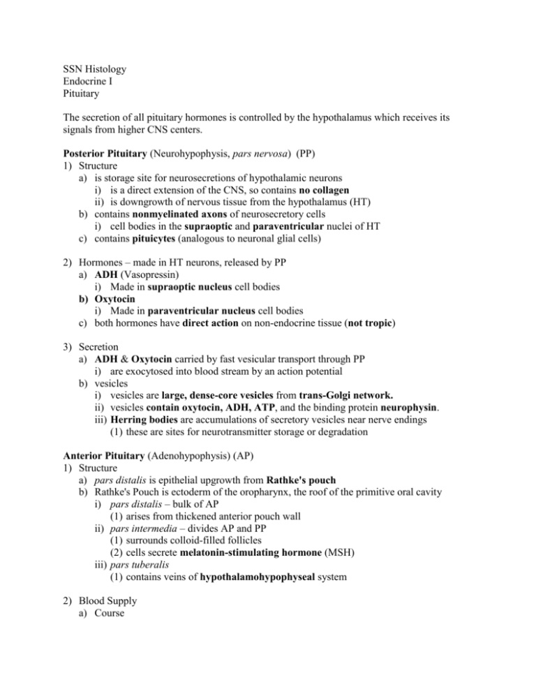
SSN Histology Endocrine I Pituitary The secretion of all pituitary hormones is controlled by the hypothalamus which receives its signals from higher CNS centers. Posterior Pituitary (Neurohypophysis, pars nervosa) (PP) 1) Structure a) is storage site for neurosecretions of hypothalamic neurons i) is a direct extension of the CNS, so contains no collagen ii) is downgrowth of nervous tissue from the hypothalamus (HT) b) contains nonmyelinated axons of neurosecretory cells i) cell bodies in the supraoptic and paraventricular nuclei of HT c) contains pituicytes (analogous to neuronal glial cells) 2) Hormones – made in HT neurons, released by PP a) ADH (Vasopressin) i) Made in supraoptic nucleus cell bodies b) Oxytocin i) Made in paraventricular nucleus cell bodies c) both hormones have direct action on non-endocrine tissue (not tropic) 3) Secretion a) ADH & Oxytocin carried by fast vesicular transport through PP i) are exocytosed into blood stream by an action potential b) vesicles i) vesicles are large, dense-core vesicles from trans-Golgi network. ii) vesicles contain oxytocin, ADH, ATP, and the binding protein neurophysin. iii) Herring bodies are accumulations of secretory vesicles near nerve endings (1) these are sites for neurotransmitter storage or degradation Anterior Pituitary (Adenohypophysis) (AP) 1) Structure a) pars distalis is epithelial upgrowth from Rathke's pouch b) Rathke's Pouch is ectoderm of the oropharynx, the roof of the primitive oral cavity i) pars distalis – bulk of AP (1) arises from thickened anterior pouch wall ii) pars intermedia – divides AP and PP (1) surrounds colloid-filled follicles (2) cells secrete melatonin-stimulating hormone (MSH) iii) pars tuberalis (1) contains veins of hypothalamohypophyseal system 2) Blood Supply a) Course i) Arterial blood runs from ICA to the superior hypophysial artery to the hypothalamus ii) blood is distributed in the primary capillary plexus in the median eminence iii) plexus drains to hypophysial portal veins, leading to secondary capillary plexus iv) portal veins supply venous blood to AP v) all capillary plexuses are fenestrated vi) finally drains to cavernous sinus b) Portal Blood Supply Importance i) HT hormones can be delivered to AP directly and in high concentration ii) HT hormones do not appear at high levels in systemic circulation iii) Most of the AP has no direct arterial supply 3) Hormones a) All AP hormones are synthesized in AP b) With one exception, all AP hormones are regulated by HT stimulating hormone i) exception: prolactin is inhibited by dopamine Tropic modulate the secretory activity of other endocrine glands TSH ACTH FSH LH Direct-Action act directly on non-endocrine tissue GH Prolactin The secretory cells of the anterior pituitary are divided into classes based on staining of their secretory vesicles with Masson's stain: Chromophils Acidophils (red/pink) Basophils (blue/black) Somatotrophs (GH) Thyrotrophs (TSH) Mammotrophs (Prl) Corticotrophs (ACTH) Gonadotrophs (FSH, LH) Chromophobes (orange/gray) smallest cell type few granules unstained cytoplasm Note that the corticotrophs have been thought to be chromophobes because they have few granules and stain very lightly. SSN Histology Endocrine I The Adrenal Gland The adrenal gland is made of 2 distinct parts that have different origins and secrete different classes of hormones: 1) adrenal cortex a) derived from the outer mesoderm b) secretes steroid hormones 2) adrenal medulla a) derived from the neural crest b) secretes peptide hormones 1) The Blood Supply of the Adrenals The arteries supplying the adrenal gland break up into a fine network of small vessels, called the capsular plexus, in the connective tissue capsule surrounding the whole gland. This plexus feeds both the cortex and the medulla: a) Cortex The vessels of the plexus feed a rich anastomosing network of cortical sinusoids. These sinusoids weave in and out between the cells of the cortex , eventually draining into small venules in the medulla. The small venules drain into the central vein of the adrenal gland. b) Medulla The capsular plexus also gives off small arteries that descend thru the cortex to the medulla where they ramify into a network of capillaries surrounding the cells of the medulla. These capillaries also eventually drain into the central vein of the adrenal gland. Note that the cells of the medulla are exposed to fresh arterial blood and blood coming from the sinusoids which have already been exposed to cortical cells. (This is important for regulation of hormone secretion.) 2) Adrenal Cortex a) Purpose: secretes steroid hormones b) Histological characteristics i) abundant lipid droplets (which are washed out during the fixation process) ii) vesicular mitochondria iii) abundant smooth ER c) Structure – 3 parts (from outer capsule inward) i) Zona Glomerulosa (ZG): (1) comprises about 15% of the cortical volume (2) the cells are arranged in twisting loops or arching cords (3) the cells have less cytoplasm than the Zona Fasciculata (ZF) so ZG appears as a darker layer (at lower powers) (4) they produce mineralocorticoids (e.g. aldosterone) (a) responsible for increased Na+ and water retention (b) secretion of these hormones are under the feedback control of the reninangiotensin system ii) Zona Fasciculata (ZF): (1) comprises about 80% of the cortical volume (2) the cells are arranged in long straight columns (3) the cells have a large amount of lipid rich cytoplasm light appearance (4) the cells should appear large and pale due to the washed out lipid vacuoles (5) they produce glucocorticoids (e.g. cortisol) (a) important in regulating carbohydrate, fat, and protein metabolism (6) secretion of hormones is under the feedback control of the CRF-ACTH system iii) Zona Reticularis (ZR): (1) comprises about 5-7% of the cortical volume (2) cells are arranged in an irregular network of branching cords and clumps (3) cells are noticeably smaller & darker than ZF cells (4) some of the cells appear brown; this is lipofuscin (a) pigment produced from normal "wear and tear" of the cells (5) principally produce a small amount of androgens, some glucocorticoids (6) secretion of hormones is under the feedback control of the CRF-ACTH system 3) Adrenal Medulla a) General i) the cells are large compared to the ZR ii) look for the large, muscular central vein b) Histology i) cells stain basophilically on H&E (they secrete peptide hormones, so they don't have the characteristic "bubbly/frothy" look of steroid secreting cells) ii) some cells may have a brownish tint due to the accumulation of chromaffin pigment (which is oxidized catecholamines) iii) cells secreting norepinephrine show a more vigorous chromaffin reaction c) Hormones i) the cells of the medulla synthesize, store, and secrete catecholamines (1) (norepinephrine and epinephrine) d) Regulation i) cells are stimulated to release their hormones into the bloodstream by preganglionic svmpathetic fibers ii) cells may also be regulated by glucocorticoids (e.g. cortisol) coming directly from the cortex via the cortical sinusoids Source zona glomerulosa zona fasciculata zona reticularis adrenal medulla Hormone mineralocorticoids (e.g. aldosterone) glucocorticoids (e.g. cortisol) sex steroids, some glucocorticoids catecholamines (NorEpi, Epi) Regulation renin-angiotensin system CRF-ACTH system CRF-ACTH system autonomic sympathetic innervation, direct adrenal cortical glucocorticoids
