lab report format handout
advertisement
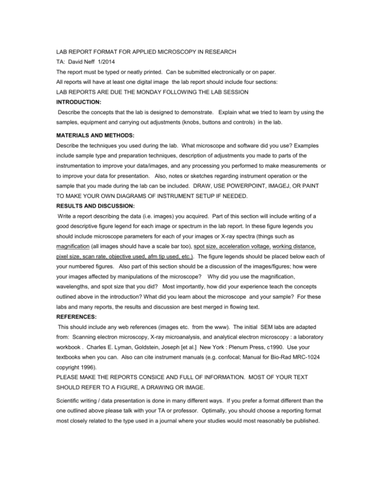
LAB REPORT FORMAT FOR APPLIED MICROSCOPY IN RESEARCH TA: David Neff 1/2014 The report must be typed or neatly printed. Can be submitted electronically or on paper. All reports will have at least one digital image the lab report should include four sections: LAB REPORTS ARE DUE THE MONDAY FOLLOWING THE LAB SESSION INTRODUCTION: Describe the concepts that the lab is designed to demonstrate. Explain what we tried to learn by using the samples, equipment and carrying out adjustments (knobs, buttons and controls) in the lab. MATERIALS AND METHODS: Describe the techniques you used during the lab. What microscope and software did you use? Examples include sample type and preparation techniques, description of adjustments you made to parts of the instrumentation to improve your data/images, and any processing you performed to make measurements or to improve your data for presentation. Also, notes or sketches regarding instrument operation or the sample that you made during the lab can be included. DRAW, USE POWERPOINT, IMAGEJ, OR PAINT TO MAKE YOUR OWN DIAGRAMS OF INSTRUMENT SETUP IF NEEDED. RESULTS AND DISCUSSION: Write a report describing the data (i.e. images) you acquired. Part of this section will include writing of a good descriptive figure legend for each image or spectrum in the lab report. In these figure legends you should include microscope parameters for each of your images or X-ray spectra (things such as magnification (all images should have a scale bar too), spot size, acceleration voltage, working distance, pixel size, scan rate, objective used, afm tip used, etc.). The figure legends should be placed below each of your numbered figures. Also part of this section should be a discussion of the images/figures; how were your images affected by manipulations of the microscope? Why did you use the magnification, wavelengths, and spot size that you did? Most importantly, how did your experience teach the concepts outlined above in the introduction? What did you learn about the microscope and your sample? For these labs and many reports, the results and discussion are best merged in flowing text. REFERENCES: This should include any web references (images etc. from the www). The initial SEM labs are adapted from: Scanning electron microscopy, X-ray microanalysis, and analytical electron microscopy : a laboratory workbook . Charles E. Lyman, Goldstein, Joseph [et al.] New York : Plenum Press, c1990. Use your textbooks when you can. Also can cite instrument manuals (e.g. confocal; Manual for Bio-Rad MRC-1024 copyright 1996). PLEASE MAKE THE REPORTS CONSICE AND FULL OF INFORMATION. MOST OF YOUR TEXT SHOULD REFER TO A FIGURE, A DRAWING OR IMAGE. Scientific writing / data presentation is done in many different ways. If you prefer a format different than the one outlined above please talk with your TA or professor. Optimally, you should choose a reporting format most closely related to the type used in a journal where your studies would most reasonably be published.


