Mitosis & Meiosis
advertisement
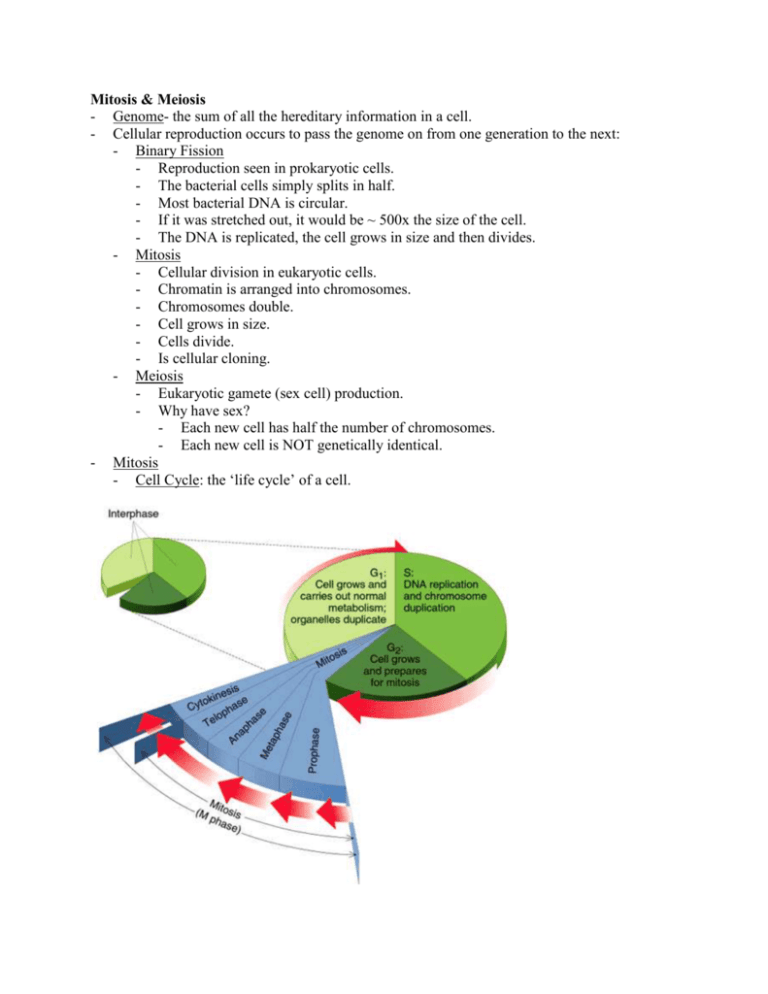
Mitosis & Meiosis - Genome- the sum of all the hereditary information in a cell. - Cellular reproduction occurs to pass the genome on from one generation to the next: - Binary Fission - Reproduction seen in prokaryotic cells. - The bacterial cells simply splits in half. - Most bacterial DNA is circular. - If it was stretched out, it would be ~ 500x the size of the cell. - The DNA is replicated, the cell grows in size and then divides. - Mitosis - Cellular division in eukaryotic cells. - Chromatin is arranged into chromosomes. - Chromosomes double. - Cell grows in size. - Cells divide. - Is cellular cloning. - Meiosis - Eukaryotic gamete (sex cell) production. - Why have sex? - Each new cell has half the number of chromosomes. - Each new cell is NOT genetically identical. - Mitosis - Cell Cycle: the ‘life cycle’ of a cell. - There are 2 phases: - M phase (mitotic phase) - - - Prophase - The nucleolus disappears. - Chromatin condenses into visible chromosomes. - There are two sister chromatids held together by a centromere. - The mitotic spindle forms in the cytoplasm. - Pro-Metaphase - The nuclear envelope disappears. - Spindle fibers extend from each pole to the cell’s equator. - Spindle fibers attach to the centromeres. - Metaphase - Chromosomes are lined up in the equator (middle) of the cell. - This is called the metaphase plate. - Anaphase - Characterized by movement. It begins when pairs of sister chromatids pull apart. - Sister chromatids move to opposite poles of the cell. - Chromosomes look like a “V” as they are pulled. - At the end of anaphase, the two poles have identical number and types of chromosomes. - Telophase & cytokinesis - Microtubules elongate the cell. - Daughter nuclei begin to form at the two poles. - Nuclear envelopes re-form. - Nucleolus reappears. - Chromatin uncoils. - Cells split their cytoplasm. - It is basically the opposite of prophase. Interphase - The non-dividing phase in a cell - Lasts about ~ 90% of the cell cycle. - The cell grows and replicates DNA preparing for Mitosis. - There are four periods: - 1. Go – a cell functioning as normal - 2. G1 phase – first growth phase - 3. S phase- synthesis of DNA - 4. G2 phase- 2nd growth phase Mitosis is a reliable process. Only one error occurs per 100,000 cell divisions. External & Internal Cues to Cell Division: - Normal growth, development and maintenance depend on the timing and rate of mitosis. - Various cell types differ in their pattern of cell division. - Human skin cells - Liver cells (only when needed) - Nerve and brain cells. - Cell density: - - - - - Crowding of cells normally inhibits cell division. Space - nutrients G1 of Interphase: - Whether a cell is destined to divide is determined at the restriction point. - If a cell is destined to divide, it proceeds to S phase. - If a cell is not destined to divide, it remains in Go. Cell Size: - The rate of cytoplasmic volume to genome size is the most important indicator of whether or not a cell will pass the restriction point. The ordered sequence of the cell cycle events is synchronized by rhythmic changes in the activity of regulatory proteins and some protein kinases. Protein kinases are enzymes that catalyze the transfer of a phosphate group from ATP to a target protein. Phosphorylation induces a conformational change that either activates or inactivates the target protein. Cyclins: - Are regulatory proteins. - Their concentration changes during the cell cycle. - Protein kinases that regulate cell cycles are called cyclin-dependent kinases (Cdks). - Even though Cdk concentration remains constant during the cell cycle, its activity changes in response to the changes in cyclin concentration. - Example: MPF - Maturation promoting factor. - Controls the progress from late interphase (G2) to mitosis. - Active MPF phosphorylates chromatin proteins, causing chromosomes to condense during prophase. - During prometaphase, the nuclear envelope disperses when its membrane proteins are phosphorylated. - Cyclin’s rhythmic activity changes in concentration regulate MPF activity. Thus it acts as a mitotic clock that regulates division. - Cyclin is produced at a uniform rate throughout the cell cycle and it accumulates during interphase. - Cyclin combines with Cdk to form active MPF. - Active MPF activates proteins that participate in mitosis and initiates prophase. - Near the end of mitosis, an enzyme that is activated by MPF destroys cyclin. - The destruction of cyclin causes the decline in active MPF at the end of mitosis. - Since MPF is activated by cyclin, it is indirectly responsible for its own decline. - Continuing cyclin synthesis raises the concentration again during interphase. - The newly formed cyclin binds to Cdk to make active MPF and mitosis begins again. Cancer Cells: - Do not respond normally to controls on cell division. - They divide excessively. - They may invade other tissues and if unchecked can kill the entire organism. - - Cancer cells in culture do not stop growing in response to cell density; they continue to grow until nutrients in the medium are exhausted. In culture, they are immortal. Normal mammalian cells divide ~ 20-50 times before they stop. What went wrong? - Immune system normally destroys cancer cells. - Enzyme f(x) may be impaired. - Oncogenes - Cancer suppressor genes - If abnormal cells evade destruction, they may become a tumor. - If cells remain at their original site, the tumor is benign. - A tumor is malignant if it has the ability to spread. - Only a malignant tumor is considered to be cancerous. - The spreading of cancer is called metastasis. Meiosis: - The formation of sex cells. - Spermatogenesis: formation of sperm cells. - Oogenesis: formation of egg cells. - Somatic cells: all cells except reproductive cells. - Gametic cells: all sex cells - Humans have 44 somatic chromosomes and 2 sex chromosomes. - Males have 44 + XY - Females have 44 + XX - Diploid: have 2 pairs of every type of chromosome (2n). - Humans have 23 pairs. - Haploid: a cell that has half the number of chromosomes for the species. All gametes are haploid (n). - When two haploid cells join, they make a diploid zygote. - Meiosis consists of separate 2 nuclear and 2 cytoplasmic divisions. - Meiosis I: creating genetic diversity - Prophase I - Homologous chromosomes lie lengthwise side by side (synapsis). This results in the formation of tetrads. - The number of tetrads per prophase I cell is equal to the haploid chromosome number. - In many species, prophase I is a very long phase, especially for oogenesis. - Crossing Over - Crossing over occurs between homologous chromatids. - Enzymes break homologous chromatids and then recombine to produce a new combination of genes. - This results in genetic diversity. - In late prophase I, homologous chromosomes are held together at specialized regions called chiasmata. - Each chiasmata is the site of crossing over. - - Similar to mitosis: Nuclear envelope disappears Chromosomes are visible - Metaphase I - Both sister chromatids are attached to spindle fibers. - These tetrads move to the metaphase plate. - Anaphase I - The tetrads separate and the pairs of sister chromatids pull to separate poles. - Each pole receives a random mixture of maternal and paternal chromosomes. - Telophase I - Each nuclear envelope may reorganize and cytokinesis occurs. - The result: two diploid cells (2n) that are now genetically different from the original parent cell. - Interkinesis - This is an interphase-like stage following Meiosis I. - It is a very short period or absent all together. - There is NO duplication of DNA. - You have 2 2n cells getting ready to divide again. - Meiosis II: reduction of chromosome #. - Prophase II - Here is no pairing of homologous chromosomes, because only one member of each pair is present in each nuclei. - There is no crossing over. - There are no tetrads. - Metaphase II - The chromosomes line up along the metaphase plate. - Anaphase II - Sister chromatids separate. - Chromosomes move to opposite poles. - Telophase II - There is one representative of each sister chromatid at each pole. - The cell undergoes cytokinesis. - The result: 4 cells, all genetically different with a haploid number (n) of chromosomes. Genetic variation has 2 sources: - During meiosis maternal and paternal chromosomes are shuffled (anaphase I). - Crossing over during prophase I.


