fellowship guidelines - Australian College of Veterinary Scientists
advertisement
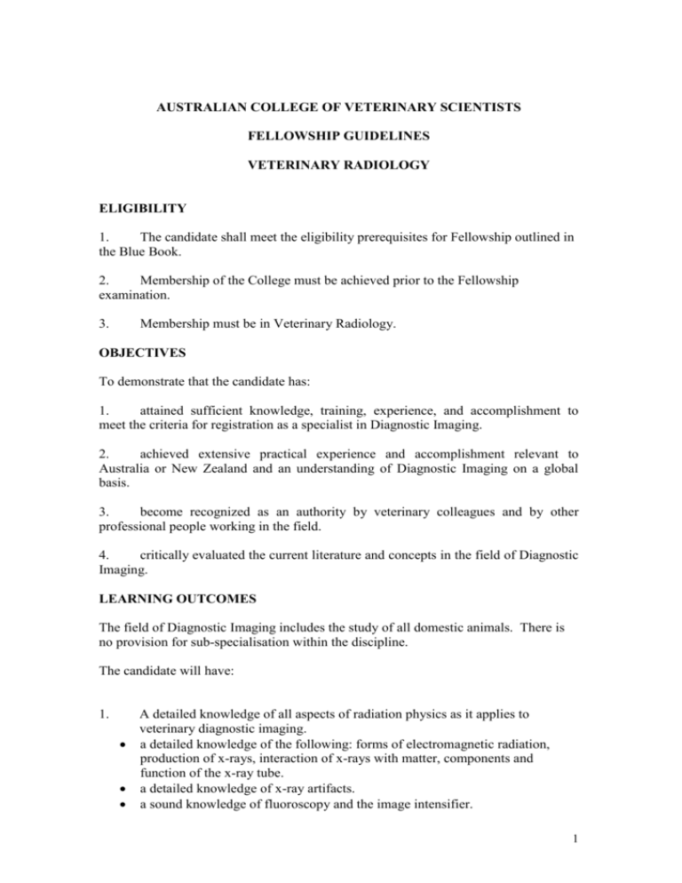
AUSTRALIAN COLLEGE OF VETERINARY SCIENTISTS FELLOWSHIP GUIDELINES VETERINARY RADIOLOGY ELIGIBILITY 1. The candidate shall meet the eligibility prerequisites for Fellowship outlined in the Blue Book. 2. Membership of the College must be achieved prior to the Fellowship examination. 3. Membership must be in Veterinary Radiology. OBJECTIVES To demonstrate that the candidate has: 1. attained sufficient knowledge, training, experience, and accomplishment to meet the criteria for registration as a specialist in Diagnostic Imaging. 2. achieved extensive practical experience and accomplishment relevant to Australia or New Zealand and an understanding of Diagnostic Imaging on a global basis. 3. become recognized as an authority by veterinary colleagues and by other professional people working in the field. 4. critically evaluated the current literature and concepts in the field of Diagnostic Imaging. LEARNING OUTCOMES The field of Diagnostic Imaging includes the study of all domestic animals. There is no provision for sub-specialisation within the discipline. The candidate will have: 1. A detailed knowledge of all aspects of radiation physics as it applies to veterinary diagnostic imaging. a detailed knowledge of the following: forms of electromagnetic radiation, production of x-rays, interaction of x-rays with matter, components and function of the x-ray tube. a detailed knowledge of x-ray artifacts. a sound knowledge of fluoroscopy and the image intensifier. 1 2. a sound knowledge of radiographic contrast media (including mechanism, side effects, and dose). Digital radiography a sound knowledge of digital radiography including image formation, different capture devices, resolution and storage, the processing of PSP plates, the advantages and disadvantages of different types of digital radiography. a basic knowledge of the artifacts associated with digital radiography 3. 4. a) b) c) d) Ultrasound A detailed knowledge of the physical principles and application of diagnostic ultrasound, including the following: a) a detailed understanding of the physical characteristics of the ultrasound beam and the interaction of ultrasound with matter (including acoustic impedance, reflection, refraction, scatter, attenuation) b) a detailed understanding of the action of a piezoelectric crystal c) a sound understanding of various transducer types, including factors that affect transducer resolution and image formation a detailed understanding of Doppler, harmonic and compound ultrasound d) a sound understanding of ultrasound artifacts. e) a sound understanding of ultrasound contrast media and their effects f) a basic understanding of the biological effects of ultrasound Radiation physics: An understanding of radiation physics as it applies to radionuclides, nuclear medicine and radiation therapy. The candidate should have a basic understanding of types of decay, the decay equation and generator systems, radiation detector systems (including those relevant to veterinary medicine, like photomultiplier tubes, scintillators and other counters. a basic knowledge of scintigraphic collimators and the construction and function of a gamma camera. a basic knowledge of factors that limit spatial and temporal resolution of scintigraphic images. A basic understanding of the indications and techniques in radiation oncology. 5. CT and MRI: physics and practical application For CT, the candidate should have a) a detailed knowledge of image formation, including CT construction and scanner types, image manipulation, and factors that affect spatial and contrast resolution of the image (factors that affect image quality). b) a sound knowledge of the factors that affect image display (including Hounsfield units). c) a sound knowledge of image artifacts and CT contrast media. For MRI, the candidate requires a) a basic knowledge of physics of magnetism as it relates to MRI. b) a detailed knowledge of the formation and display of MR images is required; including understanding of T1, T2, PD weighted images, and other common sequences (e.g. fat saturation and fluid-attenuation sequences). c) a sound knowledge of MRI contrast media, including mechanisms, dose and side effects. 2 d) A basic knowledge of magnet design, factors that affect MR safety and radiofrequency coil function. 6. Radiation protection in veterinary medicine. The candidate should have a) a basic knowledge of atomic and nuclear physics (including atomic composition, structure and binding forces, forms of decay, half-life equation). b) a sound knowledge of the forms of particulate radiation and their interactions with matter (including x-rays, gamma rays, alpha particles, electrons, protons and neutrons) c) a basic knowledge of the differences between direct and indirect ionization, and the biological effects associated with each form d) a basic knowledge of the mechanisms of acute and late radiation injury, including the difference between deterministic and stochastic effects of radiation induced injury e) a basic knowledge of the term exposure (including equivalent dose, absorbed dose) f) a detailed knowledge of radiation monitoring and safety equipment and regulations, and should be able to convey this information in a coherent matter to other veterinarians. 7. A detailed knowledge of the relevant Australian or New Zealand laws and Codes of Practice as they apply to the use of ionising radiation. 8. Anatomy and physiology: a detailed knowledge of the anatomy and physiology of domestic animals as related to veterinary diagnostic imaging. The candidate requires a) a detailed knowledge of the anatomy and physiology of the following species: canine, feline and equine b) a sound knowledge of the anatomy and physiology of birds, c) a basic knowledge of the anatomy and physiology of bovine, ovine, camelids and pocket pet species. d) A sound knowledge of the embryology of the cardiovascular, urinary and neurological systems as it relates to development of congenital conditions of these systems. 9. Clinical and pathophysiological features of the disease of domestic animals as related to veterinary diagnostic imaging. a) a detailed knowledge of canine, feline disease. b) a detailed knowledge of equine musculoskeletal disease, a sound knowledge of neurological and respiratory disease, and a basic understanding of pathophysiology of equine alimentary disease. c) a basic knowledge of avian disease d) a basic understanding of the relevant differences in clinical and pathophysiological features of disease amongst all species. 10. Image acquisition, interpretation and reporting: The candidate should have a) a demonstrable advanced skill in acquisition, interpretation and reporting of radiographic images. For advanced imaging the candidate should have 3 b) experience in both the acquisition and interpretation of CT and MRI for small animals, and some experience in interpretation of these modalities in horses. c) some experience in acquisition of scintigraphic studies and a sound knowledge of interpretation of studies in both small and large animals. The candidate should have detailed knowledge of the indications and contraindications for all studies of all modalities. 11. Demonstrable sonography and sonology skills including both practical skills required to examine patients and interpretative and communication skills that demonstrate an ability to advise referring veterinarians of findings. In particular the candidate requires a) a detailed knowledge of abdominal and thoracic sonography b) a sound knowledge of musculoskeletal, vascular and small parts sonography c) a basic knowledge of reproductive sonography (in dogs, cats and horses). d) a sound knowledge of echocardiography. e) The candidate is required to be experienced in ultrasound guided biopsy techniques, including fine needle aspiration and percutaneous biopsy, with a sound knowledge of the incidence of complications of these techniques. 12. Special radiographic and ultrasonographic procedures: The candidate should have a) a detailed knowledge of the pharmacology of contrast media and their physiological effects. b) a detailed knowledge of myelography, oesophagography, urinary contrast and fluroscopic studies. c) a sound knowledge of other less frequently performed procedures, including sinography, low-dose gastrograms and other gastrointestinal studies, selective and non-selective angiography. 13. A demonstrable skill in performing special radiologic and ultrasonographic procedures. 14. A sound knowledge of the principles of veterinary cardiology. EXAMINATIONS Refer to the Blue Book Section 7. Written Paper I: This paper is designed to test the Candidate’s knowledge of the principles of Veterinary Diagnostic Imaging as described in the Learning Outcomes. Answers may cite specific examples where general principles apply, but should primarily address the theoretical basis underlying each example. Questions may be of any type including short answer, multiple choice and essay based questions. Written Paper II: 4 This paper is designed to (a) test the Candidate’s ability to apply the principles of the Veterinary Diagnostic Imaging to particular cases/problems or tasks, and to (b) test the Candidate’s familiarity with the current practices and current issues that arise from activities within the discipline of Veterinary Diagnostic Imaging in Australia and New Zealand. Questions may be of any type including short answer, multiple choice and essay based questions. Practical examination: The practical examination will be conducted in two sessions, each approximately 3 hours (for a total of ~6 hours). The examination will cover interpretation of radiographs, ultrasound, CT, MRI, nuclear medicine, special procedures, and their artifacts. The emphasis will be on the core species (canine, feline, equine); however, cases from any species may be included. Oral examination: Refer to Blue Book TRAINING PROGRAMS Refer to the Blue Book Section 4.3 In addition to the stipulations of the Blue book: 1. The Radiology Chapter requires a three year training program (144 weeks) 2. Clinical training should include primarily exposure to dogs, cats and horses with some exposure to ruminants, camelids, exotic animals and birds. 3. Clinical training should include the following: radiography, radiology, contrast procedures, fluoroscopy, digital radiography, sonography, sonology, scintigraphy, computerised tomography and magnetic resonance imaging. 4. The candidate should interpret a minimum 3000 radiological examinations of small animals (primarily dogs and cats), 500 radiological examinations of large animals (primarily horses), 1000 sonographic examinations, and a minimum of 100 examinations that demonstrate adequate knowledge and interpretive skills in CT, MRI and nuclear medicine. The cases to be included in the case log will be those cases in which the candidate has produced a written report. If, for example, a case has an osteosarcoma of its radius and thoracic radiographs for a metastasis check then this may be counted as two cases if a report is produced for both regions. The training program should be targeted, with achievable goals set by the Supervisor and candidate for each 6 months. It is anticipated that the first year should be spent initially learning some radiography, then concentrating on radiology and ultrasound with some exposure to the other modalities. The second year is spent consolidating the first with more CT, nuclear medicine with shifting emphasis in the third year to more CT and MRI and nuclear medicine with further consolidation of radiology and 5 ultrasound. It should be expected that the candidate’s case log output is lower in the first year but that they become more independent and productive in their second and third years. Sonography and Sonology Assessment: The candidate’s supervisor will continually assess the Candidate’s development of sonography and sonology skills. If the Candidate’s skills were found to be less than satisfactory at the end of the third year of their approved training program, the Candidate will be required to undertake further training before being further assessed. The Candidate will not proceed to formal examinations until they have been determined to have adequate sonography and sonology skills. A pro-forma letter will be completed by the Candidate’s supervisor and submitted with the Fellowship training credentials documentation, to state whether the Candidate is considered technically proficient in ultrasound, and to justify the reasons for the assessment. TRAINING IN RELATED DISCIPLINES Refer to the Blue Book Section 3.4.2. Candidates for Fellowship in Veterinary Diagnostic Imaging must spend time as stipulated by the Blue Book in any four of the following related disciplines: Clinical Pathology, Small Animal Medicine, Cardiology, Small Animal Surgery, Equine Medicine, Equine Surgery, Neurology, Oncology. EXTERNSHIPS Refer to the Blue Book Section 3.4.1. ACTIVITY LOG AND ACTIVITY LOG SUMMARY Activity log categories The Activity Log should be recorded using Blue Book Appendix 8.5. The Activity Log Headings should be as follows: Date Category (Species) Patient Details Details of Imaging Study Performed Diagnosis Confirming Pathologic or Other Findings Activity log summary An Activity Log Summary should be provided for each imaging modality (Radiology, Ultrasound, Special Radiographic Procedures, CT, MRI and Nuclear Medicine) according to the attached template. Each summary should contain case numbers for a 6 month period. For each imaging modality, cases are recorded by species and the region imaged (as listed below). This allows the candidate and their supervisor to monitor their case load for each modality (e.g. numbers of canine abdominal ultrasounds, numbers of equine musculoskeletal radiographs, etc), and assess whether the targets mentioned above (section 4 in the Training Programs heading) are being achieved. 6 Radiology: Thorax Abdomen Musculoskeletal/Neurological Other Ultrasound: Thorax - non cardiac Thorax - cardiac Abdomen Musculoskeletal Small Parts (eg thyroid, eye, etc) Biopsies/FNA Special Radiologic Procedures: Myelography Urinary contrast studies Oesophagrams Other contrast studies Fluoroscopy (non-contrast) CT: Thorax Abdomen Musculoskeletal Neurological MRI: Neurological Other Nuclear Medicine: Musculoskeletal Thyroid Hepatic Other Species list for each modality: Canine Feline Equine Bovine Ovine Avian Other Note that an imaging study of a region is considered a case. If multiple regions are imaged of a single patient (e.g. radiographs of a long bone and thorax for metastasis 7 check) these would be considered two cases; one musculoskeletal and one thorax – non cardiac, provided both are reported. PUBLICATIONS Refer to the blue book Section 3.11. RECOMMENDED READING LIST The candidate is expected to research the depth and breadth of the knowledge of the discipline. This list is intended to guide the candidate to some core references and source material. The list is not comprehensive and is not intended as an indicator of the content of the examination. Candidates at Fellowship level are expected to have library search skills and maintain a watching brief over relevant literature. Physics Bushberg JT, Seibert JA, Leidholdt Jr EM, .Boone LM (1994) The Essential Physics of Medical Imaging 2nd ed. Lippincott, Williams and Wilkins Curry T.S. et al (1984) Christensen’s Introduction to the Physics of Diagnostic Radiology, 4th ed. Lea and Febiger, Philadelphia. Kremkau F.W. (2005) Diagnostic Ultrasound: Principles and instruments. 5th ed W.B Saunders CO. Philadelphia. Radiation Protection and Safety Relevant (to candidate) Australian State or New Zealand legislation and codes of practice governing the safe use of ionising radiation. Anatomy Assheuer J, Sager M. (1997) MRI and CT atlas of the dog. Blackwell Science. Berlin. Coulson A and Lewis N (2002) An Atlas of Interpretive Radiographic Anatomy of the Cat and Dog Blackwell Publishing Denoix JM (2005) The Equine Distal Limb – An Atlas of Clinical Anatomy and Comparative Imaging. Manson Publishing, London. Evans HE and Christensen CC (1993) Miller's Anatomy of the Dog. 3rd Ed. W.B. Saunders Co. Philadelphia. Getty R (1975) Sisson and Grossman’s Anatomy of Domestic Animals. 5th ed. W.B. Saunders Co. Philadelphia. 8 Schebitz H and Wilkens H (1986) Atlas of Radiographic Anatomy of the Horse. Verlag Paul Parey, Berlin. Schebitz H and Wilkens H (1986) Atlas of Radiographic Anatomy of the Dog and Cat. Verlag Paul Parey, Berlin. Silverman S and Tell L (2005) Radiology of Rodents, Rabbits, and Ferrets. Pub Elsevier Saunders, Missouri,. Smith SA and Smith BJ. (1992) Atlas of Avian Radiographic Anatomy. Saunders. Philadelphia Imaging Barr FJ and Kirberger RM. BSAVA Manual of Canine and Feline Musculoskeletal Imaging. Pub BSAVA 2006 Berry C.R. and Daniel G.B. (2006) Textbook of Veterinary Nuclear Medicine, North Carolina State University, Raleigh. Boon JA (1998) Manual of Veterinary Echocardiography, Williams and Wilkins Burk RI and Ackerman N. (1996) Small Animal Radiology and Ultrasonography 2nd edit W.B. Saunders Co Philadelphia Butler J.A. et al (2000) Clinical Radiology of the Horse. Blackwell Scientific Publications, Oxford. 2nd ed. Cartee RE, Nautrup CP, Tobias R. (2000) An atlas and textbook of diagnostic ultrasonography of the dog and cat. Manson Publishing. Dennis R, Kirberger R, Wrigley R. (2001) Handbook of Small Animal Radiological Differential Diagnoses W. B. Saunders Ettinger SJ, Feldman (2004) Textbook of Veterinary Internal Medicine. 6th ed. W.B. Saunders Co. Philadelphia. Farrow CS (1994) Radiology of the Cat. Mosby, St. Louis. Han CM and Hurd CD (2000) Practical Diagnostic Imaging for the Veterinary Technician. 2nd Ed. Mosby St Louis. Kealy JK and McAllister H. (2000) Diagnostic radiology and ultrasonography of the dog and cat. 2nd ed. WB Saunders. Philadelphia Kittleson and Keinle (1998) Small Animal Cardiovascular Medicine. Mosby, St Louis Lavin. (1999) Radiography in Veterinary Technology. Philadelphia. 2nd edition. Saunders. 9 Mannion P (2006) Diagnostic Ultrasound in Small Animal Practice. Blackwell Publishing Morgan JP Radiology of Veterinary Orthopaedics. Ventyre Press. 2nd Ed 1999. Morgan JP, Leighton RL (1995) Radiology of Small Animal Fracture Management. WB Saunders Co. Philadelphia Morgan JP, Wind A and Davidson AP (2000) Heriditary Bone and Joint Diseases in the Dog. Schlutershe & Co , Germany Nyland T.G. and Mattoon J.S. (2001) Veterinary Diagnostic Ultrasound. 2nd ed Saunders Philadelphia. O’Brien T.R. (1978) Radiographic Diagnosis of Abdominal Disorders in the Dog and Cat. W.B. Saunders Co. Philadelphia. Rantanen NW and McKinnon AO Williams and Wilkins (1998) Equine Diagnostic Ultrasonogrphy Reef VB. (1998) Equine Diagnostic Ultrasound. W. B. Saunders. Philadelphia Ross M.W., Dyson S.J. (2003) Diagnosis and Management of Lameness in the Horse (2003) Sharp NJH and Wheeler SJ. (2005) Small Animal Spinal Disorders 2nd edit Elsevier Stashak T.S. (2001) Adam’s lameness in horses. 4th ed. Lea and Febiger, Philadelphia. Suter P.R. (1984) Thoracic Radiography. A text atlas of thoracic diseases of the dog and cat. Peter F. Suter, Wettswil, Switzerland Thrall D.E. (2007) Textbook of Veterinary Diagnostic Radiology. 5th edition. Saunders Co. Philadelphia. Withrow SJ. MacEwan EG. (2001) Small animal clinical oncology. Saunders. Philadelphia 10
