The Generation and Characterization of Transgenic Mice
advertisement
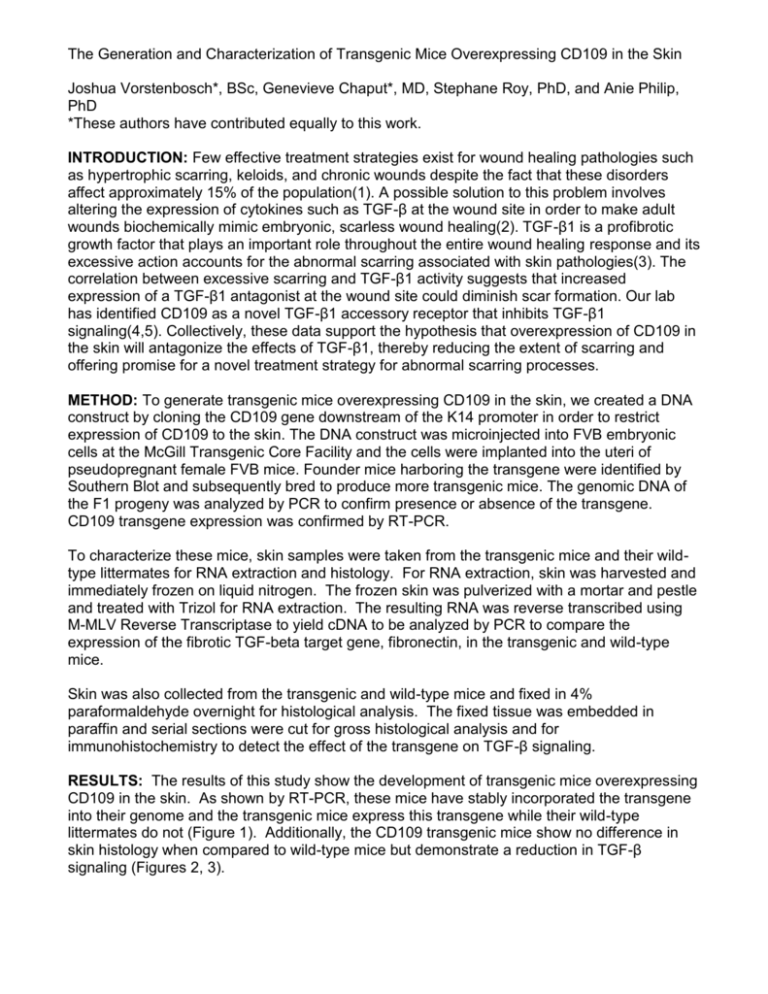
The Generation and Characterization of Transgenic Mice Overexpressing CD109 in the Skin Joshua Vorstenbosch*, BSc, Genevieve Chaput*, MD, Stephane Roy, PhD, and Anie Philip, PhD *These authors have contributed equally to this work. INTRODUCTION: Few effective treatment strategies exist for wound healing pathologies such as hypertrophic scarring, keloids, and chronic wounds despite the fact that these disorders affect approximately 15% of the population(1). A possible solution to this problem involves altering the expression of cytokines such as TGF-β at the wound site in order to make adult wounds biochemically mimic embryonic, scarless wound healing(2). TGF-β1 is a profibrotic growth factor that plays an important role throughout the entire wound healing response and its excessive action accounts for the abnormal scarring associated with skin pathologies(3). The correlation between excessive scarring and TGF-β1 activity suggests that increased expression of a TGF-β1 antagonist at the wound site could diminish scar formation. Our lab has identified CD109 as a novel TGF-β1 accessory receptor that inhibits TGF-β1 signaling(4,5). Collectively, these data support the hypothesis that overexpression of CD109 in the skin will antagonize the effects of TGF-β1, thereby reducing the extent of scarring and offering promise for a novel treatment strategy for abnormal scarring processes. METHOD: To generate transgenic mice overexpressing CD109 in the skin, we created a DNA construct by cloning the CD109 gene downstream of the K14 promoter in order to restrict expression of CD109 to the skin. The DNA construct was microinjected into FVB embryonic cells at the McGill Transgenic Core Facility and the cells were implanted into the uteri of pseudopregnant female FVB mice. Founder mice harboring the transgene were identified by Southern Blot and subsequently bred to produce more transgenic mice. The genomic DNA of the F1 progeny was analyzed by PCR to confirm presence or absence of the transgene. CD109 transgene expression was confirmed by RT-PCR. To characterize these mice, skin samples were taken from the transgenic mice and their wildtype littermates for RNA extraction and histology. For RNA extraction, skin was harvested and immediately frozen on liquid nitrogen. The frozen skin was pulverized with a mortar and pestle and treated with Trizol for RNA extraction. The resulting RNA was reverse transcribed using M-MLV Reverse Transcriptase to yield cDNA to be analyzed by PCR to compare the expression of the fibrotic TGF-beta target gene, fibronectin, in the transgenic and wild-type mice. Skin was also collected from the transgenic and wild-type mice and fixed in 4% paraformaldehyde overnight for histological analysis. The fixed tissue was embedded in paraffin and serial sections were cut for gross histological analysis and for immunohistochemistry to detect the effect of the transgene on TGF-β signaling. RESULTS: The results of this study show the development of transgenic mice overexpressing CD109 in the skin. As shown by RT-PCR, these mice have stably incorporated the transgene into their genome and the transgenic mice express this transgene while their wild-type littermates do not (Figure 1). Additionally, the CD109 transgenic mice show no difference in skin histology when compared to wild-type mice but demonstrate a reduction in TGF-β signaling (Figures 2, 3). Figure 1: Confirmation of transgene expression in skin. Figure 2: Reduction of TGF-β target gene expression in transgenic mouse skin. Figure 3: Histological assessment of the effect of CD109 transgene on skin histology and TGF-β signaling in the skin. Legend: WT=wild-type littermate mice; K14-CD109=transgenic mice; H&E=hematoxylin and eosin staining; TGF-BetaRI=TGF-β receptor I staining; TGFBetaRII=TGF-β receptor II staining. CONCLUSION: This study demonstrates the generation of transgenic mice overexpressing CD109 in the skin that reduce TGF-beta signaling and thus offers great promise for the development of novel wound healing treatment strategies. Overexpression of the transgene has been confirmed to be restricted to the transgenic mice by RT-PCR and this overexpression does not interfere with normal skin development. The overexpression of CD109 diminishes TGF-β signaling as indicated by the reduced expression of type I and type II TGF-β receptors and reduced expression of the fibrotic TGF-β target gene fibronectin. Collectively, these data illustrate that CD109 decreases the TGF-β response in the skin. As TGF-β plays a critical role in wound healing, especially in pathological scarring, CD109 has great potential as a novel therapeutic agent for modulation of the wound healing response. By making CD109 more readily available at the wound site, either by increasing its expression endogenously or via topical application, we can reduce TGF-β signaling and in turn diminish scarring in patients affected by pathologies such as keloids and hypertrophic scars. REFERENCES 1. Edriss AS, Eak J. Management of Keloid and Hypertrophic Scars. Annals of Burns and Fire Disasters 18(4):202-210, 2005. 2. Ferguson MW, O'Kane S. Scar-free healing: from embryonic mechanisms to adult therapeutic intervention. Philos Trans R Soc Lond B Biol Sci 359(1445):839-50, 2004. 3. Colwell AS, Yun R, Krummel TM, Longaker MT, Lorenz HP. Keratinocytes modulate fetal and postnatal fibroblast transforming growth factor-beta and Smad expression in co-culture. Plast Reconstr Surg 119(5):1440-5. 4. Tam BY, Germain L, Philip A. TGF-β receptor expression on human keratinocytes: a 150 kDa GPI-anchored TGF-β1 binding protein forms a heteromeric complex with type I and type II receptors. J Cell Biochem 70(4):573-86, 1998. 5. Finnson KW, Tam BY, et al. Identification of CD109 as part of the TGF-β receptor system in human keratinocytes. Faseb J 20(9):1525-7, 2006.
