Purify Protein Invitro Kinase Assay
advertisement
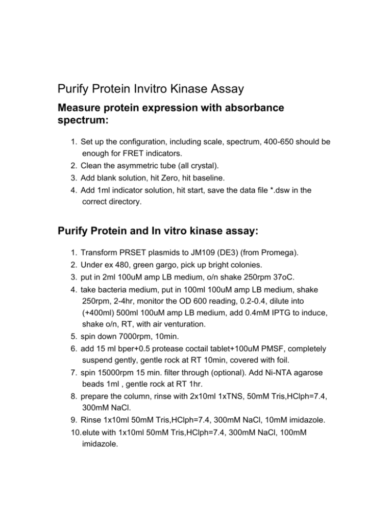
Purify Protein Invitro Kinase Assay Measure protein expression with absorbance spectrum: 1. Set up the configuration, including scale, spectrum, 400-650 should be enough for FRET indicators. 2. Clean the asymmetric tube (all crystal). 3. Add blank solution, hit Zero, hit baseline. 4. Add 1ml indicator solution, hit start, save the data file *.dsw in the correct directory. Purify Protein and In vitro kinase assay: 1. Transform PRSET plasmids to JM109 (DE3) (from Promega). 2. Under ex 480, green gargo, pick up bright colonies. 3. put in 2ml 100uM amp LB medium, o/n shake 250rpm 37oC. 4. take bacteria medium, put in 100ml 100uM amp LB medium, shake 250rpm, 2-4hr, monitor the OD 600 reading, 0.2-0.4, dilute into (+400ml) 500ml 100uM amp LB medium, add 0.4mM IPTG to induce, shake o/n, RT, with air venturation. 5. spin down 7000rpm, 10min. 6. add 15 ml bper+0.5 protease coctail tablet+100uM PMSF, completely suspend gently, gentle rock at RT 10min, covered with foil. 7. spin 15000rpm 15 min. filter through (optional). Add Ni-NTA agarose beads 1ml , gentle rock at RT 1hr. 8. prepare the column, rinse with 2x10ml 1xTNS, 50mM Tris,HClph=7.4, 300mM NaCl. 9. Rinse 1x10ml 50mM Tris,HClph=7.4, 300mM NaCl, 10mM imidazole. 10. elute with 1x10ml 50mM Tris,HClph=7.4, 300mM NaCl, 100mM imidazole. 11. dialysis the protein solution 4oC, 4hr-o/n, in 50 mM Tris pH 8, 100 mM NaCl, 10 mM MgCl2, 2mM DTT. 10x Src kinase buffer stock solution: 500 mM Tris pH 8, 1 M NaCl, 100 mM MgCl2, dilute 10x, add 1:1000 2M DTT. 12. Measure the absorbance following the above protocol. 13. YFP (extinction coefficient) EC=77000 M-1 CM-1 , CFP EC=32500 M1 CM-1, GFP EC=62000 M-1 CM-1. So Concentration=reading/EC In vitro kinase assay 1. Find the 3-way cuvette. Side line facing the emission, small aligned holes for excitation. Clean the cuvette. 2. add 100 ul of indicator solution 3. put in the holder and cover the cover. Make sure to ajust the aperture to ex=0.5-1 and em=0.5-1 4. Hit F10, select HV#950 ON and HV#440 ON. 5. Define Exp. 6. Emission Scan 7. Data acquisition paramter: Number of scan: 1 Start:470 End: 530 Incremen: 2nm Integration time: 1sec Excitation monochrome: 434 Acquisition mode: s / r 8. Correct factor file: Mcorrect.spt Measure the peak 526/476, record the number as time goes on, calculate the ratio. Add Src, Add ATP. 9. Esp, Esp, Run experiment, Go. For Absorbance: Scan, connect on line. Baseline checked Zero for the blank. Start for the samples. Save data as .csv for ascII format For Fluorometer: Other functions, output to ascII, F1 to pickup files from directory. For Platereader: Connect, move in. measurement configuration. Dynamics: 3-5 cycles without ATP. 60 cycles after ATP, 2 min interval. 30 flashes. Increasing step: 5 nm. Band width 5nm. Excitation wavelength: 420nm. Emission: 471 to 531. temperature: 30oC.
