Laboratory work 1
advertisement
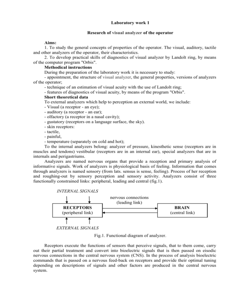
Laboratory work 1 Research of visual analyzer of the operator Aims: 1. To study the general concepts of properties of the operator. The visual, auditory, tactile and other analyzers of the operator, their characteristics. 2. To develop practical skills of diagnostics of visual analyzer by Landolt ring, by means of the computer program "Orbis". Methodical instructions During the preparation of the laboratory work it is necessary to study: - appointment, the structure of visual analyzer, the general properties, versions of analyzers of the operator; - technique of an estimation of visual acuity with the use of Landolt ring; - features of diagnostics of visual acuity, by means of the program "Orbis". Short theoretical data To external analyzers which help to perception an external world, we include: - Visual (a receptor - an eye); - auditory (a receptor - an ear); - olfactory (a receptor in a nasal cavity); - gustatory (receptors on a language surface, the sky). - skin receptors: - tactile, - painful, - temperature (separately on cold and hot); To the internal analyzers belong: analyzer of pressure, kinesthetic sense (receptors are in muscles and tendons) vestibular (receptors are in an internal ear), special analyzers that are in internals and perigastriums. Analyzers are named nervous organs that provide a reception and primary analysis of informative signals. Work of analyzers is physiological basis of feeling. Information that comes through analyzers is named sensory (from lats. sensus is sense, feeling). Process of her reception and roughing-out by sensory perception and sensory activity. Analyzers consist of three functionally constrained links: peripheral, leading and central (fig.1). INTERNAL SIGNALS nervous connections (leading link) RECEPTORS (peripheral link) BRAIN (central link) EXTERNAL SIGNALS Fig.1. Functional diagram of analyzer. Receptors execute the functions of sensors that perceive signals, that to them come, carry out their partial treatment and convert into bioelectric signals that is then passed on eisodic nervous connections in the central nervous system (CNS). In the process of analysis bioelectric commands that is passed on a nervous feed-back on receptors and provide their optimal tuning depending on descriptions of signals and other factors are produced in the central nervous system. All analyzers on the structure of the same types. They have on the periphery vehicles that perceive irritants, are receptors in that there is converting of energy of irritant into the process of excitation. From receptors on sensory(perceptible) neurons and synapses(to the contacts between: by nervous cages) impulses come in the central nervous system. The analyzer and signal kind. The latent period (Average value), ms Tactile (touch).......................................... 90...220 Acoustical (sound)................................... 110... 180 Visual (light)............................................ 150... 220 Olfactory (smell).............................. ……310... 390 Temperature (warmly, a cold).............. 280... 1600 Vestibular mechanism......................... 400 The flavoring: Salty .....................................................310 The sweet................................... 450 The sour..................................... 540 The bitter.................................... 1080 The painful.................................... 130... 890 For the description of the intensity of feeling of operator, we usually use the logarithmic dependence. The law of Вебера-Фехнера is as follows: S = с ln R where, S is intensity of feeling, c - a coefficient of proportion, R - a size of irritator. The visual analyzer The basic structure of the eye is similar to a simple camera with an aperture – iris, a lens, and a light sensitive surface – retina. Light enters the eye through the cornea, then passes through the iris and the lens and falls on the retina. Here the light stimulates the light-sensitive cells on the retina (rods and cones) and these pass small electrical impulses by way of the optic nerve to the visual cortex in the brain. Here, the electrical impulses are interpreted and an image is perceived. Fig.1. Eye structure The cornea is a clear “window” at the very front of the eye. The cornea acts as a fixed focusing device. The focusing is achieved by the shape of the cornea bending the incoming light rays. The cornea is responsible for between 70% and 80% of the total focusing ability (refrection) of the eye. The iris (the coloured part of the eye) controls the amount of light that is allowed to enter the eye. It does this by varying the size of the pupil (the dark area in the centre of the iris). The size of the pupil can be changed very rapidly to cater for changed light levels. The amount of light can be adjusted by a factor 5:1. After passing through the pupil, the light passes through the lens. Its shape is changed by the muscles (ciliary muscles) surrounding it which results in the final focusing adjustment to place a sharp image onto the retina. The change of shape of the lens is called accommodation. In order to focus clearly on a near object, the lens is thickened. To focus on a distant point, the lens is flattened. The degree of accommodation can be affected by factors such as fatigue or the ageing process. When a person is tired accommodation is reduced, resulting in less sharp vision known as visual acuity. Visual acuity is the ability of the eye to discriminate sharp detail at varying distances. Factors can affect and limit the visual acuity of the eye include: Physiological factors such as: -physical imperfections in one or both eyes; -age; -fatigue. Illumination: Visibility gets better with the increase of illumination Too much light leads to blinding; Elderly people need more light. Color sight: 8 % of men and 0.5 % (zero point five) of women are inclined to colorblindness; Distinction of green and red colors is the basic problem; Ageing leads to non recognizing of blue and yellow colors. Sight: Acceptable standard of sight is obligatory for engineers. Points or contact lenses must be used after doctor's prescription; On aviation enterprises a check up of operator’s vision should be conducted regularly; Ability to distinguish colours should be checked depending on goals that are carried out. Fig. 2. Change of sharpness of sight with age The computer program for diagnostics of visual acuity of "Orbis" The program "Orbis" is aimed to study visual acuity with the use of personal computer. The word "Orbis" means a circle or a ring. The program is fast, simple and convenient in usage. It is accessible to each owner of the personal computer, because it does not demand profound knowledge and certain skills. Unlike other similar programs "Orbis" allows to use standard devices of input-output of the information: the keyboard, the mouse, the joystick, sound and video of a card with display of manipulations surveyed, using graphic indexes and sound signals. It allows to reduce inspection time, and also to use the program practically on any personal computer. Attention! The general condition can influence the general result of research surveyed, weariness, psychological excitement, change of arterial pressure, vascular spasms. Questions 1. 2. 3. 4. 5. 6. Appointment of the program "Orbis". Name external analyzers of the operator. What latent period of the visual analyzer? What purpose of work? Name the structure of the visual analyzer. What is the blind spot?
