introduction - SurgicalCriticalCare.net
advertisement
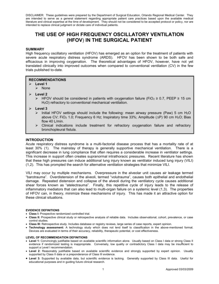
DISCLAIMER: These guidelines were prepared by the Department of Surgical Education, Orlando Regional Medical Center. They are intended to serve as a general statement regarding appropriate patient care practices based upon the available medical literature and clinical expertise at the time of development. They should not be considered to be accepted protocol or policy, nor are intended to replace clinical judgment or dictate care of individual patients. THE USE OF HIGH FREQUENCY OSCILLATORY VENTILATION (HFOV) IN THE SURGICAL PATIENT SUMMARY High frequency oscillatory ventilation (HFOV) has emerged as an option for the treatment of patients with severe acute respiratory distress syndrome (ARDS). HFOV has been shown to be both safe and efficacious in improving oxygenation. The theoretical advantages of HFOV, however, have not yet translated clinically into improved outcomes when compared to conventional ventilation (CV) in the few trials published to-date. RECOMMENDATIONS Level 1 None Level 2 HFOV should be considered in patients with oxygenation failure (FiO 2 ≥ 0.7, PEEP ≥ 15 cm H2O) refractory to conventional mechanical ventilation. Level 3 Initial HFOV settings should include the following: mean airway pressure (Paw) 5 cm H2O above CV; FiO2 1.0; Frequency 6 Hz; Inspiratory time 33%; Amplitude (P) 90 cm H2O; Bias flow 40 L/min. Clinical indications include treatment for refractory oxygenation failure and refractory bronchopleural fistula. INTRODUCTION Acute respiratory distress syndrome is a multi-factorial disease process that has a mortality rate of at least 30% (1). The mainstay of therapy is generally supportive mechanical ventilation. There is a significant decrease in lung compliance that often requires a considerable increase in ventilator settings. This increase in support often creates supranormal intrathoracic pressures. Recent literature has shown that these high pressures can induce additional lung injury known as ventilator induced lung injury (VILI) (1,2). This has prompted the search for alternative ventilation strategies that minimize VILI. VILI may occur by multiple mechanisms. Overpressure in the alveolar unit causes air leakage termed “barotrauma”. Overdistension of the alveoli, termed “volutrauma”, causes both epithelial and endothelial damage. Repeated distension and collapse of the alveoli during the ventilatory cycle causes additional shear forces known as “atelectrauma”. Finally, this repetitive cycle of injury leads to the release of inflammatory mediators that can also lead to multi-organ failure on a systemic level (1,3). The properties of HFOV can, in theory, minimize these mechanisms of injury. This has made it an attractive option for these clinical situations. EVIDENCE DEFINITIONS Class I: Prospective randomized controlled trial. Class II: Prospective clinical study or retrospective analysis of reliable data. Includes observational, cohort, prevalence, or case control studies. Class III: Retrospective study. Includes database or registry reviews, large series of case reports, expert opinion. Technology assessment: A technology study which does not lend itself to classification in the above-mentioned format. Devices are evaluated in terms of their accuracy, reliability, therapeutic potential, or cost effectiveness. LEVEL OF RECOMMENDATION DEFINITIONS Level 1: Convincingly justifiable based on available scientific information alone. Usually based on Class I data or strong Class II evidence if randomized testing is inappropriate. Conversely, low quality or contradictory Class I data may be insufficient to support a Level I recommendation. Level 2: Reasonably justifiable based on available scientific evidence and strongly supported by expert opinion. Usually supported by Class II data or a preponderance of Class III evidence. Level 3: Supported by available data, but scientific evidence is lacking. Generally supported by Class III data. Useful for educational purposes and in guiding future clinical research. 1 Approved 03/03/2009 HFOV works by maintaining lung inflation at a constant elevated mean airway pressure (Paw) while using a piston to cycle the ventilation rate at several hundred times per minute. This results in a tidal volume that is often smaller than the anatomical dead space of the lungs. Recent studies, the most prominent being by the ARDS Network group, have shown that lower tidal volumes help to reduce volutrauma injury to the lungs (4). The smaller tidal volumes of HFOV fit well with this concept. HFOV also functions to minimize the cycle of alveolar distension and collapse by maintaining airway pressures throughout ventilation. The rapid pressure changes created by the oscillating piston are attenuated at the alveolar level thereby minimizing atelectrauma and improving alveolar recruitment (3). This effect is also improved by the use of periodic recruitment maneuvers while on HFOV. All of these attributes of HFOV would seem to make it an attractive therapy for severe ARDS patients. Unfortunately, the theoretical advantages of HFOV have not yet been proven in the clinical setting. Current literature shows that HFOV is safe and improves oxygenation in ARDS patients. Current controlled trials show promising trends, but have yet to demonstrate improved outcomes with the use of this therapy. LITERATURE REVIEW In 2000, the ARDS Network published a comparison between low tidal volume (6 mL/kg) (“lung protective”) and traditional tidal volume (12 mL/kg) ventilation. The result was a significant decrease in mortality in the low tidal volume group. While the optimal tidal volume strategy has not been determined, it is evident that there is certain pathology caused by over distention and collapse of alveoli during the respiratory cycle. This disruption of the functional pulmonary unit results in both local and systemic inflammation further exacerbating hypoxemia and multi-organ failure (4). In addition, studies by Ranieri et al. showed a decrease in white blood cells and select inflammatory mediators in blood and alveolar lavage samples in patients ventilated using lung protective strategies (5). The largest of the two randomized controlled trials using HFOV in the adult population was published by Derdak et al. in 2002 (6). The purpose of this study was to evaluate the safety and efficacy of HFOV. One hundred forty-eight adult patients with ARDS were randomized to HFOV versus conventional ventilation (CV). The HFOV group was exposed to significantly higher Paw and showed a significant improvement in PaO2/FiO2 ratio over the first 16 hours. This improvement, however, did not persist past 24 hours. Thirty day mortality was 37% in the HFOV patients and 52% in CV patients. This difference only trended towards significance (6,7). Of note, there was also a significant increase in pulmonary artery occlusion pressure (PAOP) and central venous pressure (CVP) measurements in the HFOV patients. There was no significant difference in complications during therapy. There was also an insignificant difference in total ventilator days (6). This paper concluded that HFOV is safe and efficacious in the adult population; however, the study was not powered to show a significant decrease in mortality. The only other randomized controlled trial was stopped prematurely secondary to enrollment issues. No major conclusions were reached in this limited trial of 61 patients (8). Mehta and colleagues reported a retrospective review of 156 patients in which HFOV was used as rescue therapy in patients with ARDS. These patients had received CV for 5.6 ± 7.6 days prior to HFOV. Most patients showed significant improvement in both PaO 2/FiO2 and oxygenation index. These effects persisted for greater than 72 hours. Twelve percent of these patients, however, failed HFOV secondary to oxygenation, ventilation, or hemodynamic difficulties. Of patients with pulmonary artery catheters, an increase in CVP and a decrease in cardiac output (CO) to low normal values were shown. PAOP showed a transient decline that lasted 6 hours. Thirty day mortality was 62% and the pneumothorax rate was 22% in these patients. In rescue efforts, HFOV has been shown to improve oxygenation with minor alteration in pulmonary artery catheter values. One of the independent risk factors for death identified was days on conventional ventilation prior to HFOV. This indicates a possible advantage to the earlier institution of HFOV therapy. The authors also concluded that HFOV is a safe and efficacious modality (9). The alveolar recruitment accomplished by HFOV can be further augmented and maintained through the use of recruitment maneuvers. Ferguson et al. reported a pilot study of 25 patients transitioned to HFOV from CV. Recruitment maneuvers included pausing oscillations while FiO 2 was increased to 1.0 and Paw 2 Approved 03/03/2009 was increased to 5 cmH20 above baseline. This combination showed a significant decrease in FiO 2 requirements within 12 hours of transition. No increase in complication rate was observed, however, overall mortality was 44% in the group. As with all of the studies involved, death was typically attributed to multi-organ failure (10). There are several case reports describing the use of HFOV in patients with high output or refractory bronchopleural fistula (BPF). Ha and Johnson report a case of a 55 year old man developing a BPF after decortication for empyema (12). In this scenario, the BPF worsened significantly with the development of ARDS. The patient recovered after a 28 day course of HFOV therapy. Crimi et al describe treatment of six patients with unilateral lung injury treated with independent lung ventilation and HFOV therapy. Three of the six patients treated had concomitant BPF (13). HFOV is recommended as a second line of therapy for BPF. REFERENCES 1. Imai Y, Slutsky AS. High-frequency oscillatory ventilation and ventilator-induced lung injury. Crit Care Med 2005; 33(3):s129-s134. 2. Downar J, Mehta S. Bench-to-bedside review: High-frequency oscillatory ventilation in adults with acute respiratory distress syndrome. Crit Care 2006; 10(6):240. 3. Fessler HE, Hess DR. Does high–frequency ventilation offer benefits over conventional ventilation in adult patients with acute respiratory distress syndrome? Resp Care 2007; 52(5):595-608. 4. The Acute Respiratory Distress Network: Ventilation with lower tidal volumes as compared with traditional tidal volumes in acute lung injury and acute respiratory distress syndrome. New Engl J Med 2000; 342:1301-1308. 5. Ranieri VM, Suter PM, Tortorella C, et al. Effect of mechanical ventilation on inflammatory mediators in patients with acute respiratory distress syndrome: A randomized controlled trial. JAMA 1999; 281(1):54-61. 6. Derdak S, Mehta S, Stewart TE, et al. High-frequency oscillatory ventilation for acute respiratory distress syndrome in adults: a randomized, controlled trial. Am J Respir Crit Care Med 2002; 166:801-808. 7. Wunsch H, Mapstone J. High-frequency ventilation versus conventional ventilation for the treatment of acute lung injury and acute respiratory distress syndrome: a systematic review and Cochrane analysis. Anesth Analg 2005; 100:1765-1772. 8. Bollen CW, van Well GT, Sherry T, et al. High frequency oscillatory ventilation compared with conventional mechanical ventilation in adult respiratory distress syndrome: a randomized controlled trial. Crit Care 2005; 9:430-439. 9. Mehta S, Granton J, MacDonald RJ, et al. High–frequency oscillatory ventilation in adults: the Toronto experience. Chest 2004; 126:518-527. 10. Ferguson ND, Chiche JD, Kacmarek RM, et al. Combining high-frequency oscillatory ventilation and recruitment maneuvers in adults with early acute respiratory distress syndrome: The Treatment with Oscillation and an Open Lung Strategy (TOOLS) Trial pilot study. Crit Care Med 2005; 33(3):479486. 11. Fessler HE, Derdak S, Ferguson ND, et al. A protocol for high-frequency oscillatory ventilation in adults: Results from a roundtable discussion. Crit Care Med 2007; 35(7):1649-1654. 12. Ha DF, Johnson D. High frequency oscillatory ventilation in the management of a high output bronchopleural fistula: a case report. Can J Anesth 2004; 51(1):78-83. 13. Crimi G, Candiani A, Conti G, et al. Clinical applications of independent lung ventilation with unilateral high-frequency jet ventilation(ILV-UHFJV.). Intensive Care Medicine 1986;12(2):90-4,1986. 3 Approved 03/03/2009 14. 4 Approved 03/03/2009
