DISSECTING DISEASE-SPECIFIC DIFFERENCES IN RA AND OA
advertisement
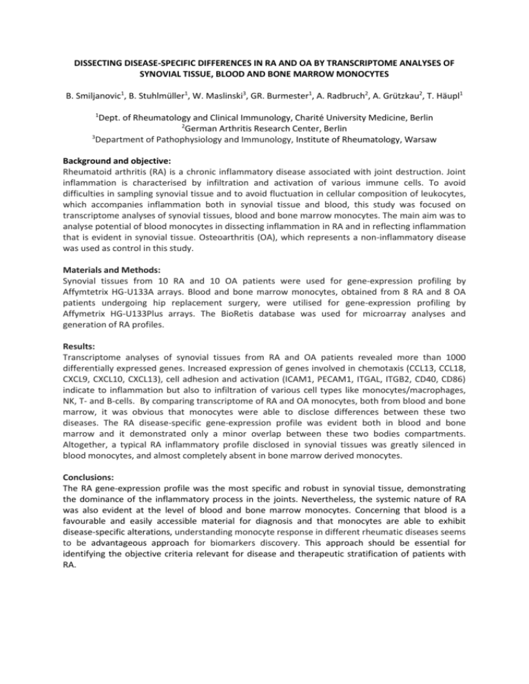
DISSECTING DISEASE-SPECIFIC DIFFERENCES IN RA AND OA BY TRANSCRIPTOME ANALYSES OF SYNOVIAL TISSUE, BLOOD AND BONE MARROW MONOCYTES B. Smiljanovic1, B. Stuhlmüller1, W. Maslinski3, GR. Burmester1, A. Radbruch2, A. Grützkau2, T. Häupl1 1 Dept. of Rheumatology and Clinical Immunology, Charité University Medicine, Berlin 2 German Arthritis Research Center, Berlin 3 Department of Pathophysiology and Immunology, Institute of Rheumatology, Warsaw Background and objective: Rheumatoid arthritis (RA) is a chronic inflammatory disease associated with joint destruction. Joint inflammation is characterised by infiltration and activation of various immune cells. To avoid difficulties in sampling synovial tissue and to avoid fluctuation in cellular composition of leukocytes, which accompanies inflammation both in synovial tissue and blood, this study was focused on transcriptome analyses of synovial tissues, blood and bone marrow monocytes. The main aim was to analyse potential of blood monocytes in dissecting inflammation in RA and in reflecting inflammation that is evident in synovial tissue. Osteoarthritis (OA), which represents a non-inflammatory disease was used as control in this study. Materials and Methods: Synovial tissues from 10 RA and 10 OA patients were used for gene-expression profiling by Affymtetrix HG-U133A arrays. Blood and bone marrow monocytes, obtained from 8 RA and 8 OA patients undergoing hip replacement surgery, were utilised for gene-expression profiling by Affymetrix HG-U133Plus arrays. The BioRetis database was used for microarray analyses and generation of RA profiles. Results: Transcriptome analyses of synovial tissues from RA and OA patients revealed more than 1000 differentially expressed genes. Increased expression of genes involved in chemotaxis (CCL13, CCL18, CXCL9, CXCL10, CXCL13), cell adhesion and activation (ICAM1, PECAM1, ITGAL, ITGB2, CD40, CD86) indicate to inflammation but also to infiltration of various cell types like monocytes/macrophages, NK, T- and B-cells. By comparing transcriptome of RA and OA monocytes, both from blood and bone marrow, it was obvious that monocytes were able to disclose differences between these two diseases. The RA disease-specific gene-expression profile was evident both in blood and bone marrow and it demonstrated only a minor overlap between these two bodies compartments. Altogether, a typical RA inflammatory profile disclosed in synovial tissues was greatly silenced in blood monocytes, and almost completely absent in bone marrow derived monocytes. Conclusions: The RA gene-expression profile was the most specific and robust in synovial tissue, demonstrating the dominance of the inflammatory process in the joints. Nevertheless, the systemic nature of RA was also evident at the level of blood and bone marrow monocytes. Concerning that blood is a favourable and easily accessible material for diagnosis and that monocytes are able to exhibit disease-specific alterations, understanding monocyte response in different rheumatic diseases seems to be advantageous approach for biomarkers discovery. This approach should be essential for identifying the objective criteria relevant for disease and therapeutic stratification of patients with RA.
