Immunoprecipitation
advertisement
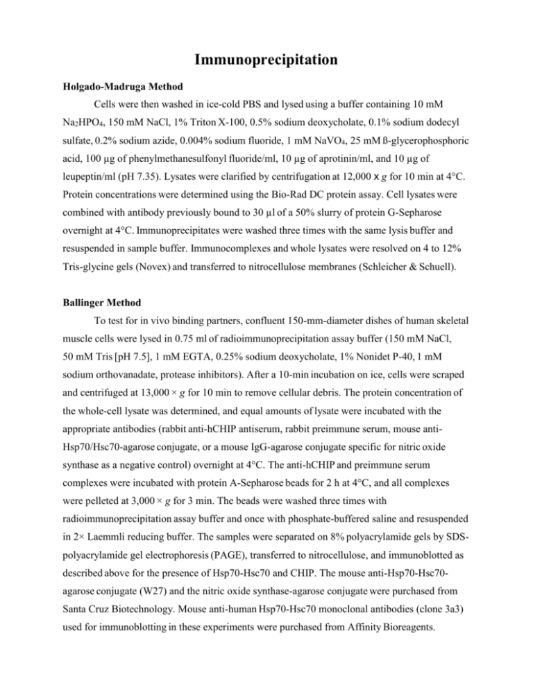
Immunoprecipitation Holgado-Madruga Method Cells were then washed in ice-cold PBS and lysed using a buffer containing 10 mM Na2HPO4, 150 mM NaCl, 1% Triton X-100, 0.5% sodium deoxycholate, 0.1% sodium dodecyl sulfate, 0.2% sodium azide, 0.004% sodium fluoride, 1 mM NaVO4, 25 mM ß-glycerophosphoric acid, 100 µg of phenylmethanesulfonyl fluoride/ml, 10 µg of aprotinin/ml, and 10 µg of leupeptin/ml (pH 7.35). Lysates were clarified by centrifugation at 12,000 x g for 10 min at 4°C. Protein concentrations were determined using the Bio-Rad DC protein assay. Cell lysates were combined with antibody previously bound to 30 µl of a 50% slurry of protein G-Sepharose overnight at 4°C. Immunoprecipitates were washed three times with the same lysis buffer and resuspended in sample buffer. Immunocomplexes and whole lysates were resolved on 4 to 12% Tris-glycine gels (Novex) and transferred to nitrocellulose membranes (Schleicher & Schuell). Ballinger Method To test for in vivo binding partners, confluent 150-mm-diameter dishes of human skeletal muscle cells were lysed in 0.75 ml of radioimmunoprecipitation assay buffer (150 mM NaCl, 50 mM Tris [pH 7.5], 1 mM EGTA, 0.25% sodium deoxycholate, 1% Nonidet P-40, 1 mM sodium orthovanadate, protease inhibitors). After a 10-min incubation on ice, cells were scraped and centrifuged at 13,000 × g for 10 min to remove cellular debris. The protein concentration of the whole-cell lysate was determined, and equal amounts of lysate were incubated with the appropriate antibodies (rabbit anti-hCHIP antiserum, rabbit preimmune serum, mouse antiHsp70/Hsc70-agarose conjugate, or a mouse IgG-agarose conjugate specific for nitric oxide synthase as a negative control) overnight at 4°C. The anti-hCHIP and preimmune serum complexes were incubated with protein A-Sepharose beads for 2 h at 4°C, and all complexes were pelleted at 3,000 × g for 3 min. The beads were washed three times with radioimmunoprecipitation assay buffer and once with phosphate-buffered saline and resuspended in 2× Laemmli reducing buffer. The samples were separated on 8% polyacrylamide gels by SDSpolyacrylamide gel electrophoresis (PAGE), transferred to nitrocellulose, and immunoblotted as described above for the presence of Hsp70-Hsc70 and CHIP. The mouse anti-Hsp70-Hsc70agarose conjugate (W27) and the nitric oxide synthase-agarose conjugate were purchased from Santa Cruz Biotechnology. Mouse anti-human Hsp70-Hsc70 monoclonal antibodies (clone 3a3) used for immunoblotting in these experiments were purchased from Affinity Bioreagents.

