Caution
advertisement
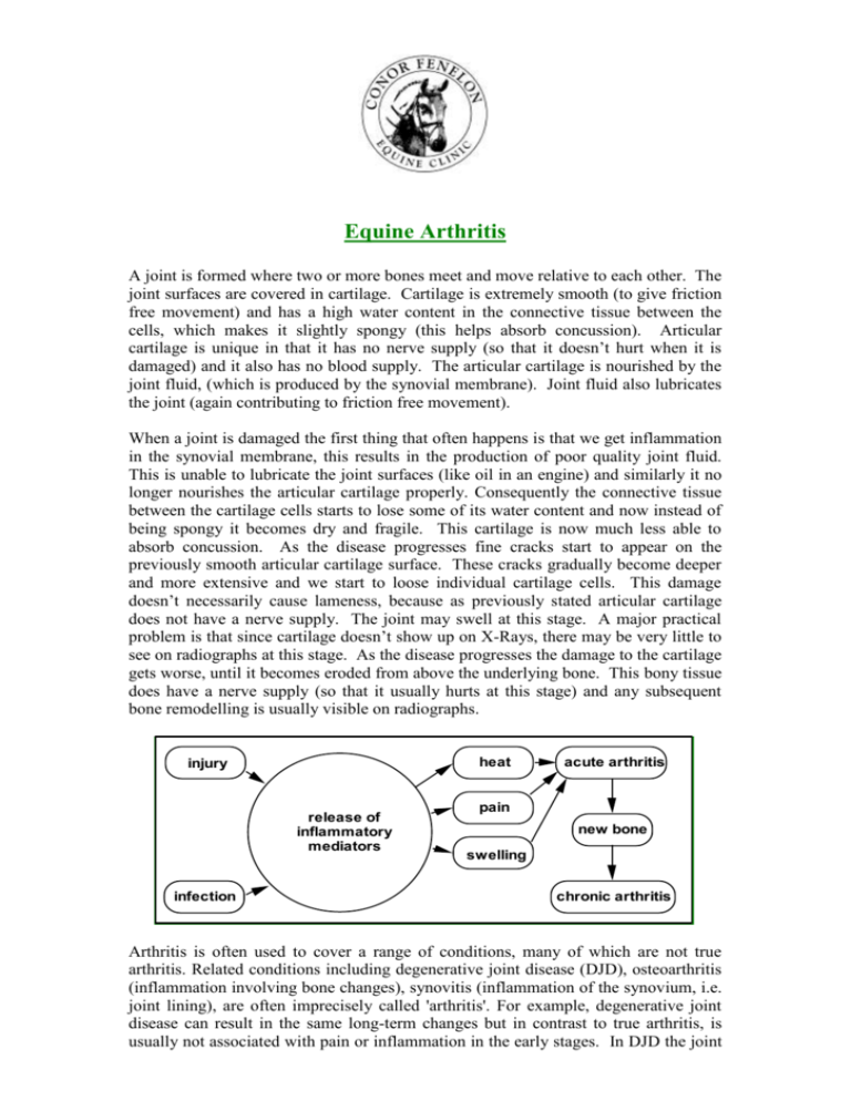
Equine Arthritis A joint is formed where two or more bones meet and move relative to each other. The joint surfaces are covered in cartilage. Cartilage is extremely smooth (to give friction free movement) and has a high water content in the connective tissue between the cells, which makes it slightly spongy (this helps absorb concussion). Articular cartilage is unique in that it has no nerve supply (so that it doesn’t hurt when it is damaged) and it also has no blood supply. The articular cartilage is nourished by the joint fluid, (which is produced by the synovial membrane). Joint fluid also lubricates the joint (again contributing to friction free movement). When a joint is damaged the first thing that often happens is that we get inflammation in the synovial membrane, this results in the production of poor quality joint fluid. This is unable to lubricate the joint surfaces (like oil in an engine) and similarly it no longer nourishes the articular cartilage properly. Consequently the connective tissue between the cartilage cells starts to lose some of its water content and now instead of being spongy it becomes dry and fragile. This cartilage is now much less able to absorb concussion. As the disease progresses fine cracks start to appear on the previously smooth articular cartilage surface. These cracks gradually become deeper and more extensive and we start to loose individual cartilage cells. This damage doesn’t necessarily cause lameness, because as previously stated articular cartilage does not have a nerve supply. The joint may swell at this stage. A major practical problem is that since cartilage doesn’t show up on X-Rays, there may be very little to see on radiographs at this stage. As the disease progresses the damage to the cartilage gets worse, until it becomes eroded from above the underlying bone. This bony tissue does have a nerve supply (so that it usually hurts at this stage) and any subsequent bone remodelling is usually visible on radiographs. heat injury release of inflammatory mediators infection acute arthritis pain new bone swelling chronic arthritis Arthritis is often used to cover a range of conditions, many of which are not true arthritis. Related conditions including degenerative joint disease (DJD), osteoarthritis (inflammation involving bone changes), synovitis (inflammation of the synovium, i.e. joint lining), are often imprecisely called 'arthritis'. For example, degenerative joint disease can result in the same long-term changes but in contrast to true arthritis, is usually not associated with pain or inflammation in the early stages. In DJD the joint structures respond to wear and tear by gradually changing shape and elasticity. Many horses with DJD move soundly. In others, these changes progress to a stage where the horse goes lame. With DJD and chronic arthritis, joint mobility is reduced but there may not be pain on flexion of the joint. There may be lameness which has gradually worsened over time or lameness which improves with exercise, i.e. the horse 'warms up'. In cases where there is multiple joint involvement, the horse may appear generally stiff at one or all gaits. In an older horse, the main sign of abnormality may be difficulty in standing up after a period of lying down. The diagnosis of arthritis is not always straightforward. In most of the cases, it is not possible to confirm the diagnosis or its significance by clinical examination alone. Further examinations may involve any or all of the following: • • • • • • Nerve block examinations, where local anaesthetic is injected around specific nerves to abolish pain from the parts of the leg that these nerves supply, confirming the site of the pain. Radiographic examinations (x-ray) to rule out fractures and to look at the bones at the joint surfaces for signs of injury, degeneration or abnormality. Radiographs are not able to demonstrate cartilage damage so are more helpful for chronic arthritis and DJD cases, where the new bone formation can be demonstrated. Ultrasound, to image the soft tissues, (cartilage, joint capsules, ligaments etc). Joint fluid collection for laboratory analysis to look specifically for signs of infection. Nuclear scintigraphy (bone scan) may be helpful in complex cases to image a specific area of increased bone metabolism. This requires the injection of a radioactive bone tracer, with a short half-life, and imaging with a gamma camera. MRI. (Magnetic Resonance Imaging). The treatment of arthritis Unfortunately there is no permanent cure for arthritis. Once a diagnosis of joint pain has been reached, treatment should serve to reduce the pain and to arrest or to slow the progression of the lesions. Individual horses sometimes do not respond as expected and very occasionally there may be complications (e.g. laminitis or joint flare /infection) associated with the individual treatments. We would often look at the way the horse is shod to see if we could make specific recommendations to improve the hoof balance or the way the horse moves in an effort to reduce the severity of the lameness. Intra-articular medication with corticosteroids (e.g. Adcortyl /Depomedrone), Hyaluronan (e.g. Hylartil, /Hyonate) and antibiotics (e.g. Amikin). Hyaluronan contains glucosamine and is a natural component of both articular cartilage and joint fluid. Hyonate can also be given intravenously. This combination of drugs is used in an attempt to reduce the inflammation, to lubricate the joint, to protect and to possibly promote regeneration of the cartilage. It appears that the combination of corticosteroid and the hyaluronan act synergistically (i.e. they seem to potentiate the effects of each other). This is most often our treatment of choice. Systemic medication with PSGAGS (e.g. Adequan). Adequan contains chondroitin sulphate, which is a structural protein in cartilage. This is believed to inhibit the enzymes which degrade the cartilage. Systemic medication with Pentosan sodium (Cartrophen). This lubricates the joint, inhibits the enzymes which degrade cartilage, promotes repair of cartilage and increases the blood flow to sub chondral bone. Although this product is currently unlicensed for use in horses in Europe it is widely used. IRAP, (Interleukin Receptor Antagonist Protein), collected from your horses own blood and injected into joints, in an attempt to affect and down regulate the degredative inflammatory process. Oral nutricuticals, (e.g. Chondroitin sulphate and pure glucosamine HCL). These are both structural proteins in the connective tissue in cartilage and have been shown to inhibit the enzymes which degrade it. It appears that they are most effective when combined with each other (e.g. as in Nutraquin or Synequin). Unfortunately it is much more expensive than pure glucosamine alone. You should always buy drugs from ‘reputable’ sources, as a survey in the USA found the almost half the nutricuticals used in humans did not contain the labelled amounts of glucosamine or chondroitin despite a label displaying a ‘guaranteed analysis’. NSAIDS (e.g. bute). These are cheap and reduce inflammation and pain. Unfortunately they have no beneficial effect on the cartilage and have side effects (e.g. intestinal ulcers, liver and kidney damage, -especially in elderly patients). We view them as an admission of defeat in that they are making no attempt to repair the damaged cartilage. When we use NSAIDS we currently prefer Danilon as we believe that it is more palatable and is possibly associated with slightly less side effects. Caution We find that it is often difficult to correlate the degree of lameness demonstrated in a clinical examination with the radiographic findings. It is important to realise that a horse can have arthritic changes which are visible on radiographs but may still move soundly and able to compete successfully. Also, it is possible for a horse to suffer from arthritis, involving the cartilage and joint fluid which will not produce demonstrable radiographic changes. Both scenarios can produce difficulties when a horse is being examined for purchase, i.e. ‘vetted’. If radiographs are taken as a requested routine part of a pre-purchase examination, the images must be carefully interpreted with reference to the findings of the clinical examination. The Acorns Equine Clinic, Pleshey, Chelmsford, Essex. CM3 1HU Telephone (01245) 231152, Fax: (01245) 231601 www.essexhorsevets.co.uk

