SupplementaryInfo2
advertisement
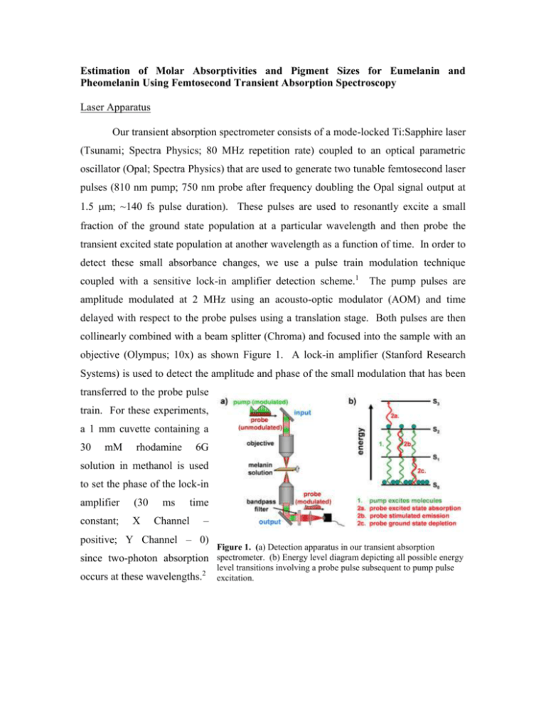
Estimation of Molar Absorptivities and Pigment Sizes for Eumelanin and Pheomelanin Using Femtosecond Transient Absorption Spectroscopy Laser Apparatus Our transient absorption spectrometer consists of a mode-locked Ti:Sapphire laser (Tsunami; Spectra Physics; 80 MHz repetition rate) coupled to an optical parametric oscillator (Opal; Spectra Physics) that are used to generate two tunable femtosecond laser pulses (810 nm pump; 750 nm probe after frequency doubling the Opal signal output at 1.5 m; ~140 fs pulse duration). These pulses are used to resonantly excite a small fraction of the ground state population at a particular wavelength and then probe the transient excited state population at another wavelength as a function of time. In order to detect these small absorbance changes, we use a pulse train modulation technique coupled with a sensitive lock-in amplifier detection scheme.1 The pump pulses are amplitude modulated at 2 MHz using an acousto-optic modulator (AOM) and time delayed with respect to the probe pulses using a translation stage. Both pulses are then collinearly combined with a beam splitter (Chroma) and focused into the sample with an objective (Olympus; 10x) as shown Figure 1. A lock-in amplifier (Stanford Research Systems) is used to detect the amplitude and phase of the small modulation that has been transferred to the probe pulse train. For these experiments, a 1 mm cuvette containing a 30 mM rhodamine 6G solution in methanol is used to set the phase of the lock-in amplifier (30 constant; X ms Channel time – positive; Y Channel – 0) Figure 1. (a) Detection apparatus in our transient absorption since two-photon absorption spectrometer. (b) Energy level diagram depicting all possible energy occurs at these wavelengths.2 level transitions involving a probe pulse subsequent to pump pulse excitation. Sepia Eumelanin Sample Preparation Since Sepia eumelanin (Sigma Aldrich Co.) is sparingly soluble in aqueous solutions, we used filtration methods to remove large undissolved particles in order to achieve viable clear samples for spectroscopic measurements. Specifically, we mixed 32 mg of Sepia in 10 mL of pH = 7 phosphate buffer solution (50 mM) and sonicated it for 1 hour to break apart larger particles. Since most of the particles still did not dissolve, we filtered the solution using a 1 m syringe filter (Whatman). The filter was held under vacuum for 18 hours to remove solvent in order to obtain the residual undissolved mass and therefore a final mass concentration. The resulting eumelanin solution had a final mass concentration of 0.84 mg/mL and remained stable for days to weeks without the appearance of undissolved particles. The background subtracted UV-Vis absorption spectrum (Figure 3 of paper) was taken using a 2 mm Hellma cuvette (QS – synthetic quartz). Synthetic Pheomelanin Sample Preparation All reagents were purchased from Sigma Aldrich Co. and used without further purification. L-dopa (0.1924 g; ~1 mmol), cysteine (0.1805 g; ~1.5 mmol) and mushroom tyrosinase (9.2 mg; 5370 units/mg) were allowed to react in a 50 mM sodium phosphate buffer solution (pH = 7) under oxygen for 4 hours. The resulting solution was acidified (pH = 3) and cooled to 4oC for 1 hour giving rise to a red/brown powder that was extracted using ultracentrifugation. We obtained 110 mg (50% yield) of synthetic pheomelanin. 20.5 mg was dissolved and diluted in a pH = 7 phosphate buffer solution (50 mM) to a final mass concentration of 0.84 mg/mL. The background subtracted UVVis absorption spectrum (Figure 3 of paper) was taken using a 0.01 mm cuvette (QS – synthetic quartz). References 1 2 D. Fu, T. E. Matthews, I. R. Piletic, and W. S. Warren, Journal of Biomedical Optics 13 (4), 0405031 (2008); D. Fu, T. Ye, T. E. Matthews, J. Grichnik, L. Hong, J. D. Simon, and W. S. Warren, Journal of Biomedical Optics 13, 054036 (2008). D. Fu, T. Ye, T. E. Matthews, G. Yurtsever, and W. S. Warren, Journal of Biomedical Optics 12 (5) (2007).


